Hand, Foot, and Mouth Disease
▶The most distinctive enteroviral exanthem.
▶Typical hand, foot, and mouth disease (HFMD) most often caused by coxsackievirus A16 but may be caused by coxsackieviruses A5, A7, A9, A10, B1, B2, B3, and B5 and enterovirus 71; more recently described atypical HFMD caused primarily by coxsackievirus A6.
▶Most commonly occurs in the summer or fall.
▶Classically seen in children younger than 5 years.
▶Incubation period is 3 to 6 days.
▶Highly contagious; may occur in epidemics.
Herpangina
▶A characteristic enanthem that clinically overlaps with the enanthem of HFMD (without features of the exanthem).
▶Most often caused by coxsackieviruses from groups A and B.
▶Most commonly seen in children between 3 and 10 years of age.
Eruptive Pseudoangiomatosis
▶An uncommon exanthem characterized by the sudden appearance of several small, angioma-like lesions in the setting of a viral prodrome or illness.
▶Most often seen in infants and children, although reported in adults (often immunocompromised) as well.
▶Associated with echovirus subtypes 25 and 32, although other viral etiologies such as cytomegalovirus have been suggested.
HFMD
▶Brief prodrome of fever, malaise may occur.
▶Cough, diarrhea noted infrequently.
▶Cervical and submandibular lymphadenopathy occasionally observed.
▶An enanthem precedes the characteristic exanthem.
▶HFMD enanthem
■Vesicles that erode to form ulcers on a red base; size ranges between 4 and 8 mm (Figure 16.1).
■Most common on buccal mucosae and tongue.
■May also involve palate, uvula, gingivae, and tonsillar pillars.
■Lesions are often quite painful, sometimes severe enough to lead to anorexia, dehydration.
▶HFMD exanthem
■Deep-seated vesicopustules with grayish white color, 3 to 7 mm in size; often the vesicles are oval (Figures 16.2 and 16.3).
■Vesicles often have surrounding erythema.
■Typically, lesions are limited to the palms and soles but also may involve lateral surfaces of hands and feet; involvement of buttocks, elbows, knees, and perineum may also be seen in younger children.
▶Atypical HFMD exanthem
■In patients with atypical HFMD, vesicles are often larger and more numerous. The lesions may enlarge into bullae, become hemorrhagic, or present as erosions (Figure 16.4).
■Distribution of lesions in atypical HFMD may be generalized but often with accentuation around the mouth (Figure 16.5), in the anogenital region, and on the extensor aspects of the extremities.
■Lesions of atypical HFMD may have a predilection for areas of eczematous dermatitis, a presentation termed eczema coxsackium, as well as areas of prior skin injury, such as a sunburn (Figure 16.6).
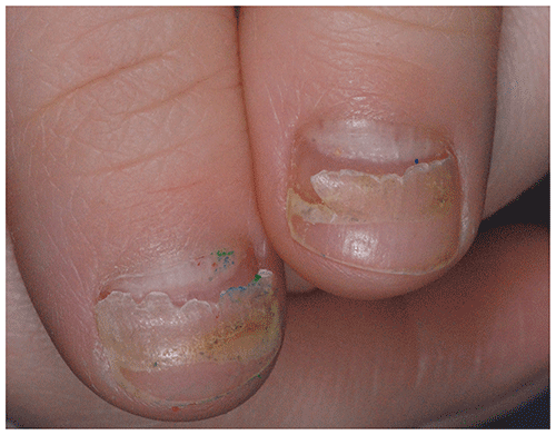
▶Temporary Beau lines (ie, transverse grooves in the nail plate) or nail shedding (onychomadesis) (Figure 16.7) may occur a few weeks to a few months after HFMD, presumably because of nail matrix arrest; these secondary changes are common after atypical HFMD.
Figure 16.1. Ulcers May Occur on the Tongue or Buccal Mucosae in Hand, Foot, and Mouth Disease.
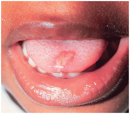
Figure 16.2. Oval Vesicles with Surrounding Erythema on the Hand of a Child Who Has Hand, Foot, and Mouth Disease.
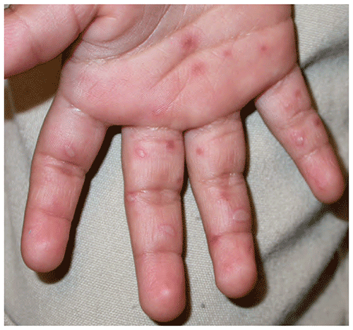
Figure 16.3. Hand, Foot, and Mouth Disease. Oval Vesicles with Mild Surrounding Erythema.
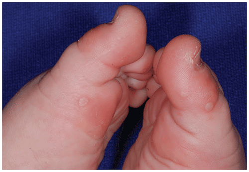
Figure 16.4. Ruptured Bullae and Large Erosions in a Young Girl with Atypical Hand, Foot, and Mouth Disease.
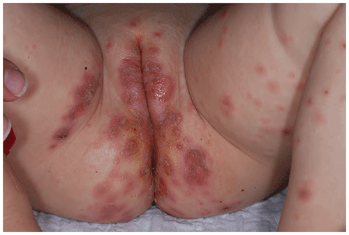
Figure 16.5. Perioral Vesicles and Erosions in a Toddler with Atypical Hand, Foot, and Mouth Disease.
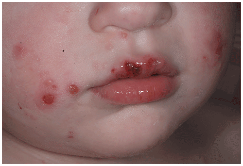
Figure 16.6. Eczema Coxsackium. Crusted Erosions and Flattened Vesicles Overlying Lichenified Plaques of Eczema on the Left Arm.
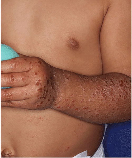
Figure 16.7. Onychomadesis (Nail Shedding) after Hand, Foot, and Mouth Disease in an Otherwise Healthy 4-Year-Old.
Herpangina
▶Enanthem with painful tiny vesicles and punched-out erosions.
▶Distributed on soft palate, uvula, tonsillar pillars, and posterior pharynx.
▶Erosions typically have a rim of erythema and a yellowish gray coating.
▶Fever and sore throat are common; 25% may have abdominal pain, vomiting.
▶Erosions persist for approximately 7 days.
Eruptive Pseudoangiomatosis
▶Acute onset of multiple, small (2–4 mm), bright red (hemangioma-like) papules with a rim of blanching.
▶Lesions blanch with pressure.
▶Preceding or concurrent fever, headache, upper respiratory symptoms may be present.
▶Lesions resolve spontaneously over 1 to 2 weeks without treatment.
▶Diagnosis of HFMD, herpangina, and eruptive pseudoangiomatosis is usually made clinically.
▶Although rarely necessary, a specific diagnosis of enteroviral infections may be made with viral culture, serological testing, or polymerase chain reaction–based testing of lesional swabs, nasopharyngeal swabs, blood, stool, or urine.
▶Viral polymerase chain reaction testing or direct fluorescent examination for herpes simplex virus is relatively rapid and may be clinically helpful to distinguish these infections from atypical HFMD, eczema coxsackium, or the oral erosions of herpangina.
▶Skin biopsy may be necessary to distinguish eruptive pseudoangiomatosis from other vascular lesions if the process is not resolving spontaneously as expected.
▶Generally, simple supportive measures (ie, oral fluids, analgesics, and antipyretics) are adequate for HFMD and herpangina.
▶Severe pain may require more aggressive pain management; hospitalization for intravenous hydration and narcotic analgesics occasionally is required.
▶Eruptive pseudoangiomatosis usually requires no therapy.
Look-alikes
| Disorder | Differentiating Features |
|---|
| Typical Hand, Foot, and Mouth Disease |
|---|
| The typical appearance and distribution of lesions usually prevents confusion with other disorders. |
| Atypical Hand, Foot, and Mouth Disease |
|---|
| Eczema herpeticum | Uniform, punched-out heme-crusted erosions in areas of atopic dermatitis predominate. Viral test result positive for herpes simplex virus.
|
| Varicella | Less common in era of universal vaccination. Crops of lesions in varying stages (papules, vesicles, crusts) are seen.
|
| Bullous impetigo | Flaccid blisters and superficial erosions with peripheral collarette of blister roof. Honey-colored crusts may or may not be present. Predominance of lesions around the nose, hands, diaper area. Bacterial culture positive for Staphylococcus aureus.
|
| Allergic contact dermatitis | Localized erythema, papules, and vesicles in a geometric pattern consistent with an external cause. Itch (often severe) very common.
|
| Autoimmune blistering disorders | |
| Herpangina |
|---|
| Herpes gingivostomatitis | |
| Aphthous ulcer (canker sores) | Usually a chronic or recurring problem. Fever and other symptoms of herpangina typically lacking. In severe disease (recurrent major aphthous ulcer), ulcers larger than 1 cm may develop.
|
| Eruptive Pseudoangiomatosis |
|---|
| Infantile hemangioma | Appears in the first weeks to month after birth and gradually involutes over several years. Typically solitary, although a diffuse pattern of numerous small lesions can occur.
|
| Pyogenic granuloma | |
| Bacillary angiomatosis | |
▶The prognosis for HFMD, herpangina, and eruptive pseudoangiomatosis is excellent; all typically resolve without sequelae.
▶Typical nail regrowth is the norm after enteroviral onychomadesis.
▶If the diagnosis is in question or lesions are persistent or recurrent.
▶Consider hospitalization if fluid intake is inadequate or dehydration is suspected or when pain control at home is inadequate.
▶Because enteroviruses are a major cause of meningitis in summer and fall, neck stiffness, lethargy, or severe irritability should prompt a thorough evaluation.