▶Also known as acute febrile mucocutaneous lymph node syndrome.
▶Acute multisystem vasculitis of children, usually younger than 5 years; involves small and medium muscular arteries.
▶Most common cause of acquired heart disease in children in high-income countries.
▶Etiology remains unknown; research has focused on infectious and environmental factors, abnormal immune responses, and genetic predisposition.
▶A marked seasonality has been observed (more cases during winter and spring).
▶No single diagnostic test exists.
▶Coronary artery aneurysms may develop in up to 25% of patients who are untreated (2%–4% with appropriate therapy).
▶Diagnosis is based on fulfillment of diagnostic criteria, which consist of fever for at least 5 days and 4 of the following 5 clinical features:
■Bilateral bulbar conjunctival injection (non-exudative), classically with perilimbic sparing (Figure 125.1).
■Oral mucosa changes, including erythematous, fissured lips (Figure 125.2); strawberry tongue (Figure 125.3); pharyngeal erythema.
■Nonsuppurative cervical lymphadenopathy (at least 1.5 cm and often unilateral).
■Edema and erythema of the hands and feet (early) (Figure 125.4); periungual desquamation in subacute phase.
■Polymorphous rash.
▶Fevers are usually high (≥39 °C [102.2 °F]).
▶Irritability is common and may reflect cerebral vasculitis or aseptic meningitis.
▶Rash may be exanthematous (macular, papular), urticarial, scarlet fever–like, erythrodermic, or erythema multiforme–like in character; pustular presentations have also been observed.
▶Accentuation of skin eruption frequently noted in perineal and genital regions (Figure 125.5); may be desquamative and is considered an important clue to the diagnosis.
▶Bacille Calmette-Guérin vaccination site erythema and induration may be noted.
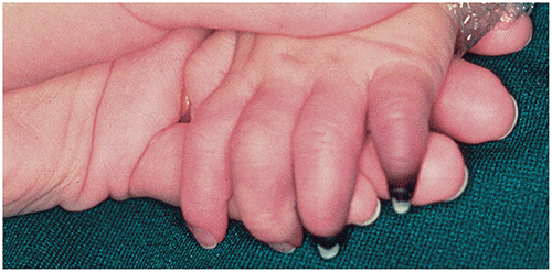
▶Vesicles, bullae, and purpura are usually not seen; peripheral gangrene may rarely occur (Figure 125.6).
▶A psoriasis-like skin eruption may be present, especially during the convalescent phase.
▶In addition to coronary artery aneurysms, other cardiac complications may include myocarditis, valvulitis, pericardial effusion, pericarditis, and myocardial infarction.
▶Other features may include gastrointestinal symptoms, lethargy, uveitis, arthralgias, gangrene.
▶Incomplete or atypical Kawasaki disease may occur, especially in infants, in which patients do not fulfill classic diagnostic criteria; in a child with unexplained fever and some diagnostic features, this diagnosis should be considered.
Figure 125.1. In Kawasaki Disease, Non-Exudative Conjunctival Injection is Present, Often with Perilimbic Sparing.
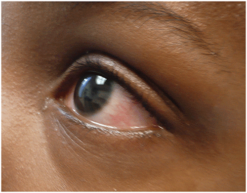
Figure 125.2. Kawasaki Disease. Hyperemia, Edema, and Fissuring of the Lips.
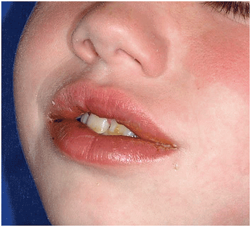
Figure 125.3. Kawasaki Disease. Strawberry Tongue Was Present in This Child with Severe Coronary Artery Aneurysms.
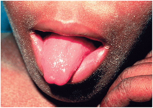
Figure 125.4. Kawasaki Disease. Erythematous Patches and Plaques with Foot Swelling.
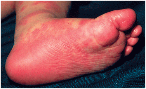
Figure 125.5. Perineal Accentuation in Kawasaki Disease. (A) Accentuation of Erythema in the Perineum and Genital Region is a Frequent Finding, as Seen in This Young Child. (B) Erythema Was Followed by Thick Desquamation in This 5-Year-Old with Skin of Color Who Had Diagnostic Features of Kawasaki Disease.
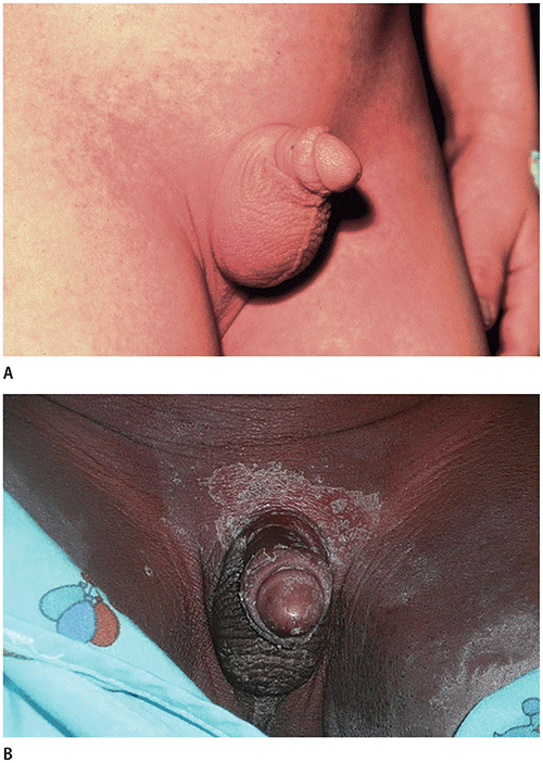
Figure 125.6. Kawasaki Disease. Peripheral Gangrene Involving the Fourth and Fifth Digits Occurred in This Toddler.
Look-alikes
| Disorder | Differentiating Features |
|---|
| Serum sickness–like eruption | History of preceding drug ingestion. Mucous membrane and eye findings usually absent. Purple urticaria most characteristic skin finding, in conjunction with periarticular swelling.
|
| Viral exanthem (ie, adenovirus, measles) | Exudative conjunctivitis, Koplik spots, severe cough present (measles). Rash begins behind ears, does not accentuate in perineum (measles). Conjunctivitis does not spare perilimbic area. Inflammatory marker levels (eg, C-reactive protein) minimally elevated (measles and adenovirus).
|
| Scarlet fever | Exudative pharyngitis present. Conjunctivae normal. When facial rash present, circumoral pallor is often present. Positive test result for Streptococcus pyogenes.
|
| Toxic shock syndrome (TSS) | Hypotension present. Renal involvement (elevated serum creatinine level), elevation of creatine phosphokinase level. Disseminated intravascular coagulopathy and adult respiratory distress syndrome–like illness may be present (streptococcal TSS). Rash appears as diffuse erythema. Primary focus of Staphylococcus aureus or streptococcal infection may be present. Note: Kawasaki disease shock syndrome may present with hypotension and shock and is differentiated from TSS by echocardiographic findings suggestive of Kawasaki disease, higher platelet counts, and more severe anemia.
|
| Systemic-onset juvenile idiopathic arthritis | Hepatosplenomegaly often present. Rash is evanescent, salmon colored; often correlates with presence of fever. Quotidian (daily) or double quotidian (twice daily) fever curve with relative wellness between spikes.
|
| Stevens-Johnson syndrome (SJS)/Mycoplasma pneumoniae–induced rash and mucositis (MIRM), reactive infectious mucocutaneous eruption (RIME) | Skin blisters, denudation (SJS > MIRM or RIME). Mucosal blistering and erosions (mouth, eyes, genitals) (MIRM or RIME > SJS). MIRM classically associated with M pneumoniae infection; RIME associated with several potential etiologic agents (Chlamydia pneumoniae, influenza, parainfluenza, others); SJS often triggered by drug exposure but may also be associated with infection.
|
▶The diagnosis is suggested by the presence of prolonged fever and other diagnostic criteria; incomplete Kawasaki disease is diagnosed when patient has fever for 5 or more days and 2 or 3 diagnostic criteria together with other supportive findings.
▶Elevation of inflammatory marker levels (eg, erythrocyte sedimentation rate, C-reactive protein) is nearly universal in Kawasaki disease; conversely, normal levels of inflammatory markers argue strongly against this diagnosis.
▶Other laboratory findings that support diagnosis include sterile pyuria, elevation of serum transaminase levels, cerebrospinal fluid pleocytosis, and increased N-terminal pro–brain natriuretic peptide levels.
▶Thrombocytopenia, anemia, and hypoalbuminemia may be present during the acute phase; thrombocytosis develops during the second to third week of disease.
▶White blood cell count may be normal to elevated, with a neutrophil predominance.
▶Cardiac imaging with 2-dimensional echocardiography is recommended; other investigative modalities include multisection spiral computed tomography and coronary magnetic resonance angiography.
▶Patients with fever for 5 days or longer and fewer than 4 principal features can have Kawasaki disease diagnosed when coronary artery disease is detected with 2-dimensional echocardiography or coronary angiography.
▶Treatment is most effective at decreasing risk of development of coronary artery aneurysms when administered within the first 10 days of the illness.
▶Intravenous immunoglobulin (IVIG), 2 g/kg as a single infusion over 10 to 12 hours.
▶Moderate-dose (30–50 mg/kg per day) or high-dose (80–100 mg/kg per day) aspirin (divided into 4 doses) during acute phase.
▶After 14 days or minimum of 3 days of patient being afebrile, dose of aspirin reduced (3–5 mg/kg once daily); if no echocardiographic variations present, aspirin is discontinued when laboratory study results are appropriate (usually 4–6 weeks).
▶Some recommend a second infusion of IVIG for those in whom it did not work the first time (or those with only transient improvement). Systemic steroids and infliximab are typically the next steps in the United States if 2 rounds of IVIG have failed. These treatments should be performed in consultation with a Kawasaki disease expert.
▶Other options for refractory Kawasaki disease can be considered via consultation with an infectious diseases or Kawasaki disease expert and may include plasmapheresis, interleukin-1 inhibitors (anakinra and canakinumab), interleukin-6 inhibitors (tocilizumab), methotrexate, and the anti-CD20 monoclonal antibody rituximab.
▶If the disease is untreated, 20% to 25% of patients develop coronary artery aneurysms; giant lesions entail the greatest risk of long-term morbidity.
▶Prognosis in those with coronary artery aneurysms is variable, and patients require lifelong follow-up.
▶Patients receiving a diagnosis when younger than 6 months or older than 9 years appear to have poorer outcomes.
▶Patients with a history of Kawasaki disease may have a more adverse cardiovascular risk profile, predisposing them to premature atherosclerosis. Consultation with a Kawasaki disease specialist or center is recommended to determine optimal long-term cardiac monitoring and disease management.
▶Referral to an experienced Kawasaki disease specialist or center should be considered for any patient presenting who has prolonged fever and clinical features that are on the diagnostic spectrum and for whom an alternative diagnosis cannot be confirmed.