Introduction
Several procedures can assist the clinician in diagnosing skin problems. Discussed here are the potassium hydroxide (KOH) preparation, fungal culture, mineral oil preparation for scabies, and Wood lamp examination.
KOH Preparation
Used to identify fungal elements (eg, spores, hyphae, pseudohyphae) in skin, hair, or nail samples. The procedure is as follows:
▶Using the edge of a glass microscope slide or #15 scalpel blade, scrape the skin and collect fragments or hair remnants on a second glass microscope slide. Preparing the area first with alcohol may be useful in helping debris stick to the blade or slide.
▶If sampling a nail, use a scalpel blade to scrape the underside of the nail (or its surface if superficial infection is suspected) and collect the debris obtained.
▶Cover the specimen on the glass slide with a coverslip.
▶Apply 1 to 2 drops of 10% to 20% KOH to the edge of the coverslip. Capillary action will draw the liquid under the entire coverslip.
▶Gently heat the slide with an alcohol lamp or match, taking care to avoid boiling, which causes the KOH to crystallize and makes interpretation of the preparation difficult.
▶Gently compress the coverslip to separate skin fragments further.
▶Scan the preparation initially under low power (using the 10× objective lens).
▶Examine any suspicious areas under higher power (using a 40× objective lens) for
■Branching hyphae or spores: characteristic of dermatophyte infections of the skin or nails (eg, tinea corporis, tinea pedis, tinea cruris, onychomycosis) (Figure 2.1).
■Spores within hair fragments (ie, an endothrix infection): characteristic of the most common form of tinea capitis in the United States caused by Trichophyton tonsurans (Figure 2.2). If tinea capitis is caused by Microsporum canis (approximately 5% of all cases), hyphae or spores will be seen on the outside of hair shafts (ie, an ectothrix infection).
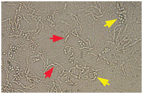
■Pseudohyphae and spores: seen in infections with Candida species (Figure 2.3).
■Spores and short hyphae (ie, “spaghetti and meatballs”): seen in tinea versicolor (Figure 2.4).
Figure 2.1. Potassium Hydroxide Preparation Showing Branching Hyphae (Arrows).
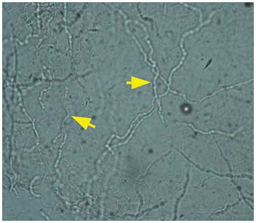
Figure 2.2. Potassium Hydroxide Preparation in Tinea Capitis Caused by Trichophyton Tonsurans. The Hair Fragment is Filled with Small Spheres (Ie, Arthrospores).
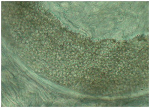
Figure 2.3. Pseudohyphae (Red Arrow) and Spores (Yellow Arrows) are Characteristic of Infection Caused by Candida Species.
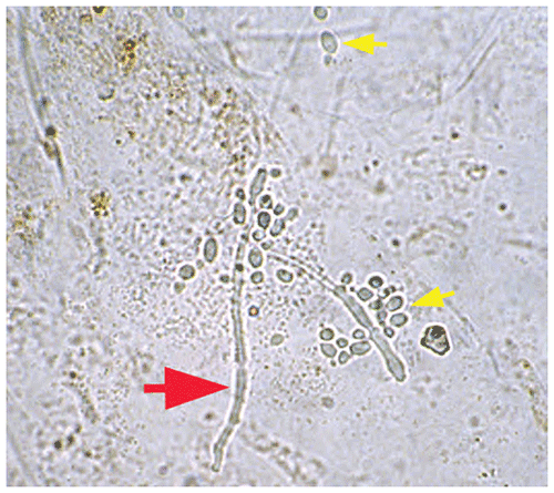
Figure 2.4. In Tinea Versicolor, the Potassium Hydroxide Preparation Reveals Short Hyphae (Red Arrows) and Spores (Yellow Arrows) (Ie, “spaghetti and Meatballs”).
Fungal Culture
▶Sampling techniques
■If sampling the skin, use the edge of a glass microscope slide or #15 scalpel, scrape the lesion, and collect scale on a glass microscope slide.
■If sampling a nail, use a scalpel blade to scrape the underside of the nail (or its surface if superficial infection is suspected), and collect the debris on a glass microscope slide or folded sheet of paper; alternatively, use a nail clipper to obtain nail clippings.
■If sampling the scalp, moisten a cotton-tipped applicator with tap water, rub the affected area of the scalp, and inoculate the fungal culture medium with the swab. If fungal culture medium is not available, a Culturette swab system (or other system) may be used to collect and transport the specimen to the laboratory.
▶Transfer the material collected to the fungal medium (typically dermatophyte test medium or Mycosel agar) and process appropriately.
■Leave the cap slightly loose to permit air entry.
■If fungal culture medium is not available, transfer the specimen in a sterile glass tube or other container to the laboratory.
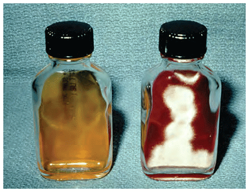
▶In the presence of a pathogenic fungus, dermatophyte test medium will change from yellow to red in 1 to 2 weeks (Figure 2.5).
Figure 2.5. Uninoculated Dermatophyte Test Medium is Yellow (Left). In the Presence of a Pathogenic Fungus, the Medium Becomes Red (Right).
Mineral Oil Preparation for Scabies
▶Place a small drop of mineral oil on a suspicious burrow, papule, or vesicle that has not been traumatized by the patient. Recent data suggest that debris from under fingernails (if present and accessible) may also be high yield for mineral oil examination.
▶Using a #15 scalpel blade oriented parallel to the skin surface, scrape the lesion. Because scabies mites live in the epidermis, it is not necessary to scrape deeply; however, some bleeding is common with the procedure. For fingernail debris, a Hyfrecator tip, curet, or small cerumen extractor can be used to collect the specimen.
▶Transfer the material to a drop of mineral oil on a glass microscope slide.
▶Repeat the process for several other suspicious lesions.
▶Cover the sample on the glass slide with a coverslip (add a few more drops of mineral oil if necessary for uniform distribution).
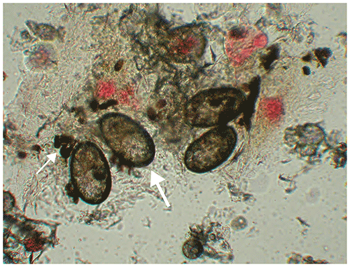
▶Examine at low power for the presence of mites, eggs, or fecal material (Figures 2.6 and 2.7).
Figure 2.6. Newly Hatched Mite (Red Arrow) and Fecal Material (Yellow Arrows) on a Mineral Oil Preparation.
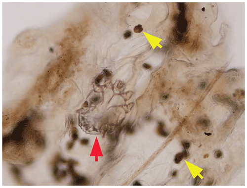
Figure 2.7. A Mineral Oil Preparation in a Patient Who Has Scabies Reveals e.g. (Large Arrow) and Mite Fecal Material (Ie, Scybala) (Small Arrow).
Wood Lamp Examination
Examination of the skin with a Wood lamp in a darkened room may assist in the diagnosis of several conditions.
▶Erythrasma (a superficial Corynebacterium infection): affected areas fluoresce coral red.
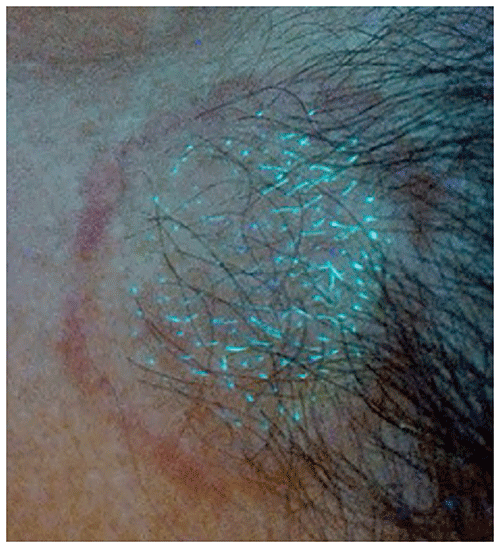
▶Tinea capitis: Wood lamp examination is useful in the recognition of only a minority of cases (perhaps 5%) of tinea capitis caused by Microsporum species (Figure 2.8). Green fluorescence does not occur when infections are caused by T tonsurans.
▶Tinea versicolor (caused by yeasts of the genus Malassezia [formerly Pityrosporum]): affected areas may fluoresce yellow gold.
▶Diseases characterized by hypopigmentation or depigmentation: in individuals who are lightly pigmented, examining the skin with a Wood lamp may assist in identifying lesions of vitiligo or ash-leaf macules of tuberous sclerosis.
Figure 2.8. Wood Lamp Examination in Tinea Capitis Caused by Microsporum Canis. There is Green Fluorescence of Affected Hairs.