▶Lentigines are usually small (≤5 mm), hyperpigmented macules that mimic ephelides (ie, freckles).
■Lentigines may be brown to black, are well-defined, and may be widely distributed (ie, not limited to sun-exposed areas).
■Lesions do not become more apparent after sun exposure.
■Isolated lentigines have no clinical significance.
▶Multiple lentigines may be associated with systemic disorders; the most common of these are as follows:
■LEOPARD syndrome: LEOPARD is an acronym for the major defects in this autosomal dominantly inherited disorder: lentigines, electrocardiographic abnormalities, ocular hypertelorism, pulmonic stenosis, abnormal genitalia, retarded (delayed) growth, and sensorineural deafness (Figure 73.1).
■Peutz-Jeghers syndrome: autosomal dominant disorder consisting of face, lip, and oral mucosa lentigines associated with benign intestinal polyposis (Figure 73.2). These patients may also have lentigines on the fingers, toes, palms, and soles.
■Lentiginosis with cardiocutaneous myxomas consists of multiple lentigines associated with cardiac and subcutaneous myxomas. Disorders considered within this disease category include the following:
– Carney complex: lentigines, cardiac and other myxomas, and endocrine tumors.
– LAMB syndrome: lentigines, atrial myxomas, mucocutaneous myxomas, and blue nevi.
– NAME syndrome: nevi, atrial myxomas, myxoid neurofibromas, ephelides, and endocrine neoplasia.
Figure 73.1. Patient with Leopard Syndrome. Multiple Lentigines with Relative Sparing of the Mucous Membranes. Reproduced with Permission from Cohen Ba. Pediatric Dermatology. 3rd Ed. Mosby; 2005:144.
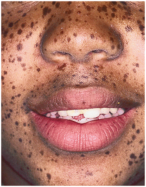
Figure 73.2. In Peutz-Jeghers Syndrome, Lentigines Appear on the Face, Lips, and Oral Mucosa. They are Associated with Gastrointestinal Polyposis.
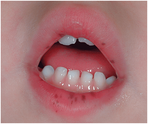
Look-alikes
| Disorder | Differentiating Features |
|---|---|
| Ephelides |
|
| Café au lait macules |
|
| Labial melanotic macule |
|
| Melanocytic nevi |
|
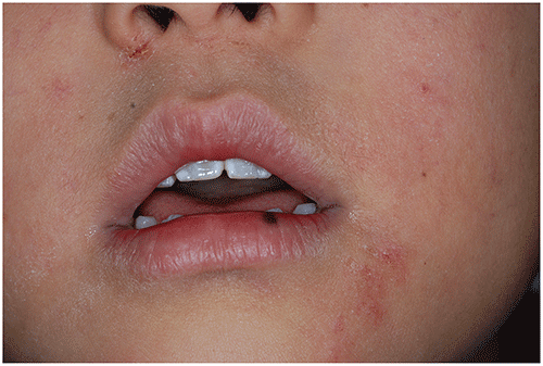
Figure 73.3. Labial Melanotic Macule. Small, Dark Brown, Solitary Macule on the Left Side of the Lower Lip of a Young Child with Eczema.