▶Vitiligo represents an acquired complete depigmentation of skin due to melanocyte destruction that is thought to be autoimmune in nature.
▶Two main forms have been described: generalized and segmental (ie, involves one area of the body and typically does not cross the midline).
▶Vitiligo develops in childhood or adolescence in about half of patients.
▶Vitiligo presents as well-defined macules or patches of complete depigmentation (ie, the skin is completely white) with typical texture (Figure 67.1).
■It may begin with speckled areas of hypopigmentation that continue to lose pigment and coalesce over time.
■Lesions may be faintly erythematous (especially the periphery) early in the course.
■Areas prone to trauma or pressure (eg, knees, elbows, small joints such as metacarpophalangeal joints, hips) are most frequently involved (Figure 67.2); this distribution may represent the Koebner phenomenon (ie, appearance of lesions at sites of injury).
■Other common locations include eyelids, perioral regions (Figure 67.3), axillae, and the groin.
▶Generalized vitiligo often starts symmetrically on the arms, legs, or periorbital areas and may progress to involve large areas.
▶Localized segmental vitiligo often follows a dermatomal distribution.
▶Trichrome vitiligo is a variant seen in children; unaffected, hypopigmented, and depigmented patches are present simultaneously in an involved area.
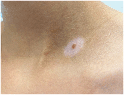
▶Halo nevi (Figure 67.4) are more common in children with vitiligo and may precede the diagnosis by months to years.
Figure 67.1. Vitiligo Appears as Well-Defined Areas of Complete Loss of Pigmentation (Ie, Depigmentation).
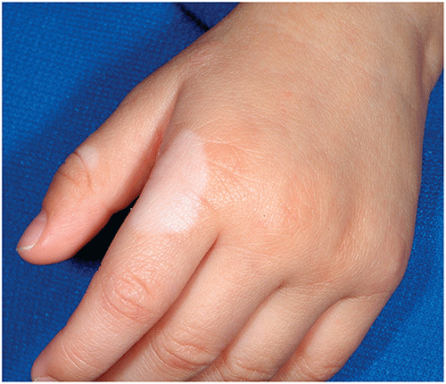
Figure 67.2. Vitiligo. This Young Patient Had Depigmented Macules and Patches on Bilateral Knees, Elbows, and Hips.
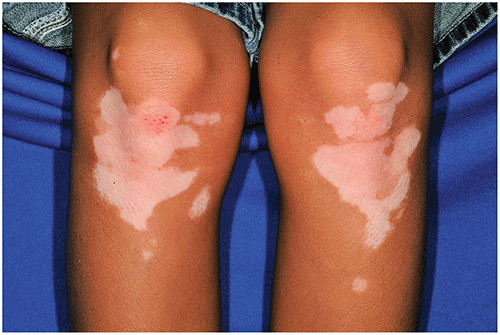
Figure 67.3. Segmental Vitiligo. This Young Patient Had Right-Sided Depigmented Patches around the Mouth and on the Upper Lip. Notice the Sharp Midline Demarcation as Well as the Islands of Repigmentation (Associated with the Patient’s Response to Topical Therapy).
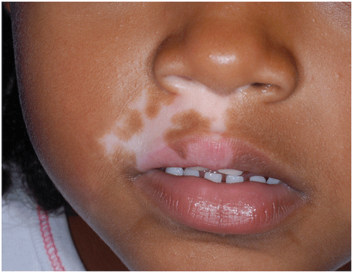
Figure 67.4. Halo Nevus is a Melanocytic Nevus that Develops a Surrounding Rim of Hypopigmentation or Depigmentation. They are More Common in Children with Vitiligo.
Look-alikes
| Disorder | Differentiating Features |
|---|
| Pityriasis alba | |
| Tinea versicolor | Well-defined hypopigmented (not depigmented) macules and patches located on the trunk, upper arms, or neck. Lesions often have associated fine scale and may be pruritic.
|
| Piebaldism | |
| Waardenburg syndrome | Pigmentary dilution of skin in conjunction with other characteristic features. Sensorineural deafness common. White forelock may be present. Other differences may include synophrys, heterochromia iridis, pseudo-hypertelorism (related to dystopia canthorum, lateral displacement of the medial canthi).
|
▶The diagnosis of vitiligo is made clinically based on typical features (well-defined macules or patches of depigmentation).
▶The distinction between hypopigmentation and depigmentation may be enhanced by examining the patient with a Wood lamp in a darkened room. Depigmented areas are well defined and strikingly prominent, while hypopigmented areas are less well defined.
▶Spontaneous repigmentation occurs in a few patients.
▶Treatment options to date are variably effective, with numerous anecdotal topical and systemic agents having limited value.
▶Patients who have vitiligo and desire therapy are best managed by or in consultation with a dermatologist. Some treatment options might include
■Topical corticosteroids.
■Topical calcineurin inhibitors (eg, tacrolimus, pimecrolimus): most useful for facial lesions.
■Topical ruxolitinib cream (Janus kinase inhibitor now US Food and Drug Administration approved for treatment of nonsegmental vitiligo in patients 12 years and older).
■Photochemotherapy using psoralens plus UV-A: often used in adolescents older than 12 years (rarely used in younger children).
■Narrowband UV-B phototherapy.
■Excimer laser therapy.
▶For patients who do not desire specific medical therapy, options include application of a camouflage cream matched to the child’s skin color and application of sunscreen (to protect depigmented skin and reduce tanning of unaffected skin).
▶Treatment options generally limited to adult patients with vitiligo include autologous skin grafting, mini-grafting with UV light exposure, and treatment of remaining pigmented areas with 20% monobenzyl ether of hydroquinone (which results in permanent skin depigmentation).
▶Generalized (nonsegmental) vitiligo is associated with an increased risk of autoimmune disease in the affected individual and first-degree relatives. While most children who have vitiligo have no other associated conditions, it is important to gather a family history and be observant for the development of symptoms suggestive of inflammatory eye disease (light sensitivity, change in vision) or other autoimmune disease (eg, type 1 diabetes, pernicious anemia, hypothyroidism, hypoparathyroidism, celiac disease, Addison disease, autoimmune hepatitis).
▶Most experts recommend thyroid function screening and antithyroid antibody levels for patients with vitiligo, with other testing performed only if indicated based on clinical signs or symptoms.
▶Variable and unpredictable.
▶Repigmentation, whether spontaneous or therapeutic, appears as perifollicular macules that coalesce to gradually fill in the area of depigmentation or as radial repigmentation in a centripetal pattern (ie, from outside toward the center).
▶Vitiligo is widespread or rapidly progressive, and phototherapy is being considered.
▶Vitiligo develops along with inflammatory eye disease or another autoimmune disorder (in which case consultation with the appropriate pediatric subspecialist is warranted). Consultation also may be of value if the patient has a first-degree relative with 2 autoimmune disorders. Rarely, vitiligo may be associated with autoimmune polyglandular syndromes, most notably type 1.