Table 101.1. Common Forms of Diaper Dermatitis
| Condition | Cause | Clinical Features | Treatment |
|---|
| Irritant dermatitis (Figure 101.1) | | Erythematous patches that involve the lower abdomen, buttocks, and thighs. Convex surfaces involved; inguinal folds often spared.
| |
| Candidiasis (Figure 101.2) | | Erythematous patches that involve the convexities and inguinal creases. Satellite papules and pustules. Scaling at the margins of involved areas.
| |
| Seborrheic dermatitis (Figure 101.3) | | Begins at 3 to 4 weeks of age and resolves by the end of the first year after birth. Salmon-pink patches with greasy scale that involve the convexities and inguinal creases. Involvement of the scalp, face, postauricular creases, umbilicus, or chest may be present.
| Skin: topical low-potency corticosteroid or antifungal preparation (eg, nystatin, clotrimazole, or other azole antifungal agent). Scalp: oil massage and brushing or antiseborrheic shampoo (eg, containing pyrithione zinc or selenium sulfide).
|
| Bullous impetigo (Figure 101.4) | | Flaccid blisters filled with clear or purulent fluid. Blisters rupture rapidly, leaving round or oval crusted erosions with a rim of scale.
| |
| Folliculitis (Figure 101.5) | | | Many lesions: oral antistaphylococcal antibiotic (the agent selected depends on local antibiotic resistance patterns). Few lesions: topical antibiotic (eg, mupirocin, clindamycin, retapamulin, ozenoxacin). Bleach baths (or cleansers containing sodium hypochlorite) may be useful for patients with persistent or recurrent infections.
|
| Intertrigo (Figure 101.6) | | Erythema and superficial erosions located in the inguinal creases. May become secondarily infected with Candida species or Streptococcus pyogenes, less commonly S aureus.
| |
| Jacquet erosive diaper dermatitis (Figure 101.7) | Multiple factors, including moisture, friction, enzymes in stool. Considered a variant of irritant dermatitis.
| | |
Figure 101.1. Irritant Diaper Dermatitis. Erythematous Patches Sparing the Skinfolds.
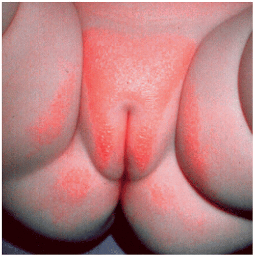
Figure 101.2. Erythematous Patches that Involve the Creases and Convexities are Characteristic of Candidal Diaper Dermatitis. Satellite Lesions and Scaling are Present.
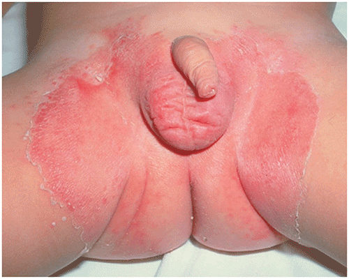
Figure 101.3. Salmon Pink Patches with Greasy Scale Involve the Creases and Convexities in Seborrheic Dermatitis.
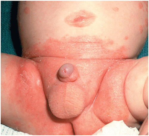
Figure 101.4. Flaccid Bullae that Rupture Easily Leaving Round, Crusted Erosions Occur in Bullous Impetigo.
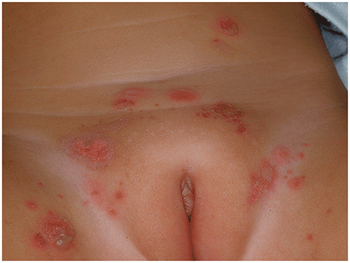
Figure 101.5. Folliculitis Often Involves the Buttocks. There are Erythematous Papules Centered on Hair Follicles. Frequently, Patients Also Have Pustules.
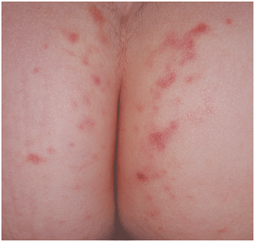
Figure 101.6. Intertrigo, Shown Here Involving the Neck, Produces Superficial Erosions in Areas Where Moist Skin Surfaces are in Apposition.
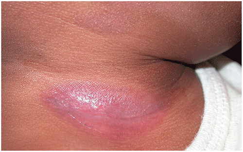
Figure 101.7. Jacquet Erosive Diaper Dermatitis. Well-Defined Shallow Ulcers and Ulcerations with Some Healed Areas.
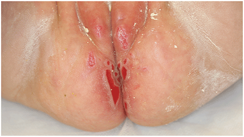
Table 101.2. Uncommon Forms of Diaper Dermatitis
| Condition | Cause | Clinical Features | Treatment |
|---|
| Psoriasis (Figure 101.8) | | Erythematous scaling papules or plaques (scaling of the scalp and umbilicus may be present). Lesions in the diaper area often lack scale characteristic of lesions located elsewhere. May be difficult to distinguish from seborrheic dermatitis.
| |
| Acrodermatitis enteropathica (Figure 101.9) | | Often begins when infants are weaned from human to cow milk formula. Scaling, erosive erythematous eruption located around the mouth and in the diaper area. Infants may have sparse hair, diarrhea, or failure to gain weight.
| |
| Langerhans cell histiocytosis (Figure 101.10) | | Lesion types: vesicles or pustules (often with a hemorrhagic crust); erythematous, orange, or yellowish brown papules or nodules; petechiae; erosions (in the diaper area). Areas affected: scalp, palms and soles, skinfolds, diaper area. Other features: infants may have hepatosplenomegaly, lymphadenopathy.
| |
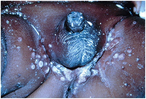
| Congenital syphilis (Figure 101.11) | | Symptoms: rash, bloody diarrhea, rhinorrhea, irritability, pain with movement (Parrot pseudoparalysis). Skin lesions: condylomata lata (ie, flat-topped papules and plaques located in the diaper area or at the angles of the mouth), scaling copper-colored papules and plaques on the trunk and extremities, or vesicles and bullae.
| |
Figure 101.8. Psoriasis in the Diaper Area Produces Erythematous Patches or Plaques. Unlike Lesions Elsewhere, Scale May be Absent.
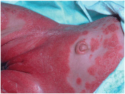
Figure 101.9. Acrodermatitis Enteropathica Causes Erythematous Patches in the Diaper Area and around the Mouth.
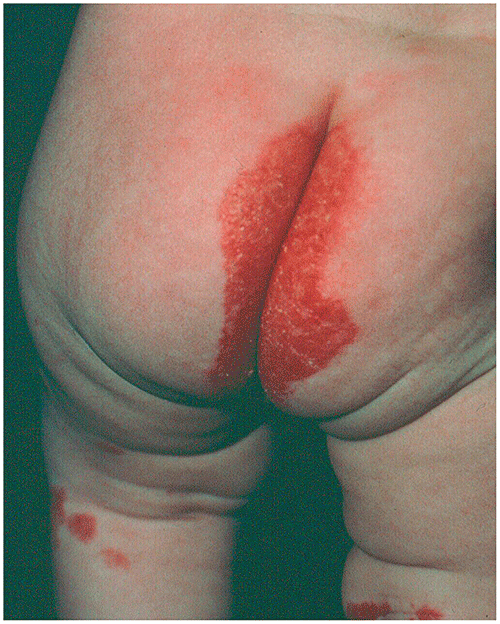
Figure 101.10. Eroded and Crusted Erythematous Papules of Langerhans Cell Histiocytosis.
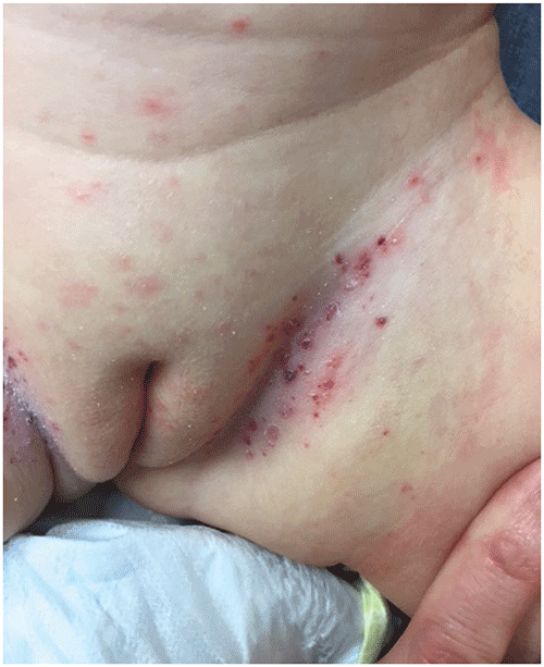
Figure 101.11. Condylomata Lata, Flat-Topped Papules and Plaques, Occur in the Diaper Area in Congenital Syphilis.
▶Infants should be referred if appropriate therapy fails (to consider alternate diagnoses and possibly perform skin biopsy).
▶Infants suspected of having Langerhans cell histiocytosis should be referred to a pediatric dermatologist or pediatric oncologist for evaluation.
▶Consultation with a pediatric infectious disease specialist is warranted for infants suspected of having congenital syphilis.