▶Spitz nevus (also called spindle cell nevus) is considered a benign, acquired form of a melanocytic nevus that usually presents in childhood or adolescence. It rarely presents in adults.
▶It was previously called a “benign juvenile melanoma” because it shares some histologic features with melanoma but does not have the same aggressive behavior.
▶The etiology and pathogenesis are unknown.
▶Spitz nevi tend to be solitary and appear on the head, neck, or extremities. Uncommonly, they can be clustered (agminated) or, rarely, disseminated.
▶Spitz nevus usually presents as a smaller than 1 cm, dome-shaped, round, pink (Figure 83.1), red (Figure 83.2), brown, or brown-black papule. It grows rapidly in the first few months and then plateaus with the possibility of regressing over time.
▶Pink or hypopigmented Spitz nevi usually have a dotted or polymorphous vascular pattern at dermoscopy (an illuminated magnification tool often used by dermatologists). Pigmented Spitz nevi typically have a very dark-brown to black color and, at dermoscopy, have a globular, negative network or starburst pattern.
Figure 83.1. Spitz Nevus. This Patient Had a Rapidly Growing Pink Papule with a Crescent of Brown Pigmentation on the Upper Extremity. The Lesion Was Excised, and Histologic Examination Revealed a Spitz Nevus Without Atypia.
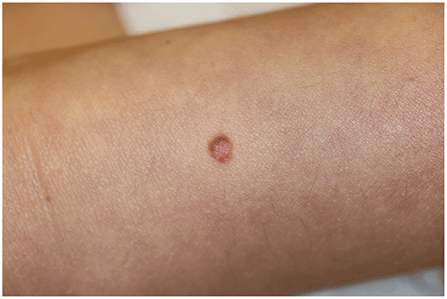
Figure 83.2. Spitz Nevus of Shoulder Presenting as a Bright Red, Well-Circumscribed Papule.
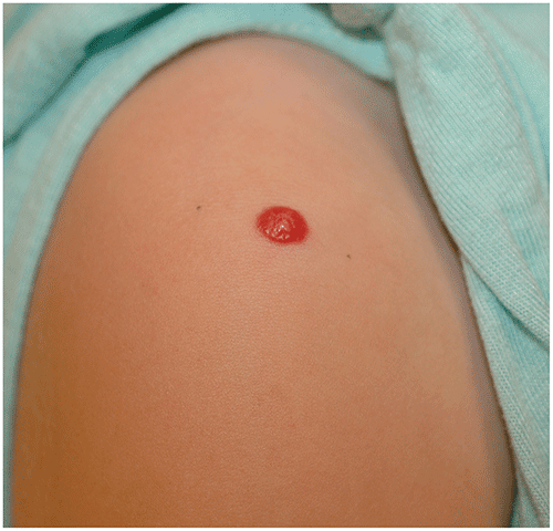
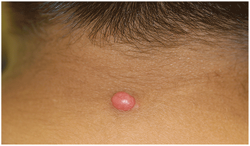
Figure 83.3. This Patient Had a Rapidly Enlarging Pink Papule on the Posterior Aspect of the Neck. The Lesion Was Excised, and Histologic Examination Revealed a Spitz Nevus with Atypical Features. As a Result, re-Excision Was Performed with Adequate Margins and Without Recurrence to Date.
Look-alikes
| Disorder | Differentiating Features |
|---|
| Pyogenic granuloma | Typically of acute onset. Bright red, friable papule, which may be pedunculated. Peripheral collarette typically present around base. Bleeds easily; surface may be eroded. Vascular lacunae are noted at dermoscopy with no melanocytic features.
|
| Intradermal melanocytic nevus | Pink, skin-colored, brown, or dark-brown papule. Typically slow growing. May see brown pigment with diascopy (compressing lesion and viewing through a glass slide).
|
| Juvenile xanthogranuloma | Orange to yellow-brown papule; early lesions may be more pink to red in appearance, sometimes mimicking a Spitz nevus. Slowly grows and then involutes over time. No melanocytic features at dermoscopy.
|
| Verruca vulgaris | Surface of the papule is rough (ie, verrucous). Thrombosed capillaries (ie, purple or black specks) may be seen within the lesion. No melanocytic features at dermoscopy.
|
▶The history, physical examination, and, when used, appearance at dermoscopy can help in making a diagnosis.
▶Definitive diagnosis often depends on excisional biopsy with histologic confirmation.
▶Typical Spitz nevi are benign, but uniform management recommendations are lacking; many advocate for excision of all lesions, while some follow the lesions clinically and excise only if atypical clinical features develop.
▶Importantly, pediatric melanoma often presents in an amelanotic fashion, which clinically translates into a red papule that may mimic Spitz nevus.
▶When lesions are excised, most experts recommend complete excision to prevent recurrence that can be misinterpreted as a melanoma and to allow for accurate dermatopathic interpretation.
▶Classic Spitz nevi are considered benign, and excision is curative.
▶“Atypical spitzoid tumors” occasionally present in children, and the prognosis for these lesions is variable (Figure 83.3).
▶Patients who have a suspected Spitz nevus should be referred for pediatric dermatologic evaluation.
▶For any such lesion that is rapidly growing, ulcerating, or bleeding, urgent referral should be requested because prompt surgical excision is indicated.