▶Discrete papular eruption commonly seen in infants and children.
▶Spread through skin-to-skin contact.
▶Caused by a poxvirus.
▶Although this infection is classically associated with immunodeficiency or sexual transmission in adults, it is not typically associated with this history in infants and children.
▶May occur as a sexually transmitted infection in sexually active adolescents and young adults.
▶Usually without symptoms.
▶Lesions are 1- to 6-mm, discrete, skin-colored, pink (Figure 12.1) to pearly (Figure 12.2) papules that may mimic vesicles; some lesions are umbilicated (ie, have a central dell or depression) or a central hyperkeratotic core.
▶Widespread or sometimes “giant” (8–15 mm) lesions may occur in individuals who are immunosuppressed.
▶Extensive lesions often are observed in children with atopic dermatitis.
▶Can occur on any cutaneous location but commonly seen on face, eyelids, neck, chest, axillae, folds of extremities, and genital region.
▶Eyelid lesions may be associated with chronic conjunctivitis or keratitis.
▶May be associated with a mild to moderate dermatitis occurring in the vicinity of the papules, known as molluscum dermatitis (Figure 12.3).
▶Linear arrangement of lesions may be present due to autoinoculation (Koebner phenomenon) (Figure 12.4).
▶Genital location most common when occurring as a sexually transmitted infection in sexually active adolescents or young adults but may also be seen in younger children.
▶Enlargement of lesions with erythema is often noted in association with the host immune response against the virus (Figure 12.5) and often heralds involution of the lesions. This has been termed the beginning of the end, or BOTE sign.
Figure 12.1. Skin-Colored to Pink Translucent Papules are Typical of Molluscum Contagiosum. Note Umbilication (Ie, Central Depression) of Larger Lesions.
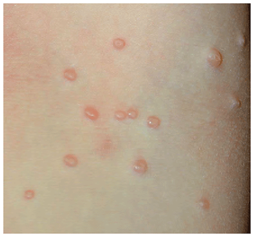
Figure 12.2. Pearly Papules of Molluscum Contagiosum. Note the Central Hyperkeratotic Core in Some of the Lesions.
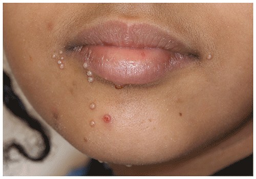
Figure 12.3. Molluscum Dermatitis. Erythematous Patches with Scale Surround Lesions of Molluscum Contagiosum.
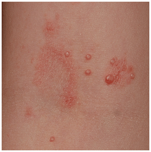
Figure 12.4. Molluscum Contagiosum with Koebnerization, Manifested as Several Lesions in a Linear Distribution.
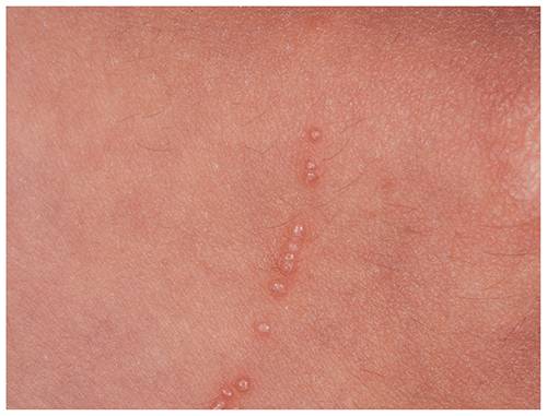
Figure 12.5. Molluscum Lesions with Enlargement and Erythema, Signifying the Host Immune Response Against the Virus. These Lesions Resolved Shortly Thereafter.
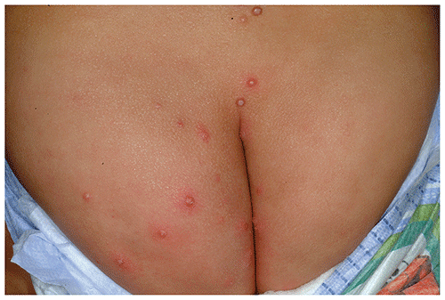
Look-alikes
| Disorder | Differentiating Features |
|---|
| Milia | |
| Frictional lichenoid dermatitis | Uniformly spaced, skin-colored to pink to hypopigmented papules on elbows, knees. No central keratin plug or umbilication.
|
| Closed comedones | Small white papules that lack central umbilication. Occur in older children or adolescents. Most often limited to facial distribution.
|
| Flat warts | |
| Cryptococcosis | Molluscum-like lesions possible in patients who are immunocompromised. May have systemic symptoms. May have ulcers in addition to papules.
|
| Histoplasmosis | Molluscum-like lesions possible in patients who are immunocompromised. May have systemic symptoms. May have ulcers in addition to papules.
|
▶The diagnosis is made clinically based on the appearance of lesions.
▶A skin scraping of a characteristic papule can be used for a crush preparation and stained with Giemsa or methylene blue, revealing numerous characteristic molluscum (Henderson-Paterson) bodies at direct microscopy; this is rarely necessary.
▶Skin biopsy occasionally is used for large or atypical lesions.
▶Lesions often resolve spontaneously over several months or years. In 1 study, the mean time to resolution was 13 months.
▶Watchful waiting is an acceptable management plan, although treatment is often requested by parents.
▶Application of cantharidin (blister beetle extract) is a painless and effective procedure; however, it must be performed by an experienced clinician and only in the office setting (not for home use). Although cantharidin had previously been an off-label therapy, the US Food and Drug Administration has now approved cantharidin 0.7% in a single-use device for the treatment of molluscum contagiosum in those aged 2 years or older. Blistering is an expected outcome of treatment with cantharidin and can be quite significant in some patients, although most patients tolerate the treatment quite well.
▶Berdazimer 10.3% topical gel was also recently approved by the US Food and Drug Administration for patients aged 1 year or older. This nitric oxide–releasing agent is applied once daily to lesions and is approved for application at home by patients or caregivers.
▶Curettage of individual lesions is effective, but limitations include pain, fear (in young children), and risks of spread and scarring.
▶Lesions near the eyelid margin, causing chronic conjunctivitis, may require surgical excision.
▶Cryotherapy with liquid nitrogen also is effective but may be traumatic for young children in whom it is poorly tolerated.
▶Imiquimod cream is an off-label option for treatment. Although applied 3 times weekly for genital condylomata, treatment of molluscum generally requires daily application and may require many weeks of application; irritant contact dermatitis is a common limiting side effect of this therapy.
▶Topical retinoids (eg, tretinoin) have been used, mainly for facial lesions; mechanism of action is probably induction of irritant dermatitis, which may be a limiting side effect. This is an off-label indication for retinoids.
▶Immunotherapy using intralesional injection of skin test antigens (most often Candida) may be effective, but published data are limited, and the associated pain, anxiety, and need for repeated injections make this option less feasible for young children.
▶When treatment is requested but is not available in your practice.
▶When diagnosis is uncertain.
▶When extensive disease is present.
▶When molluscum is associated with poorly controlled atopic dermatitis.