▶Acrodermatitis enteropathica (AE) is an autosomal recessive disorder that results in a characteristic acral and periorificial eruption, diarrhea, and alopecia.
■AE is caused by variations in the SLC39A4 gene that encodes an intestinal transporter required for zinc absorption from the small intestine.
▶Acquired zinc deficiency (eg, due to excessive losses [eg, chronic diarrhea], dietary restriction, administration of total parenteral nutrition with inadequate zinc supplementation) results in symptoms and signs analogous to those of AE. An unusual form of acquired zinc deficiency may occur in infants who are breastfed whose mothers produce zinc-deficient milk.
▶In neonates and infants who are fed formula, the symptoms of AE typically appear days to weeks after birth when zinc stores become depleted. In infants who are breastfed, AE becomes manifest shortly after weaning. This delay in onset is thought to be the result of enhanced bioavailability of zinc in human milk.
▶Rash is often the first sign of disease.
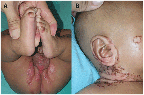
■The rash of AE is acral (ie, on the extremities) and periorificial (ie, around the mouth, nose, eyes, and anus) (Figures 121.1–121.4). The digits may be involved with periungual erythema and swelling.
■Lesions are erythematous patches with well-defined borders. Scaling, erosions, crusting, vesicles, and bullae may occur.
▶The most common associated symptoms are alopecia and diarrhea. Others include anorexia, behavioral changes (eg, irritability, apathy), failure to thrive, ocular symptoms (eg, blepharitis, conjunctivitis), and recurring bacterial or fungal infections.
Figure 121.1. Acrodermatitis Enteropathica. Erythema, Scaling, and Crusting on the Hand.
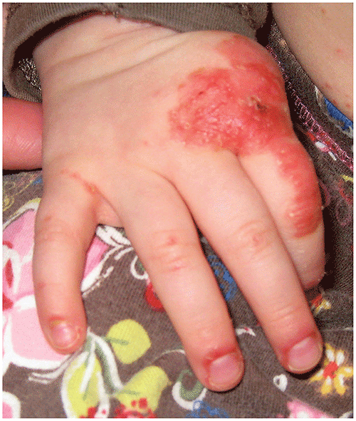
Figure 121.2. Acrodermatitis Enteropathica. Erythema and Crusting around the Mouth of the Patient Shown in Figure 121.1.
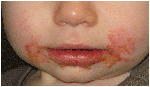
Figure 121.3. Erythema and Crusting in the Diaper Area of an Infant Who Has Acrodermatitis Enteropathica.
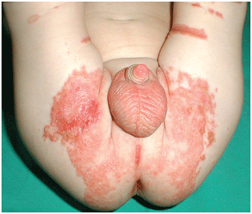
Figure 121.4. Acrodermatitis Enteropathica. (A) Multiple Well-Demarcated Erosions and Patches in a 3-Month-Old, Involving the Toes and Genital and Perineal Areas. (B) Well-Defined, Crusted Erosions on the Neck and Right Auricle. Patchy Alopecia of the Scalp is Also Visible. Used with Permission from Leung Akc, Leong Kf, Lam Jm. Acrodermatitis Enteropathica in a 3-Month-Old Boy. Cmaj. 2021;193(7):e243.
Look-alikes
Kwashiorkor, essential fatty acid deficiency (including the rash seen in cystic fibrosis), vitamin B12 deficiency, isoleucine deficiency, certain organic acid disorders, and biotin deficiency may produce an eruption similar to that of AE. The acral and periorificial distribution of the rash of AE may help to distinguish it from other disorders.
| Disorder | Differentiating Features |
|---|
| Atopic dermatitis | Diaper area usually spared. Trunk often affected in infants (uncommon in acrodermatitis enteropathica [AE]). Facial involvement (eg, cheeks) occurs frequently, but perioral and periorbital involvement uncommon. Pruritus usually present.
|
| Crusted impetigo | |
| Irritant diaper dermatitis | |
| Psoriasis | |
| Seborrheic dermatitis | Scalp involvement common (ie, cradle cap). Skin lesions often have greasy scale. Perioral and periorbital involvement uncommon. Erosive changes absent.
|
▶The presence of AE is suggested by the acral and periorificial distribution of the erosive rash and the presence of lesions with well-defined borders.
▶A serum zinc level 50 mcg/dL or lower supports the clinical diagnosis; care should be exercised to collect blood in correct tubes, given potential zinc contamination of some materials.
▶Low serum alkaline phosphatase level is often present.
▶Rapid response of the rash and other symptoms to zinc supplementation supports the diagnosis.
▶Oral zinc supplementation (usually lifelong in AE). Supplementation should be provided at a dose of 1 to 3 mg/kg per day of elemental zinc. Several formulations exist, but zinc sulfate is the preferred oral agent; the dose is 5 to 15 mg/kg per day. This may be divided twice or 3 times a day and is best administered 1 to 2 hours before a feeding or meal.
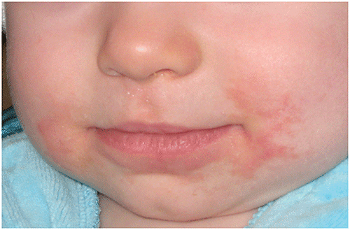
▶Irritability, anorexia, diarrhea, and the rash improve within days (Figure 121.5).
▶Monitor zinc level periodically (every 3–6 months) and adjust dose accordingly.
▶Adverse effects include the following:
■Zinc may cause nausea or vomiting.
■Because zinc may interfere with the absorption of copper, some recommend periodic measurement of the serum copper concentration.
Figure 121.5. The Patient Shown in Figure 121.2 10 Days after Beginning Zinc Supplementation. The Perioral Eruption Has Improved Greatly.
▶Excellent, although lifelong zinc supplementation generally is required for neonates or infants who have AE.
▶In patients who are zinc deficient due to defective mammary zinc secretion, discontinuation of zinc supplementation is typically possible at the time of weaning.
▶Consider consultation or referral if the zinc therapy fails. If the patient has an acquired form of zinc deficiency, identification and treatment of the underlying cause are necessary.