section name header

Basics ⬇

Outline


BASICS

Definition!!navigator!!

Causes in order of frequency—(1) external abscesses, (2) internal infection, (3) ulcerative lymphangitis.

Pathophysiology!!navigator!!

  • Soil-borne, Gram-positive, pleomorphic, intracellular facultative anaerobic rod
  • Likely transmitted by mechanical vectors (flies), horse-to-horse contact, and contaminated soil
  • Portals of entry—traumatized mucous membranes or skin, including insect-induced ventral midline dermatitis
  • Incubation time 3–4 weeks
  • Exotoxin phospholipase D increases vascular permeability to promote spread of infection through tissue and lymphatics and causes local edema and pain. Internal abscesses result from hematogenous or lymphatic spread
  • Most immunocompetent horses mount a strong immune response and have a single bout of infection

Systems Affected!!navigator!!

External Abscesses

  • Skin
  • Lymphatic
  • Musculoskeletal

Internal Infection

  • Hepatobiliary
  • Renal
  • Respiratory
  • Other (nervous, reproductive)

Ulcerative Lymphangitis

  • Musculoskeletal
  • Lymphatic

Incidence/Prevalence!!navigator!!

Endemic farms—sporadic. Naive populations—higher incidence. Mortality rate—<1% for external abscesses, 30–40% for internal infection.

Geographic Distribution!!navigator!!

Most common—southwestern USA; seen throughout North America.

Signalment!!navigator!!

All ages affected. Foals <6 months old are rarely affected, suggesting passive transfer of immunity when born to mares in endemic areas. No sex predilection.

Signs!!navigator!!

External Abscesses

  • Historic cases on the property
  • Edema of pectorals, axillae, ventrum, or inguinal area
  • Progress to larger localized, painful and firm swellings in which small, multifocal abscesses develop
  • Mature abscesses may have a palpable soft depression (Web Figure 1)
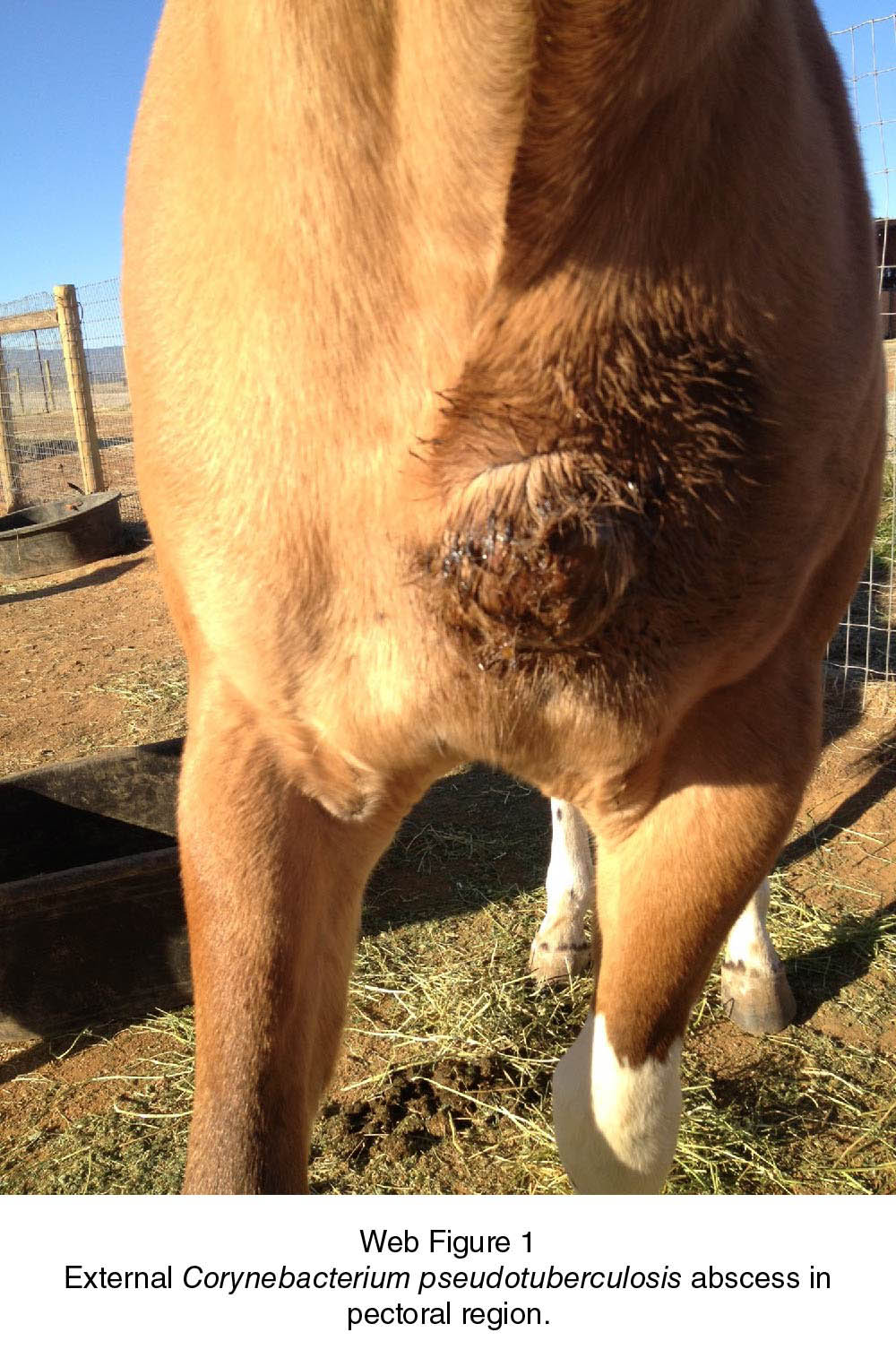
  • Purulent material—thick, non-odorous, and tan
  • Limb edema or lameness (triceps or inguinal areas)
  • Variable signs of systemic inflammation/discomfort (fever, lethargy, anorexia, tachycardia)

Internal Infection

  • History of external abscess weeks prior
  • Anorexia
  • Lethargy
  • Weight loss
  • Fever
  • Dependent edema
  • Tachycardia
  • Tachypnea, nasal discharge (respiratory disease)
  • Abdominal discomfort
  • Abnormal urination (urinary tract disease)

Ulcerative Lymphangitis

  • Limb edema (cellulitis/lymphangitis)
  • Multiple draining lesions
  • Lameness
  • Variable signs of systemic inflammation/discomfort

Other

  • Lameness (osteomyelitis, septic arthritis, or tenosynovitis)
  • Neurologic signs (meningitis)
  • Stridor (arytenoid chondritis)
  • Nasal discharge (sinusitis, guttural pouch empyema)
  • Vaginal discharge, abortion (metritis, placentitis)

Causes!!navigator!!

  • Corynebacterium pseudotuberculosis biovar equi (nitrate reductase positive)
  • Biovar ovis (nitrate negative) causes caseous lymphadenitis in small ruminants
  • Natural cross-species transmission does not occur

Risk Factors!!navigator!!

  • Endemic disease
  • High ambient temperatures or drought. Most cases are diagnosed in summer and fall. Increased insect vectors after a wet, mild winter
  • Poor vector control
  • Immunocompromised horses

Diagnosis ⬆ ⬇

Outline


DIAGNOSIS

Differential Diagnosis!!navigator!!

External Abscesses

  • Other bacterial infections (e.g. Streptococcus equi ssp. equi and S. equi ssp. zooepidemicus)
  • Hematoma/seroma
  • Neoplasia
  • Other causes for ventral edema (e.g. hypersensitivity, hypoproteinemia, vasculitis)
  • Other causes for lameness

Internal Infection

  • Other infectious organisms (e.g. S. equi ssp. equi and S. equi ssp. zooepidemicus, Coccidioides immitis)
  • Foreign body
  • Hepatopathy, splenitis, pyelonephritis, peritonitis, respiratory disease of other etiologies
  • Neoplasia

Ulcerative Lymphangitis

  • Other infectious causes of cellulitis/lymphangitis
  • Immune-mediated vasculitis
  • Exotic infectious disease (e.g. Burkholderia mallei, Histoplasma capsulatum var. farciminosum)
  • Other—osteomyelitis, septic arthritis, meningitis, upper airway disease of other etiologies

CBC/Biochemistry/Urinalysis!!navigator!!

  • Anemia of chronic inflammation
  • Leukocytosis with neutrophilia
  • Hyperfibrinogenemia
  • Hyperglobulinemia
  • Biochemical abnormalities relate to the organs affected and systemic illness
  • Pyuria—renal abscess

Other Laboratory Tests!!navigator!!

  • Gram stain—Gram-positive, pleomorphic rods
  • Bacterial culture
  • PCR—may be more sensitive in cases that are culture negative (e.g. previous treatment with antimicrobials, low quantities of bacteria)
  • Synergistic hemolysin inhibition test—measures immunoglobulin G to exotoxin. Titers—negative ≤16, exposure or acute infection = 16–128, internal infection ≥512 provided no concurrent external abscess. Exposure, active disease, and recovery titers overlap. May stay increased after infection for months

Imaging!!navigator!!

Ultrasonography

  • Early external infection—diffuse edema. Mature abscesses have thick capsules with heterogeneous contents and variable loculations
  • Internal infection—thoracic: consolidation, pleural changes/effusion; abdominal: increased peritoneal fluid, abnormalities in the viscera or lymph nodes
  • Abscesses are singular or multifocal within organ parenchyma, often thinly encapsulated with hypoechoic contents, and the organ may be enlarged

Radiography

  • Thoracic radiographs—diffuse or patchy interstitial and/or alveolar patterns with pneumonia
  • Limb radiographs—rule out other causes of lameness or osteomyelitis

Other Diagnostic Procedures!!navigator!!

Abdominocentesis—peritonitis. Transtracheal wash—septic bronchopneumonia; culture and antimicrobial sensitivity. Endoscopy—guttural pouch or laryngeal involvement. Blood culture—in severe sepsis.

Pathologic Findings!!navigator!!

Abscesses are encapsulated with purulent contents and may distort surrounding tissues.

Treatment ⬆ ⬇

Outline


TREATMENT

Appropriate Health Care!!navigator!!

  • Most noncomplicated external abscesses or ulcerative lymphangitis can be dealt with in the field or as an outpatient
  • Biosecurity to prevent spread of exudate is advised for draining external abscesses

Nursing Care!!navigator!!

  • External abscesses—hot packs/poultices encourage abscess maturation. Mature abscesses may rupture spontaneously or be surgically lanced and lavaged with saline and/or antiseptic solutions
  • Internal infection—supportive care
  • Ulcerative lymphangitis—topical antimicrobials and frequent bandage changes to provide wound coverage and compression. Free-choice exercise and hand-walking
  • Application of insect repellent to prevent vectoring of infectious material

Client Education!!navigator!!

  • Nondraining lesions—not directly infectious
  • Draining lesions—confine to a single area; exudate carries high bacterial load and should be collected, disinfected, and disposed of in a closed system

Surgical Considerations!!navigator!!

  • Mature external abscesses can be lanced at a dependent site, with or without ultrasonography guidance. Lancing immature abscesses may increase tissue inflammation
  • Abscesses deep to large muscle groups (e.g. triceps)—drainage through insertion of a chest tube
  • Septic peritonitis may require an abdominal drain and lavage

Medications ⬆ ⬇

Outline


MEDICATIONS

Drug(s) of Choice!!navigator!!

  • Uncomplicated external abscesses—antimicrobials often not indicated
  • Horses with external abscesses and concurrent systemic illness, recurrent infections, or poor immunity and horses with internal infection or ulcerative lymphangitis require antimicrobials until evidence of infection has completely resolved
  • Susceptible in vitro to many antimicrobials and does not appear to be developing widespread resistance
  • Effectiveness in vivo depends on penetration through the abscess capsule, activity in exudate, intracellular bacterial location, and drug bioavailability
  • External abscesses—trimethoprim–sulfamethoxazole (30 mg/kg PO every 12 h), trimethoprim–sulfadiazine (24 mg/kg PO every 12 h), or minocycline (4 mg/kg PO every 12 h). Sulfas and beta-lactams may be inactivated in presence of large, acidic abscess
  • Internal abscesses—minocycline (4 mg/kg PO every 12 h), doxycycline (10 mg/kg PO every 12 h), enrofloxacin (7.5 mg/kg PO every 24 h), trimethoprim–sulfas (doses above), and potassium penicillin (20 000–40 000 IU/kg IV every 4–6 h). Rifampin (rifampicin) (5 mg/kg PO every 12 h) is highly effective when added to another antimicrobial (e.g. trimethoprim–sulfa). Ceftiofur is less effective due to a high minimum inhibitory concentration
  • Ulcerative lymphangitis—treat aggressively with long-term antimicrobials
  • NSAIDs—control discomfort and inflammation

Contraindications!!navigator!!

  • Enrofloxacin—contraindicated in growing horses
  • Chloramphenicol and doxycycline have variable bioavailability in horses and may be less effective in vivo

Alternative Drugs!!navigator!!

Immunostimulants—not critically evaluated or currently recommended.

Follow-up ⬆ ⬇

Outline


FOLLOW-UP

Prevention/Avoidance!!navigator!!

Insect Vector Control

  • Prompt disposal of organic debris, fly repellent systems, fly predators, or feed-through insect growth regulators
  • Apply topical insect repellents (particularly ventral midline and wounds)

Biosecurity

  • Disposable gloves and handwashing
  • Isolate horses with draining external abscesses
  • Collect, disinfect, and dispose of exudate
  • Conditionally licensed bacterin vaccine available (Boehringer Ingelheim International GmbH)

Possible Complications!!navigator!!

Disseminated disease and purpura haemorrhagica

Expected Course and Prognosis!!navigator!!

  • External abscesses—simple abscesses: 14–30 days to fully resolve; additional abscesses may develop from lymphatic spread. Prognosis: good
  • Internal infection—>30 days, often 90–120 days. Prognosis: fair to good if treated
  • Serially monitor leukogram, globulins and ultrasound findings
  • Ulcerative lymphangitis—≥30 days. Prognosis: good

Miscellaneous ⬆ ⬇

Outline


MISCELLANEOUS

Zoonotic Potential!!navigator!!

Rarely occurs; 1 reported student developed pneumonia after exposure to an infected horse, likely from bacterial inhalation in a contaminated environment.

Pregnancy/Fertility/Breeding!!navigator!!

Abortion from placentitis or fetal infection may occur.

Synonyms!!navigator!!

  • Pigeon fever
  • Dryland distemper

Abbreviations!!navigator!!

  • NSAID = nonsteroidal anti-inflammatory drug
  • PCR = polymerase chain reaction

Internet Resources!!navigator!!

Center for Equine Health, University of California, Davis. http://viewer.zmags.com/publication/67e69b2d#/67e69b2d/1

Suggested Reading ⬆ ⬇

Pratt SM, Spier SJ, Carroll SP, et al. Evaluation of clinical characteristics, diagnostic test results, and outcome in horses with internal infection caused by Corynebacterium pseudotuberculosis: 30 Cases (1995–2003). J Am Vet Med Assoc 2005;227:441–448.

Author(s) ⬆

Authors: Sharon J. Spier and Emily H. Berryhill

Consulting Editor: Ashley G. Boyle

Acknowledgment: The authors and editor acknowledge the prior contribution of Mathilde Leclère.