| Author: Diego Acero
|
| Author: Diego Acero
|
A Look at Acute Pancreatitis
Pancreatitis is an inflammation of the pancreas. An accessory organ of the gastrointestinal (GI) tract, the pancreas lies in the left upper abdomen behind the stomach and between the spleen and duodenum. It functions as both an exocrine and endocrine gland, producing enzymes that promote digestion and hormones that aid in glucose balance. (See Functions of the pancreas.)
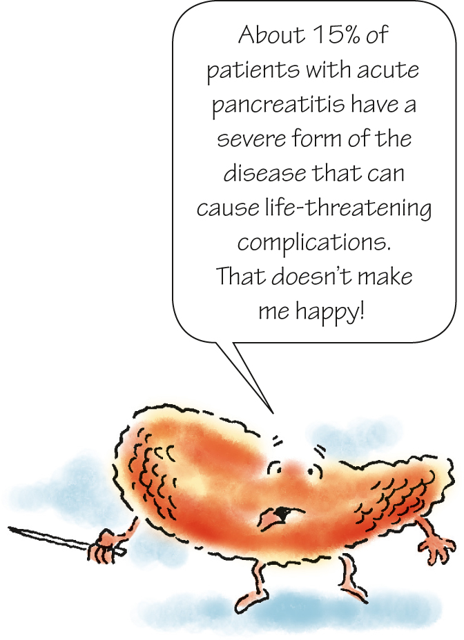
Adults of any age can develop acute pancreatitis; however, children rarely develop the disorder. It can occur in either edematous (interstitial) or necrotizing form. Both types cause inflammation that begins in the acinar cells. (See Edematous vs. necrotizing.) Most cases of acute pancreatitis are mild; however, about 15% of patients experience a severe form of the disease that can cause life-threatening complications, including fluid and electrolyte disturbances.
Acute pancreatitis that occurs on two or more occasions is classified as acute recurrent pancreatitis. When progressive recurring episodes of inflammation cause structural damage and loss of glandular function, pancreatitis becomes chronic. (See Chronic pancreatitis.)
How it happens
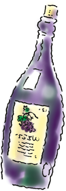
The two most common causes of acute pancreatitis are gallstones and alcohol consumption. Gallstones are the most common cause of acute pancreatitis. They pass into the bile duct and temporarily block the opening into the duodenum at the point where it's joined by the pancreatic duct. This obstructs pancreatic juices from flowing out of the pancreas into the duodenum. The backflow of these digestive juices causes lysis (dissolving) of pancreatic cells and subsequent pancreatitis. This typically mild type of pancreatitis resolves with the passage or removal of the gallstones.
Evil spirits
It's believed that ethyl alcohol, the second most common cause of acute pancreatitis, can cause the disease in several ways. Alcohol can affect the motility of the sphincter of Oddi, has direct toxic and metabolic effects on the pancreas, and forms protein plugs that obstruct small ducts in the pancreas. It's also believed that alcohol produces a temporary but significant reduction in blood flow to the pancreas. When these episodes occur repeatedly, they result in ischemic damage to the cells.
These effects can occur from long-term excessive consumption or from a single episode of binge drinking. A person sensitive to the effects of alcohol may have an attack of acute pancreatitis a few hours to a day or two after drinking; such a person may not need to drink very much alcohol to precipitate an attack. Alcohol typically results in a more severe type of pancreatitis because of the cell necrosis that occurs. With repeated attacks, pancreatitis becomes chronic.
Cause and exocrine effect
Acute pancreatitis can result from several other, less common, causes. (See Causes of acute pancreatitis.) But no matter what the cause, it's the exocrine functions that fail during acute pancreatitis. The enzymes that the pancreas normally excretes into the duodenum are instead activated within the pancreas or its ducts and begin to digest the pancreatic tissue itself. The inflammation that results causes intense pain, third-space shift of large volumes of fluids (which results in hypovolemia), pancreatic fat necrosis (with accompanying con sumption of serum calcium), and, occasionally, hemorrhage.
Imbalances caused by acute pancreatitis
Acute pancreatitis—whether mild or severe—can cause fluid and electrolyte imbalances, including hypovolemia, hyponatremia, hypocalcemia, hypomagnesemia, and hypokalemia.
Turn up the (blood) volume!
Hypovolemia is a major cause of death in patients with acute pancreatitis. Severe pancreatic damage triggers the release of systemic inflammatory mediators that produce increased capillary permeability and vasodilation. This, in turn, produces massive fluid shifts from the intravascular spaces to the interstitial spaces and the retroperitoneum, resulting in hypovolemia. Vomiting, diarrhea, excessive sweating, and, possibly, hemorrhage also contribute to hypovolemia.
The case of the lost electrolytes
Acute pancreatitis can also result in the loss of calcium, magnesium, and potassium. Vomiting and diarrhea can result in hyponatremia, hypokalemia, and (when severe) hypomagnesemia. Hyponatremia can also result from excessive sweating and increased antidiuretic hormone secretion caused by hypovolemia.
Hypocalcemia in acute pancreatitis usually results from concomitant hypoalbuminemia. Fat necrosis—caused by lipase necrosing the fat tissue in pancreatic interstitium and peripancreatic spaces—may result in the release of free fatty acids and intraperitoneal saponification, further decreasing serum calcium levels. Fat necrosis can also lead to hypomagnesemia because magnesium is deposited in areas of fat necrosis, which reduces serum levels. Because hypomagnesemia can contribute to hypocalcemia, hypomagnesemia should be corrected first.
What to look for
Often, the only symptom of mild pancreatitis is steady epigastric pain centered near the navel that's unrelieved by vomiting. In severe pancreatitis, the patient will likely report severe, persistent, piercing abdominal pain, usually in the midepigastric region, although the pain may generalize or occur in the left upper quadrant and radiate to the back or other areas. The abdominal pain is usually sudden in onset, and gradually becomes more severe until reaching a constant ache. The patient typically describes the pain as boring or penetrating and may report recent consumption of a large meal or alcohol. The pain may ease when the patient leans forward or lies on side with knees drawn toward the chest.
Sign language
Other possible signs and symptoms include nausea; vomiting; fever; mild jaundice; tachycardia; tachypnea; muscle spasms; and fatty, foul-smelling stools. Depending on the severity of the illness and degree of fluid loss and hemorrhage, the patient may also be hypotensive. Assessment may reveal Grey Turner sign (flank ecchymosis); Cullen sign (periumbilical ecchymosis); Chvostek and Trousseau signs (hypocalcemia); and abdominal distention, rigidity, and tenderness with hypoactive bowel sounds.

Pancreatitis can lead to severe, life-threatening complications. The patient will need close monitoring for signs and symptoms that may indicate such complications. (See Complications of acute pancreatitis.)
What tests show
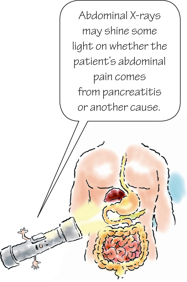
Several types of diagnostic tests can help identify acute pancreatitis, including imaging and blood studies. Severity scoring can help predict the severity of the disease and the patient's prognosis.
Imaging studies
Blood studies
Severity scoring
Several tools help predict the severity of acute pancreatitis and the patient's prognosis. It's crucial to use these tools early on because early recognition of severe pancreatitis can significantly improve the outcome. Tools include Ranson criteria, the acute physiology and chronic health evaluation (APACHE II) score, the multi organ system failure (MOSF) score, the modified Glasgow prognostic criteria, and the Balthazar score.
Ranson criteria look at several factors on admission (such as the patient's age and laboratory values) and then again 48 hours later (laboratory values and systemic effects). If the patient meets three or more criteria, the criteria predict a severe course and an increased mortality risk. (See Ranson criteria.)
The APACHE II score uses a point score calculated from routine physiologic measurements to provide a statistical prediction of mortality. However, the scoring system is time-consuming—a minus for assessing severe acute pancreatitis.
Extra sensitive
More sensitive than Ranson criteria and the APACHE II, the MOSF score predicts the severity of the disease at admission and can be recalculated daily. It's typically used on intensive care units. The scoring system evaluates seven organ systems, with a higher score indicating more severe disease. All patients with an MOSF score greater than 3 have severe disease.
Similar to Ranson criteria but with fewer criteria, the modified Glasgow criteria allows for daily scoring. The Balthazar score uses CT scan results to arrive at a prognosis. (See The Balthazar score: A CT scan severity index.)
How it's treated
Medical management for acute pancreatitis is fairly straightforward. The patient stays nothing by mouth and IV fluid is given. Analgesics help with pain management. Antibiotics are generally not required.
Keep those fluids flowing
Patients with evidence of significant third-space fluid shift and those who have a large amount of fluid in the retroperitoneal space need aggressive fluid replacement. These fluid shifts deplete intravascular volume and may lead to tachycardia, hypotension, kidney failure, hemoconcentration, and generalized circulatory collapse. Patients who develop hemoconcentration are also at increased risk for pancreatic necrosis and organ failure.
Most cases of pancreatitis respond well to colloids or lactated Ringer solution to treat hypovolemia and shock. Patients require close monitoring for electrolyte abnormalities, particularly hypo calcemia, hypokalemia, and hypomagnesemia. Fluid replacement aims to correct these imbalances and replace volume. If a patient develops necrotizing pancreatitis or experiences a sudden, severe attack with hemorrhaging, the patient may also need packed red blood cells to maintain hemodynamic stability.
Pushing back at pancreatic pain
The severe pain that typically accompanies pancreatitis calls for pain relief. For mild to moderate pain relief, acetaminophen (Tylenol) is effective. Tramadol is an analgesic for moderately severe pain. It inhibits the ascending pain pathways, altering perception of and response to pain. Meperidine (Demerol) is a synthetic opioid narcotic analgesic for the relief of severe pain. It has multiple actions similar to morphine. Due to the depressing effects on the respiratory system with opioids, careful observation of patients must be maintained with the administration of any opioid. Positioning patients in the knee-to-chest position may also help relieve pain.
No food, please—just rest
Part of treatment includes letting the pancreas rest—and that means giving the pancreas a break from food, which stimulates enzyme secretion. Initially, the patient with pancreatitis should receive nothing by mouth. As pain subsides and the patient's condition improves, the patient can begin oral intake, with careful monitoring. The patient should at first receive small amounts low in fat and protein.
If needed, antacids can help neutralize gastric secretions, and histamine antagonists can decrease hydrochloric acid production. Anticholinergic medications can reduce vagal stimulation, decrease GI motility, and inhibit pancreatic enzyme secretion. If the patient has severe pancreatitis with significant pancreatic necrosis, the patient may need insulin to correct hyperglycemia.
A patient with recurrent vomiting, gastric distention, or intestinal ileus may need a nasogastric (NG) tube inserted. An NG tube with suctioning also suppresses pancreatic secretions by decreasing gastric fluids.
Although controversial, antibiotic therapy for acute pancreatitis is indicated if secondary infection or necrotizing pancreatitis is present.
The oral route is out
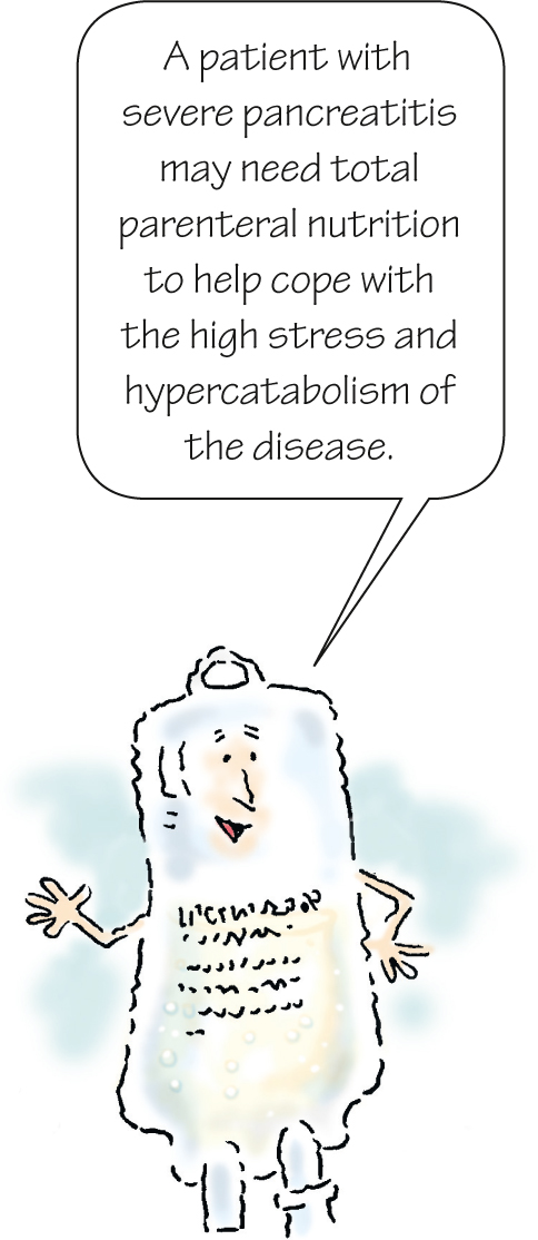
Until a patient with pancreatitis can tolerate oral intake, the patient must receive nutrition by an alternate method. In milder cases, food should be withheld for no more than 7 days. A patient with severe pancreatitis requires nutritional support because of the inherently high level of stress and hypercatabolism. Such a patient may receive total parenteral nutrition (without lipids if triglycerides are increased) or enteric feedings to maintain nutrition.
Eradicating infections
An inactive bowel allows intestinal flora to cross the colonic wall and infect the pancreas, particularly necrotic areas, which are the most vulnerable to infection.
Antibiotics can help prevent infection or treat one that's already present. If infected pancreatic necrosis arises, the patient may need surgical debridement and drainage. The health care provider may also use a CT scan to guide aspiration of necrotic areas. This allows identification of the infecting organism, leading to more effective treatment.
To remember some of the key treatments for acute pancreatitis, just think of the word PANCREAS:
Pain control
Arresting shock with IV fluids
Nasogastric intubation
Calcium monitoring
Renal evaluation
Ensuring pulmonary function
Antibiotic administration
Surgery or special procedures as needed.
Coping with complications
To prevent the disease from progressing, the patient may need surgery or ERCP to remove gallstones or other biliary tract obstructions. A patient with a pancreatic abscess or pseudocyst may need surgical drainage. If a patient develops complications in the cardiovascular, renal, pulmonary, GI, or neurologic system, the patient will need treatment for the specific complication.
How you intervene
Patients with acute pancreatitis need careful monitoring, thorough assessments, and diligent nursing care. Follow these guidelines:
Dangerous drop-offs
 If you answered all five questions correctly, bravo! You're not only acute, you're pancreatitis proficient.
 If you answered four questions correctly, that's cool! You're on your way to glandular glory.
 If you answered fewer than four questions correctly, that's okay! Just review the chapter until you can fully digest the information.
References
Medscape. (2018). Pancreatitis prognosis criteria. https://reference.medscape.com/calculator/pancreatitis-prognosis-criteria
Tang, J. (2021). Acute pancreatitis. https://emedicine.medscape.com/article/181364-overview