| Author: Fawn O. Workman
|
| Author: Fawn O. Workman
|
Phosphorus is the primary anion, or negatively charged ion, found in intracellular fluid (Willis, 2018). It is contained in the body as phosphate. (The two words—phosphorus and phosphate—are commonly used interchangeably [Alexander et al., 2014].) About 85% of phosphorus exists in bone and teeth, combined in a 1:2 ratio with calcium. About 14% is in soft tissue, and less than 1% is in extracellular fluid.
Why it's important
An essential element of all body tissues, phosphorus is vital to various body functions. It plays a crucial role in cell membrane integrity (phospholipids make up the cell membranes); muscle function; neurologic function; and the metabolism of carbohydrates, fat, and protein. Phosphorus is a primary ingredient in 2,3-diphosphoglycerate (2,3-DPG), a compound in red blood cells (RBCs) that promotes oxygen delivery from RBCs to the tissues.
Phosphorus also helps buffer acids and bases. It promotes energy transfer to cells through the formation of energy-storing substances such as adenosine triphosphate (ATP). It is also essential for white blood cell (WBC) phagocytosis and platelet function. Finally, with calcium, phosphorus is necessary for healthy bones and teeth.
The lowdown on low phosphorus levels
Normal serum phosphorus levels in adults range from 2.5 to 4.5 mg/dl (or 1.8 to 2.6 mEq/L). In comparison, the normal phosphorus level in the cells is 100 mEq/L. Plasma phosphorus (mostly as inorganic phosphate) totals about 0.2% of the body's phosphate (Cho, 2016). Because phosphorus is located primarily within the cells, serum levels may not always reflect the total amount of phosphorus in the body. For example, it's important to distinguish between a decrease in the level of serum phosphate (hypophosphatemia) and a decrease in total body storage of phosphate (phosphate deficiency).
How the body regulates phosphorus
The total amount of phosphorus in the body is related to dietary intake, hormonal regulation, kidney excretion, and transcellular shifts. For adults, the range for the recommended daily requirement of phosphorus is 800 to 1,200 mg. Phosphorus is readily absorbed through the gastrointestinal (GI) tract, facilitated by active vitamin D (Cho, 2016). The amount absorbed is proportional to the amount ingested. (See Dietary sources of phosphorus.)
Most ingested phosphorus is absorbed through the jejunum. The kidneys excrete about 90% of phosphorus as they regulate serum levels. (The GI tract excretes the rest.) If dietary intake of phosphorus increases, the kidneys increase excretion to maintain normal levels of phosphorus. A low-phosphorus diet causes the kidneys to conserve phosphorus by reabsorbing more of it in the proximal tubules.
Balancing it out with PTH
The parathyroid gland controls the hormonal regulation of phosphorus levels by affecting the activity of parathyroid hormone (PTH). (See PTH and phosphorus.) Changes in calcium levels, rather than changes in phosphorus levels, affect the release of PTH. You may recall that phosphorus balance is closely related to calcium balance.
Calcium and phosphorus have an inverse relationship. For instance, when the serum calcium level is low, the phosphorus level is high. As a result, the parathyroid gland releases PTH, which causes an increase in calcium and phosphorus resorption from bone, raising both calcium and phosphorus levels. Phosphorus absorption from the intestines also increases. (Activated vitamin D—calcitriol—also enhances phosphorus absorption in the intestines.)
Kidneys enter the equation
PTH also acts on the kidneys to increase excretion of phosphorus. The renal effect of PTH outweighs its other effects on the serum phosphorus level particularly that of returning the phosphorus level to normal. Reduced PTH levels allow for phosphorus reabsorption by the kidneys resulting in a rise in serum levels.
Shifty business
Certain conditions cause phosphorus to move, or shift, in and out of cells. Insulin moves not only glucose but also phosphorus into the cell—alkalosis results in the same kind of phosphorus shift. Those shifts affect serum phosphorus levels. (See Older adult patients at risk.)
 <2.5 mg/dl or <1.8 mEq/L
Hypophosphatemia occurs when the serum phosphorus level falls below 2.5 mg/dl (or 1.8 mEq/L). Although this condition generally indicates a deficiency of phosphorus, it can occur under various circumstances when total body phosphorus stores are normal. Severe hypophosphatemia occurs when serum phosphorus levels are less than 1 mg/dl, and the body cannot support its energy needs with decreased oxygenation of tissue and impaired cell metabolism. The condition may result in muscle weakness, paresthesias, encephalopathy, respiratory failure or difficulty weaning from ventilator due to diaphragm weakness, hematologic impairments, coma, dysrhythmias, and rhabdomyolysis (Alexander et al., 2014; Cho, 2016).
How it happens
Three underlying mechanisms can lead to hypophosphatemia: a shift of phosphorus from extracellular fluid to intracellular fluid, a decrease in intestinal absorption of phosphorus, and an increased loss of phosphorus through the kidneys. Some causes of hypophosphatemia may involve more than one mechanism.
Several factors may cause phosphorus to shift from extracellular fluid into the cell. Here are the most common causes.
When hyperventilation happens
Respiratory alkalosis, one of the most common causes of hypophosphatemia, can stem from several conditions that produce hyperventilation, including sepsis, alcohol withdrawal, heatstroke, pain, anxiety, diabetic ketoacidosis (DKA), hepatic encephalopathy, and acute salicylate poisoning. Although the mechanism that prompts respiratory alkalosis to induce hypophosphatemia is unknown, the response is a shift of phosphorus into the cells and a resulting decrease in serum phosphorus levels.
Sugar high
Hyperglycemia, an elevated serum glucose level, causes the release of insulin, which transports glucose and phosphorus into the cells. The same effect may occur in a patient with diabetes who's receiving insulin or in a significantly malnourished patient; at particular risk for malnourishment are older adults, those with alcohol use disorders, and those with eating disorders including anorexia nervosa.
Failure to add phosphorus
After initiation of enteral or parenteral feeding without enough phosphorus supplementation, phosphorus shifts into the cells. This shift—called refeeding syndrome—usually occurs 3 or more days after feedings begin. Patients recovering from hypothermia can also develop hypophosphatemia as phosphorus moves into the cells.
Abnormal absorption
Malabsorption syndromes, starvation, and prolonged or excessive use of phosphorus-binding antacids or sucralfate are among the many causes of impaired intestinal absorption of phosphorus. Because vitamin D contributes to intestinal absorption of phosphorus, inadequate vitamin D intake or synthesis can inhibit phosphorus absorption. Chronic diarrhea or laxative abuse can also result in increased GI loss of phosphorus. Decreased dietary intake rarely causes hypophosphatemia because phosphate is found in most foods.
Calling the kidneys to account
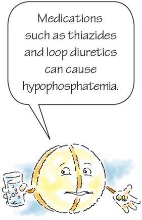
Diuretic use is the most common cause of phosphorus loss through the kidneys. Thiazides, loop diuretics, and acetazolamide are the diuretics that most commonly cause hypophosphatemia. (See Medications associated with hypophosphatemia.)
The second most common cause is DKA in patients with diabetes who have poorly controlled blood glucose levels. In DKA, high glucose levels induce an osmotic diuresis resulting in significant loss of phosphorus from the kidneys. Ethanol affects phosphorus reabsorption in the kidneys so that more phosphorus is excreted in urine. Chronic alcohol use–related renal tubular dysfunction may be reversed after a month of abstinence (Cho, 2016).
A buildup of PTH, which occurs with hyperparathyroidism and hypocalcemia, also leads to hypophosphatemia because PTH stimulates the kidneys to excrete phosphate. Finally, hypophosphatemia occurs in patients who have extensive burns. Although the mechanism is unclear, the condition seems to happen in response to the extensive diuresis of salt and water that typically occurs during the first 2 to 4 days after a burn injury. Respiratory alkalosis and carbohydrate administration may also play a role here.
What to look for
Mild to moderate hypophosphatemia doesn't usually cause symptoms. Noticeable effects of hypophosphatemia typically occur only in severe cases. The characteristics of severe hypophosphatemia are apparent in many organ systems. Signs and symptoms may develop acutely because of rapid decreases in phosphorus or gradually, as the result of slow, chronic reductions in phosphorus.
Hypophosphatemia affects the musculoskeletal, central nervous, cardiac, and hematologic systems. Because phosphorus is required to make high-energy ATP, many of the signs and symptoms of hypophosphatemia are related to low energy stores.
Weak and weary
With hypophosphatemia, muscle weakness is the most common symptom. Other symptoms may include diplopia (double vision), malaise, and anorexia. The patient may experience a weakened hand grasp, slurred speech, or dysphagia. The patient may also develop myalgia (tenderness or pain in the muscles).
Respiratory failure may result from weakened respiratory muscles and poor contractility of the diaphragm. Respirations may appear shallow and ineffective. In later stages, the patient may be cyanotic. Keep in mind that it may be difficult to wean a mechanically ventilated patient with hypophosphatemia from the ventilator.
With severe hypophosphatemia, rhabdomyolysis (skeletal muscle destruction) can occur with altered muscle cell activity. Muscle enzymes such as creatine kinase are released from the cells into the extracellular fluid. Loss of bone density, osteomalacia (softening of the bones), and bone pain may also occur with prolonged hypophosphatemia. Fractures can result.
Logical neurologic effects
Without enough phosphorus, the body cannot make enough ATP, a cornerstone of energy metabolism. As a result, central nervous system cells can malfunction, causing paresthesia, irritability, apprehension, memory loss, and confusion. The neurologic effects of hypophosphatemia may progress to seizures or coma.

When the heart isn't hardy
The heart's contractility decreases because of low energy stores of ATP. As a result, the patient may develop hypotension and low cardiac output. Severe hypophosphatemia may lead to cardiomyopathy, which treatment can reverse.
Oxygen delivery drop-off
A drop in the production of 2,3-DPG causes a decrease in oxygen delivery to tissues. Because hemoglobin has a stronger affinity for oxygen than for other gases, oxygen is less likely to be given up to the tissues because it circulates through the body. As a result, less oxygen is delivered to the myocardium, which can cause chest pain.
Hypophosphatemia may also cause hemolytic anemia because of changes in the structure and function of RBCs. Patients with hypophosphatemia are more susceptible to infection because of the effect of low levels of ATP in WBCs. Lack of ATP results in a decreased functioning of leukocytes. Chronic hypophosphatemia also affects platelet function, resulting in bruising and bleeding, particularly mild GI bleeding.
What tests show
These diagnostic test results may indicate hypophosphatemia or a related condition:
How it is treated
Treatment varies with the severity and cause of the condition. It includes addressing the underlying cause and correcting the imbalance with phosphorus replacement and a high-phosphorus diet. The route of replacement therapy depends on the severity of the imbalance.
Milder measures
Oral/dietary replacement of phosphate is preferred for mild to moderate acute hypophosphatemia and chronic hypophosphatemia (Cho, 2016). Treatment includes a diet high in phosphorus-rich foods, such as eggs, nuts, whole grains, organ meats, fish, poultry, and milk products. However, if calcium is contraindicated or the patient can't tolerate milk, the patient should instead receive oral phosphorus supplements. Oral supplements include Neutra-Phos and Neutra-Phos-K and can be used for moderate hypophosphatemia. Dosage limitations are related to the adverse effects, most notably nausea and diarrhea. (See When dietary changes aren't working.)
Sterner steps
For patients with severe hypophosphatemia or a nonfunctioning GI tract, IV phosphorus replacement is the recommended choice. Two preparations are used: IV potassium phosphate and IV sodium phosphate. Dosage is guided by the patient's response to treatment and serum phosphorus levels.
Potassium phosphate requires slow administration (no more than 10 mEq/hour). Adverse effects of IV replacement for hypophosphatemia include hyperphosphatemia and hypocalcemia. Once serum phosphorus levels exceed 1 mg/dl, the patient can usually be transitioned to oral replacement therapy (Cho, 2016).
How you intervene
If your patient begins total parenteral nutrition or is otherwise at risk for developing hypophosphatemia, monitor for signs and symptoms of this imbalance. If the patient has already developed hypophosphatemia, your nursing care should focus on careful monitoring, safety measures, and interventions to restore normal serum phosphorus levels. (See Teaching about hypophosphatemia.) Alert the practitioner to any changes in the patient's condition and take these other actions:
Monitor for hypotension, which may occur with a too rapid infusion rate. Serum calcium, phosphate, and potassium should be assessed every 6 hours during repletion. Magnesium levels should also be monitored (Cho, 2016).
 >4.5 mg/dl or >2.6 mEq/L
Hyperphosphatemia occurs when serum phosphorus levels exceed 4.5 mg/dl (or 2.6 mEq/L) and usually reflects the kidneys' inability to excrete excess phosphorus. The condition commonly occurs along with an increased release of phosphorus from damaged cells. Severe hyperphosphatemia occurs when the serum phosphorus levels reach 6 mg/dl or higher.
How it happens
Hyperphosphatemia can result from several underlying mechanisms, including impaired renal excretion of phosphorus, a shift of phosphorus from the intracellular fluid to the extracellular fluid, and an increase in dietary intake of phosphorus.
Kidney filter failure
Usually, renal excretion of phosphorus equals the amount the GI tract absorbs daily. Hyperphosphatemia most commonly results from kidney failure due to the kidneys' inability to excrete excess phosphorus.
When glomerular filtration rate drops below 30 ml/minute, the kidneys can't filter excess phosphorus adequately. Because the kidneys are responsible for most of the excretion of phosphorus, their inability to filter phosphorus leads to an elevated serum phosphorus level.
PTH problem
A risk after thyroid or parathyroid surgery, hypoparathyroidism impairs synthesis of PTH. When less PTH is synthesized, less phosphorus is excreted from the kidneys. The result? Elevated serum phosphorus levels.
Shift work
Several conditions can cause phosphorus to shift from the intra cellular fluid to the extracellular fluid. Acid-base imbalances, such as respiratory acidosis and DKA, are typical examples. Anything that causes cell destruction can also result in a transcellular shift of phosphorus.
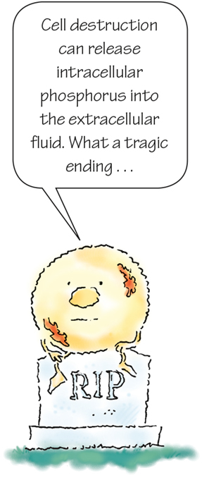
Destruction of cells can trigger the release of intracellular phosphorus into the extracellular fluid, causing serum phosphorus levels to rise. Chemotherapy, for example, causes significant cell destruction, as do muscle necrosis and rhabdomyolysis, conditions that can stem from infection, heatstroke, and trauma.
Increased intake
Excessive intake of phosphorus can result from overadministration of phosphorus supplements or of laxatives or enemas that contain phosphorus (such as Fleet enemas). (See Medications associated with hyperphosphatemia.)
In infants, excessive intake of vitamin D can result in increased absorption of phosphorus and lead to elevated serum phosphorus levels. (See Cow's milk and hyperphosphatemia.)
What to look for
Hyperphosphatemia causes few clinical problems by itself. However, phosphorus and calcium levels have an inverse relationship: If one is high, the other is low. Because of this seesaw relationship, hyperphosphatemia may lead to hypocalcemia, which can be life-threatening. Signs and symptoms of acute hyperphosphatemia are usually caused by the effects of hypocalcemia.
The patient may develop paresthesia in the fingertips and around the mouth, which may increase in severity and spread proximally along the limbs and to the face. Severe muscle spasm, cramps, pain, and weakness may prevent the patient from performing normal activities. The patient may exhibit hyperreflexia and positive Chvostek and Trousseau signs. These signs are due to low calcium levels and may progress to tetany and neurologic disorders.
Neurologic signs and symptoms include decreased mental status, delirium, and seizures. Electrocardiogram (ECG) changes include a prolonged QT interval and ST segment. The patient may also experience hypotension, heart failure, anorexia, nausea, and vomiting. Bone development may also be affected.
To remember some of the signs and symptoms of hyperphosphatemia, think of the word CHEMO (keeping in mind that chemotherapy can lead to hyperphosphatemia):
Cardiac irregularities
Hyperreflexia
Eating poorly
Muscle weakness
Oliguria.
Calcification cues
When phosphorus levels rise, phosphorus binds with calcium, forming an insoluble compound called calcium phosphate. Organ dysfunction can result when calcium phosphate precipitates or is deposited, in the heart, lungs, kidneys, or other soft tissues. This process, called calcification, usually occurs as a result of chronically elevated phosphorus levels. (See A look at calcification.)
With calcification, the patient may experience arrhythmias, an irregular heart rate, and decreased urine output. Corneal haziness, conjunctivitis, cataracts, and impaired vision may occur, and papular eruptions may develop on the skin.
What tests show
The following diagnostic tests results may indicate hyperphosphatemia or a related condition such as hypocalcemia:
How it's treated
An elevated serum phosphorus level may be treated with medications and other therapeutic measures. Treatment aims to correct the underlying disorder, if one exists, and correct hypocalcemia.
Going low phospho
If a patient's elevated serum phosphorus level results from excessive phosphorus intake, the condition may be easily remedied by reducing phosphorus intake. Therapeutic measures include reducing dietary intake of phosphorus and eliminating the use of phosphorus-based laxatives and enemas. (See When dietary changes aren't enough.)
Altering absorption
Medication therapy may help decrease absorption of phosphorus in the GI system. Such treatment may include aluminum, magnesium, or calcium gel or phosphate-binding antacids. To avoid the risk of aluminum accumulation and associated neurotoxicity, limit use of aluminum-containing products to no more than a few days (Cho, 2016). Although widely used, calcium salts such as calcium carbonate and calcium acetate may cause hypercalcemia, and the patient will need careful dosing. Polymeric phosphate binders such as sevelamer hydrochloride may also be given. Anyone with underlying renal insufficiency or kidney failure should not receive magnesium antacids because they may cause hypermagnesemia. A patient with end-stage renal disease may receive lanthanum carbonate, a noncalcium, nonaluminum phosphate binder.
Keep in mind that a mildly elevated phosphorus level may benefit a patient with kidney failure. Higher phosphorus levels (on the higher side of the normal range) allow more oxygen to move from the RBCs to tissues, which can help prevent hypoxemia and limit the effects of chronic anemia on oxygen delivery.
Treat what's underneath
Treatment of the underlying cause of hyperphosphatemia, including conditions such as respiratory acidosis or DKA, can lower serum phosphorus levels. In a patient with diabetes, administering insulin causes phosphorus to shift back into the cells, which can result in decreased serum phosphorus levels.
Situation: Severe
Patients with severe hyperphosphatemia may receive IV saline solution to promote renal excretion of phosphorus. However, this treatment requires the patient to have functional kidneys and the ability to tolerate the increased load of sodium and fluid. Patients may also receive proximal diuretics such as acetazolamide to increase renal excretion of phosphorus.
As a final therapeutic intervention, hemodialysis or peritoneal dialysis may be initiated if the patient has chronic kidney failure or an extreme case of acute hyperphosphatemia with symptomatic hypocalcemia.
How you intervene
Keep an eye out for patients at risk for hyperphosphatemia and monitor them carefully. Also, use care when administering phosphorus in IV infusions, enemas, and laxatives because the extra phosphorus may cause hyperphosphatemia.
If your patient has already developed hyperphosphatemia, your care should focus on careful monitoring, safety measures, and interventions to restore normal serum phosphorus levels. Follow these steps to provide care for the patient:
 If you answered all five questions correctly, you're phosphabulous! Keep up the good work.
 If you answered four questions correctly, way to go! You're a natural when it comes to phosphorus balance.
 If you answered fewer than four questions correctly, that's okay. Take a break to renew your mental power and then review the chapter again.
References
Alexander, M., Corrigan, A. M., Gorski, L. A., & Phillips, L. (2014). Core curriculum for infusion nursing (4th ed.). Lippincott Williams & Wilkins.
Blaine, J., Chonchol, M., & Levi, M. (2015). Renal control of calcium, phosphate, and magnesium homeostasis. Clinical Journal of the American Society of Nephrology, 10(7), 1257–1272. https://www.ncbi.nlm.nih.gov/pmc/articles/PMC4594074/
Cho, K. C. (2016). Electrolyte & acid-base disorders. In M. A. Papadakis, S. J. McPhee, & M. W. Rabow (Eds.), Current medical diagnosis & treatment 2017 (56th ed., pp. 901–912). McGraw-Hill Education.
Chapter 6: Disruptions in homeostasis. (2018). In Willis, L. (Ed.), Lippincott certification review: Medical-surgical nursing (6th ed., pp. 59–88). Wolters Kluwer.
Kidney Disease: Improving Global Outcomes CKD-MBD Update Work Group. (2011). KDIGO 2017 Clinical Practice Guideline update for the diagnosis, evaluation, prevention, and treatment of chronic kidney disease-mineral and bone disorder (CKD-MBD). Kidney International Supplements, 7(1), 1–59. https://www.kisupplements.org/article/S2157-1716(17)30001-1/fulltext
Shaman, A. M., & Kowalski, S. R. (2016). Hyperphosphatemia management in patients with chronic kidney disease. Saudi Pharmaceutical Journal, 24(4), 494–505. https://www.ncbi.nlm.nih.gov/pmc/articles/PMC4908098/
St-Jules, D. E., Goldfarb, D. S., Pompeii, M. L., & Sevick, M. A. (2017). Phosphate additive avoidance in chronic kidney disease. Diabetes Spectrum, 30(2), 101–106. https://www.ncbi.nlm.nih.gov/pmc/articles/PMC5439363/
Stremke, E. R., & Hill Gallant, K. M. (2018). Intestinal phosphorus absorption in chronic kidney disease. Nutrients, 10(10), 1364. https://www.ncbi.nlm.nih.gov/pmc/articles/PMC6213936/