section name header

Disease Prologue

Summary

Cancers Staged Using This Staging System

This classification applies to carcinomas of the lung, including non-small cell and small cell carcinomas, and bronchopulmonary carcinoid tumors.

Cancers Not Staged Using This Staging System

This classification does not apply to sarcomas or other rare tumors of the lung.

Summary of Changes

The changes introduced in the AJCC Cancer Staging Manual, 8th Edition of the TNM classification for lung cancer derive from analyses of the new retrospective and prospective databases of the International Association for the Study of Lung Cancer (IASLC). These databases contain information on patients diagnosed with lung cancer from 1999 to 2010 originating from 35 different databases in 16 countries around the world.1

ChangeDetails of ChangeLevel of Evidence
Definition of Primary Tumor (T)Tis: Adenocarcinoma in situ (AIS), Tis (AIS), added in addition to squamous carcinoma in situ (SCIS), Tis (SCIS)II2
Definition of Primary Tumor (T)T1mi: Addition of a new T category: minimally invasive adenocarcinomaII2
Definition of Primary Tumor (T)T1: Subdivision into T1a, T1b, and T1c at 1-cm intervals from less than or equal to 1 cm to less than or equal to 3 cmII3
Definition of Primary Tumor (T)T2: Subdivision into T2a and T2b at 1-cm intervals from greater than 3 cm to less than or equal to 5 cmII3
Definition of Primary Tumor (T)T2: Tumors with endobronchial location less than 2 cm from the carina, but without involving the carina, are now included in this T category.II3
Definition of Primary Tumor (T)T2: Tumors with complete atelectasis or pneumonitis are now included in this category.II3
Definition of Primary Tumor (T)T3: Tumors greater than 5 cm but less than or equal to 7 cm are now included in this T category.II3
Definition of Primary Tumor (T)T3: Invasion of the mediastinal pleura is no longer used as a T descriptor.II3
Definition of Primary Tumor (T)T4: Tumors greater than 7 cm are now included in this T category.II3
Definition of Primary Tumor (T)T4: Tumors with invasion of the diaphragm are now included in this T category.II3
Definition of Distant Metastasis (M)M1b: The revised M1b category now includes tumors with a single extrathoracic metastasis in a single organ.II4
Definition of Distant Metastasis (M)M1c: This new M1 category includes tumors with multiple extrathoracic metastases in one or multiple organs.II4
AJCC Prognostic Stage GroupsStage IA now is divided into three stages—IA1, IA2, and IA3—to include T1aN0M0, T1bN0M0, and T1cN0M0 tumors, respectively.II5
AJCC Prognostic Stage GroupsStage IIB now includes T1aN1M0, T1bN1M0, T1cN1M0, and T2aN1M0 tumors.II5
AJCC Prognostic Stage GroupsStage IIIB now includes T3N2M0 tumors.II5
AJCC Prognostic Stage GroupsStage IIIC: This new stage includes T3N3M0 and T4N3M0 tumors.II5
AJCC Prognostic Stage GroupsStage IVA includes tumors with any T and any N, but with M1a or M1b disease.II5
AJCC Prognostic Stage GroupsStage IVB includes tumors with any T and any N, but with M1c disease.II5

ICD-O-3 Topography Codes

CodeDescription
C34.0Main bronchus
C34.1Upper lobe, lung
C34.2Middle lobe, lung
C34.3Lower lobe, lung
C34.8Overlapping lesion of lung
C34.9Lung, NOS

WHO Classification of Tumors

This list includes histology codes and preferred terms from the WHO Classification of Tumors and the International Classification of Diseases for Oncology (ICD-O). Most of the terms in this list represent malignant behavior. For cancer reporting purposes, behavior codes /3 (denoting malignant neoplasms), /2 (denoting in situ neoplasms), and in some cases /1 (denoting neoplasms with uncertain and unknown behavior) may be appended to the 4-digit histology codes to create a complete morphology code.

CodeDescription
8000Neoplasm, malignant
8010Carcinoma, NOS
8012Large cell carcinoma
8013Large cell neuroendocrine carcinoma
8013Combined large cell neuroendocrine carcinoma
8022Pleomorphic carcinoma
8023NUT carcinoma
8031Giant cell carcinoma
8032Spindle cell carcinoma
8033Pseudosarcomatous carcinoma
8040Diffuse idiopathic pulmonary neuroendocrine cell hyperplasia
8041Small cell carcinoma
8042Oat cell carcinoma
8045Combined small cell carcinoma
8070Squamous cell carcinoma
8070Squamous cell carcinoma in situ
8071Keratinizing squamous cell carcinoma
8072Non-keratinizing squamous cell carcinoma
8082Lymphoepithelioma-like carcinoma
8083Basaloid squamous cell carcinoma
8140Adenocarcinoma
8140Adenocarcinoma in situ
8144Enteric adenocarcinoma
8200Adenoid cystic carcinoma
8230Solid adenocarcinoma
8240Typical carcinoid
8246Neuroendocrine carcinoma, NOS
8249Atypical carcinoid
8250Adenocarcinoma in situ, non-mucinous
8250Lepidic adenocarcinoma
8252Bronchiolo-alveolar carcinoma, non-mucinous
8253Invasive mucinous adenocarcinoma
8253Adenocarcinoma in situ, mucinous
8254Mixed invasive mucinous and non-mucinous adenocarcinoma
8255Adenocarcinoma with mixed subtypes
8256Minimally invasive adenocarcinoma, non-mucinous
8257Minimally invasive adenocarcinoma, mucinous
8260Papillary adenocarcinoma
8265Micropapillary adenocarcinoma
8333Fetal adenocarcinoma
8430Mucoepidermoid carcinoma
8480Colloid adenocarcinoma
8481Mucin-producing adenocarcinoma
8551Acinar adenocarcinoma
8560Adenosquamous carcinoma
8562Epithelial-myoepithelial carcinoma
8972Pulmonary blastoma
8980Carcinosarcoma

Histology is not ideal for clinical use in patient care, as it describes an unspecified or outdated diagnosis. Data collectors may use this code only if there is not enough information in the medical record to document a more specific diagnosis.

Travis WD, Brambilla E, Burke AP, Marx A, Nicholson AG, eds. World Health Organization Classification of Tumours of the Lung, Pleura, Thymus and Heart. Lyon: IARC; 2015. Used with permission.

International Agency for Research on Cancer, World Health Organization. International Classification of Diseases for Oncology. ICD-O-3-Online.http://codes.iarc.fr/home. Accessed August 16, 2017. Used with permission.

Introduction

Lung cancer is the most frequent cancer diagnosed and the leading cause of cancer mortality in the world. The World Health Organization (WHO) reported that in 2012, more than 1.8 million lung cancers were diagnosed, for an incidence of 13%, the highest among all cancers except nonmelanoma skin cancers. In addition, more than 1.5 million patients died from the disease, for a 19.4% cancer mortality rate, again number one among all neoplasms.8 Lung cancer is classified according to the TNM system, which codes the anatomic extent of the disease and is the most important prognosticator we have to date. As such, the classification does not include clinical, biological, molecular, or genetic descriptors, although they may be used in combination with the TNM classification to build prognostic groups, different from stage groups, which are combinations of tumors with TNMs of similar prognosis.

For the second consecutive time, the revision of the TNM classification for lung cancer has derived from analyses of the new retrospective and prospective databases collected by the IASLC.1 Detailed analyses of the T, N, and M components of the classification; of the stages; and of the applicability of the TNM classification to small cell lung cancer already have been published and are the basis for the recommendations for changes in the 8th Edition of the TNM classification.3-5,9,10

Whereas there is no alternative way to classify non-small cell lung cancer anatomically, small cell lung cancer may be classified by the dichotomous “limited and extensive disease” system and by the TNM classification. However, the analyses performed in revising the AJCC Cancer Staging Manual, 6th Edition of the classification for the AJCC Cancer Staging Manual, 7th Edition clearly showed the advantages of classifying small cell lung cancer with the TNM system at clinical and pathological staging.11,12 The new analyses performed for the 8th Edition of the classification confirm that the TNM system works for small cell lung cancer and that its use increases our capacity to indicate prognosis, as limited disease may be subdivided in anatomic stages from IA1 to IIIC, with a wide range of 5-year survival rates (93% for Stage IA1 and 19% for Stage IIIC), progressively worsening as tumor stage increases. This prognostic refinement is lost if the dichotomous classification is used.10 Therefore, the recommendation to use the TNM to classify small cell lung cancer is emphasized in the 8th Edition, especially to indicate prognosis and to stratify tumors in future clinical trials.

Although the TNM classification does not explain all the prognostic variability found in lung cancer, it is the strongest prognosticator. The anatomic classification may be determined by a variety of noninvasive and invasive methods. It is universally applicable in different medical settings and constitutes the basis for planning therapy and therapeutic clinical trials. Therefore, even if prognostic groups are built in the future to refine prognosis for individual patients, the TNM classification system will remain a fundamental component among other variables. Its periodic revision is relevant, because the more robust the TNM classification, the more important its contribution to prognostic groups.13

Anatomy

Primary Site(s)

For the purpose of TNM classification, the lungs are not paired organs but a single organ.14 Basically, they are formed by the bronchi and the lung parenchyma. Lung cancer is a bronchogenic neoplasm arising from the epithelial cells of the bronchial mucosa or from the cells lining the alveoli. The right lung has three lobes—upper, middle, and lower—with three, two, and five segments, respectively. The left lung has two lobes—upper and lower—with five and four segments, respectively. The segment is considered the smallest anatomic unit of the lung.

Although all lung cancers may be located in any part of the lung, squamous cell and small cell carcinomas tend to arise from the mucosa of the more central bronchi, involving the lobar origins and the main bronchi. This central location often causes bronchial obstruction and atelectasis, either lobar or complete. The natural progression of these central tumors is to invade the bronchial wall and the mediastinal structures, such as the pericardium, the phrenic nerve, the superior vena cava, and more rarely, the esophagus, the aorta, and the heart. On the other hand , adenocarcinomas tend to locate in the periphery of the lung, with extension to the visceral pleura, often causing pleural dissemination and malignant pleural effusion, and to the chest wall. The earlier adenocarcinomas, such as adenocarcinoma in situ and minimally invasive adenocarcinoma, also tend to be located peripherally. The fact that lung lesions do not generate pain and that lung compliance allows tumors to grow within the lung parenchyma accounts for the late diagnosis of the disease. Only when the tumor causes bronchial obstruction and subsequent atelectasis, pneumonia or dyspnea, bleeding from the bronchial mucosa, or pain due to invasion of the parietal pleura, do patients present with symptoms, and the diagnostic process begins. A high index of suspicion is needed to avoid minimizing the nonspecific symptoms and attributing them to benign diseases.

Regional Lymph Nodes

Spread to the regional lymph nodes is a common feature in lung cancer. The natural progression from the primary tumor to the intrapulmonary, hilar, mediastinal, and supraclavicular lymph nodes is not found in every patient with lung cancer and nodal disease. Some patients have mediastinal nodal disease without intrapulmonary or hilar nodal involvement, which is referred to as skip metastases. Figure 36.1 shows the regional pulmonary, mediastinal, and supraclavicular lymph nodes, and Table 36.1 describes the anatomic limits of the nodal stations and their grouping in nodal zones.15

Lymph node station number (#)DescriptionAnatomic limits
Supraclavicular zone
1Low cervical, supraclavicular, and sternal notch nodes
  • Upper border: lower margin of cricoid cartilage
  • Lower border: clavicles bilaterally and , in the midline, the upper border of the manubrium. 1R designates right-sided nodes, 1L left-sided nodes in this region.
  • For lymph node station 1, the midline of the trachea serves as the border between 1R and 1L.
Upper zone
2Upper paratracheal nodes

2R

  • Upper border: apex of the right lung and pleural space and , in the midline, the upper border of the manubrium
  • Lower border: intersection of caudal margin of the innominate vein with the trachea
  • Like lymph node station 4R, 2R includes nodes extending to the left lateral border of the trachea

2L

  • Upper border: apex of the lung and pleural space and , in the midline, the upper border of the manubrium
  • Lower border: superior border of the aortic arch
3Prevascular and retrotracheal nodes

3a: Prevascular

  • On the right
    • Upper border: apex of chest
    • Lower border: level of carina
    • Anterior border: posterior aspect of sternum
    • Posterior border: anterior border of superior vena cava
  • On the left
    • Upper border: apex of chest
    • Lower border: level of carina
    • Anterior border: posterior aspect of sternum
    • Posterior border: left carotid artery

3p: Retrotracheal

  • Upper border: apex of chest
  • Lower border: carina
4Lower paratracheal nodes

4R—includes right paratracheal nodes, and pretracheal nodes extending to the left lateral border of the trachea

  • Upper border: intersection of caudal margin of innominate vein with the trachea
  • Lower border: lower border of the azygos vein

4L—includes nodes to the left of the left lateral border of the trachea, medial to the ligamentum arteriosum

  • Upper border: upper margin of the aortic arch
  • Lower border: upper rim of the left main pulmonary artery
Aortopulmonary zone
5Subaortic (aortopulmonary window)

Subaortic lymph nodes lateral to the ligamentum arteriosum

  • Upper border: lower border of the aortic arch
  • Lower border: upper rim of the left main pulmonary artery
6Para-aortic nodes (ascending aorta or phrenic)

Lymph nodes anterior and lateral to the ascending aorta and aortic arch

  • Upper border: a line tangential to the upper border of the aortic arch
  • Lower border: lower border of the aortic arch
Subcarinal zone
7Subcarinal nodes
  • Upper border: carina of the trachea
  • Lower: border: upper border of the lower lobe bronchus on the left; lower border of the bronchus intermedius on the right
Lower zone
8Paraesophageal nodes (below carina)

Nodes lying adjacent to the wall of the esophagus and to the right or left of the midline, excluding subcarinal nodes

  • Upper border: upper border of the lower lobe bronchus on the left; lower border of the bronchus intermedius on the right
  • Lower border: diaphragm
9Pulmonary ligament nodes

Nodes lying within the pulmonary ligament

  • Upper border: inferior pulmonary vein
  • Lower border: diaphragm
Hilar/interlobar zone
10Hilar nodes

Includes nodes immediately adjacent to the mainstem bronchus and hilar vessels, including proximal portions of the pulmonary veins and main pulmonary artery

  • Upper border: lower rim of the azygos vein on the right; upper rim of the pulmonary artery on the left
  • Lower border: interlobar region bilaterally
11Interlobar nodes

Between the origin of the lobar bronchi; optional notations for subcategories of station:

  • 11s: between the upper lobe bronchus and bronchus intermedius on the right
  • 11i: between the middle and lower bronchi on the right
Peripheral zone
12Lobar nodesAdjacent to the lobar bronchi
13Segmental nodesAdjacent to the segmental bronchi
14Subsegmental nodesAdjacent to the subsegmental bronchi

Adapted from Rusch et al.,15 with permission.

36.1 International Association for the Study of Lung Cancer lymph node map (From Rusch et al.15 Copyright © 2008 Aletta Ann Frazier, MD).

Metastatic Sites

Although any organ may be the site of metastasis from primary lung cancer, the brain, bones, adrenal gland s, contralateral lung, liver, pericardium, kidneys, and subcutaneous tissue are the most common sites of metastatic spread. In the absence of specific clinical findings, the staging process should focus on ruling out metastasis in these common sites.

Classification Rules

Clinical Classification

Clinical classification or pretreatment clinical classification, designated TNM or cTNM, is essential for selecting and evaluating therapy. It is based on the evidence found before treatment, including the results of history and physical examination, imaging studies (e.g., computed tomography [CT] and positron emission tomography [PET]), laboratory tests, and staging procedures such as bronchoscopy or esophagoscopy with or without ultrasound-guided biopsy (e.g., using endobronchial ultrasound [EBUS] or endoscopic ultrasound [EUS]), mediastinoscopy, mediastinotomy, extended cervical mediastinoscopy, thoracentesis, pleural biopsy, pericardioscopy, thoracoscopy, and video-assisted thoracoscopic surgery, as well as exploratory thoracotomy.

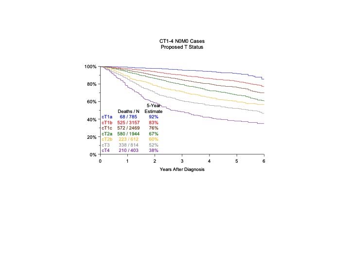
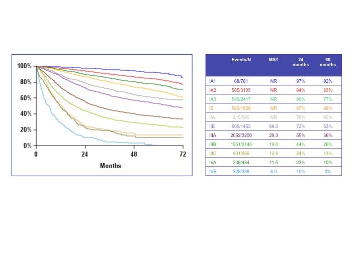
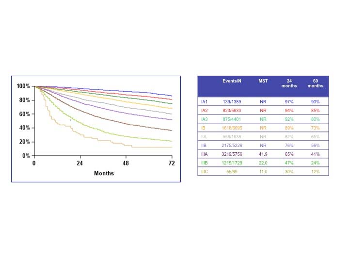
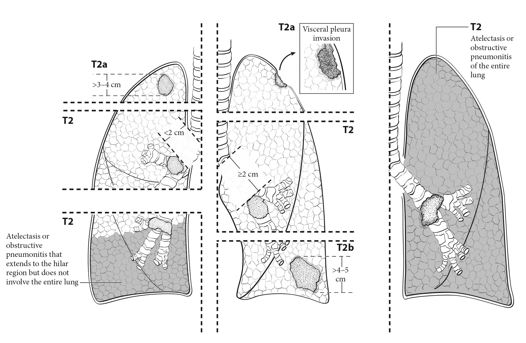
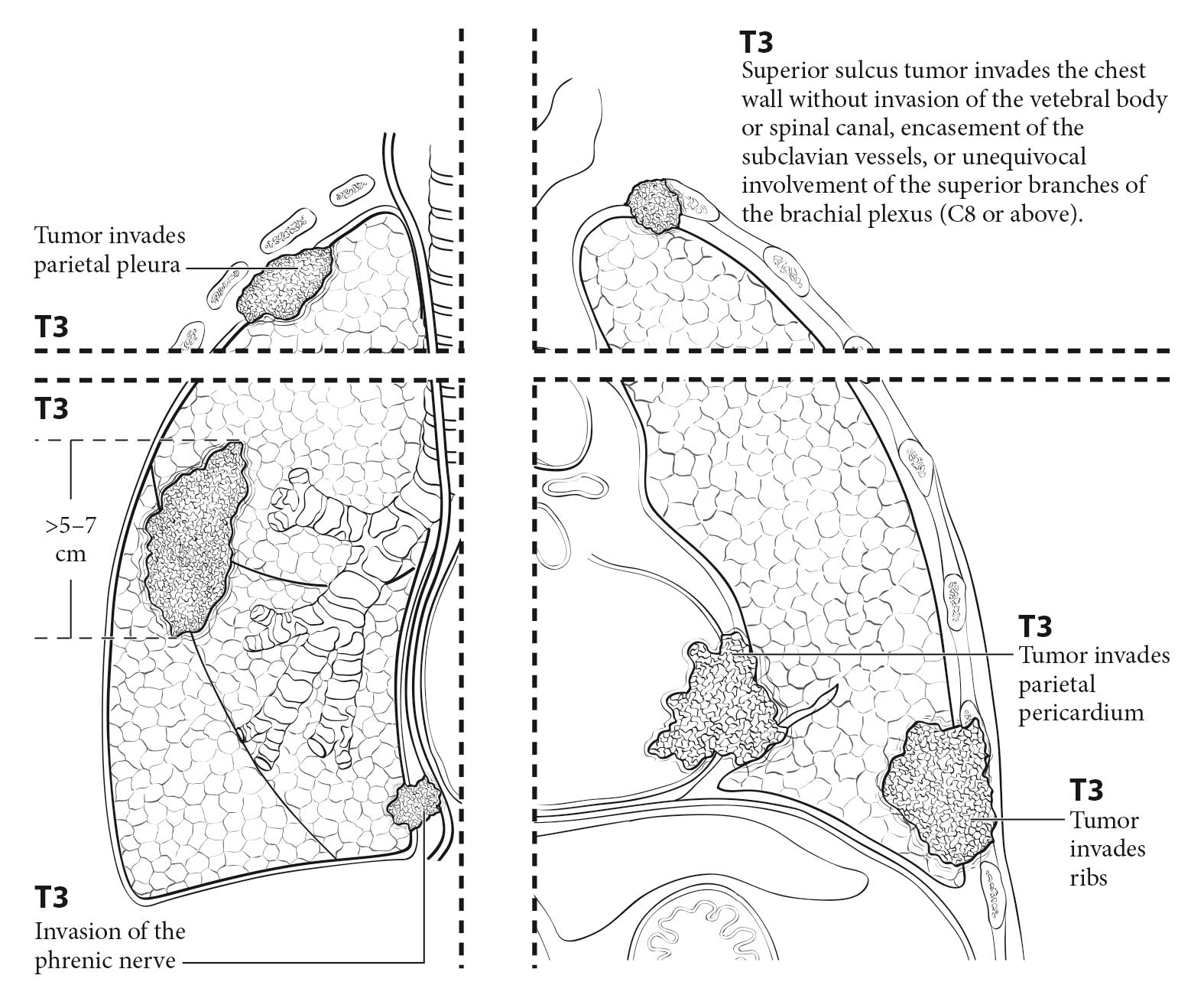
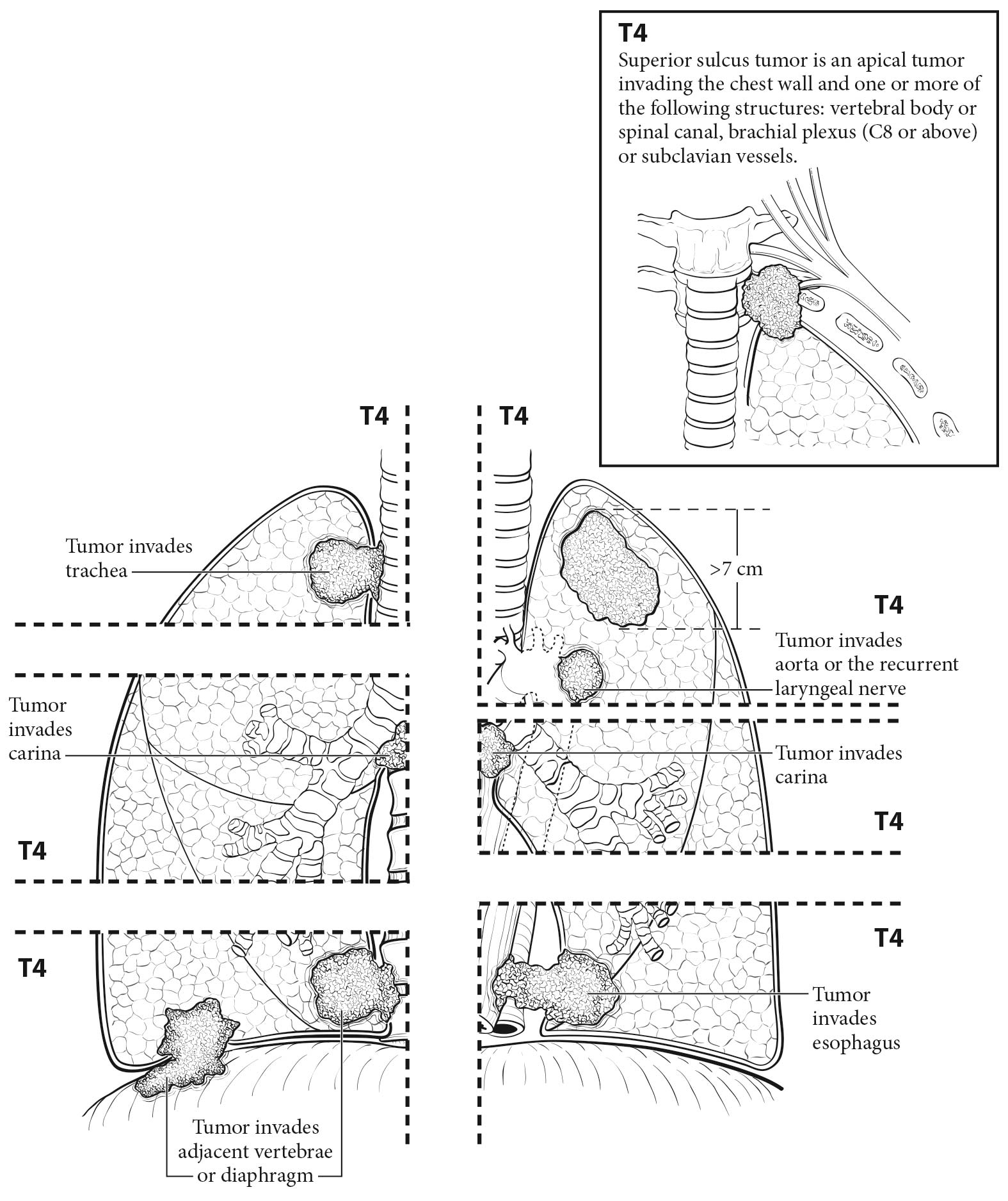
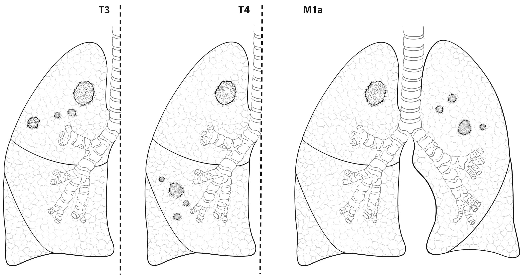
The analyses of the new IASLC database concerning the primary tumor (T) component of the TNM classification revealed that tumor size has more prognostic relevance than was shown in previous editions. Each centimeter increase in size, from less than 1 cm to up to 5 cm, separates tumors of significantly different prognosis, and larger tumors have a worse prognosis than their assigned T categories in the 7th Edition. Regarding the descriptors of tumor invasion, the prognosis worsens if tumor invasion is more central. On the other hand , endobronchial tumors less than or equal to 2 cm from the carina, but not involving the carina, and those causing complete atelectasis or pneumonitis do not have a worse prognosis than those more than 2 cm from the carina or those causing partial atelectasis and pneumonitis. Figure 36.2 shows the survival curves of the new T categories for clinically staged tumors with no nodal involvement and no metastases, and Figure 36.3 shows the survival curves for completely resected tumors with no nodal involvement and no metastases. In these graphs, the survival curves separate completely, with no overlapping or crossing; and survival differences between T3 and T4 tumors with no nodal involvement or metastasis are statistically significant, which was not the case in previous editions of the TNM classification.3

36.2 Overall survival according to the proposed T categories for the 8th Edition of the TNM classification for clinically staged T1-T4N0M0 tumors. p <0.001 (From Rami-Porta et al.3).

36.3 Overall survival according to the proposed T categories for the 8th Edition of the TNM classification for pathologically staged T1-T4N0M0R0 tumors. p < 0.001 (From Rami Porta et al.3).

The analyses performed for the 8th Edition of the TNM classification validate the present N categories at clinical and pathological staging. Figures 36.4 and 36.5 show the survival curves according to the N categories for clinically and pathologically staged tumors, respectively. These analyses also reveal that quantification of nodal disease has prognostic impact. In the recent data analysis, quantification was based on the number of involved lymph node stations. The progressive worsening of survival is as follows: single-station N1 has the best prognosis; multiple-station N1 disease follows, but it has the same prognosis as single-station N2 disease without N1 disease (skip metastasis); single-station N2 disease with N1 disease follows; and finally, multiple-station N2 disease has the worst prognosis. These findings derived from pathological staging and could not be validated at clinical staging.9 Therefore, they could not be used to modify the present N categories. However, knowing the prognostic impact of the number of involved nodal stations is clinically relevant, as postoperative prognosis can be refined for patients with pathological nodal disease, and it also may be useful for stratifying tumors in future clinical trials focusing on nodal disease. The subclassification of nodal disease presented in Table 36.2 is recommended for prospective registration of clinical and pathological data.

36.4 Overall survival according to the N descriptors for clinically staged tumors (From Asamura et al.9).

36.5 Overall survival according to the N descriptors for pathologically staged tumors including all types of resection (R0, R1 and R2) (From Asamura et al.9).

36.2 Subclassification of nodal disease8

NXRegional lymph nodes cannot be assessed
N0No regional lymph node metastasis
N1aSingle-station N1
N1bMultiple-station N1
N2a1Single-station N2 without N1 (skip metastasis)
N2a2Single-station N2 with N1
N2bMultiple-station N2
N3As defined in the 8th Edition

Figure 36.6 shows the survival curves according to the number of involved nodal stations in pathologically staged tumors.

36.6 Overall survival according to number of involved lymph node stations for pathologically staged tumors including all types of resection (R0, R1 and R2). N1a: single N1 station; N1b: multiple N1 stations; N2a1: single N2 station without N1 disease (skip metastasis); N2a2: single N2 station with N1 disease; N2b: multiple N2 stations (From Asamura et al.9).

The prospectively collected IASLC database through the electronic data capture (EDC) online system had nearly 4,000 patients. Their data had sufficient detail to validate the M1a categories established in the 7th Edition and to analyze extrathoracic metastatic disease (M1b) according to organ location and number of metastases. The results of these analyses show that number of metastases had more prognostic relevance than organ location and led to the recommendation to separate single extrathoracic metastasis in one organ (new M1b) from multiple extrathoracic metastases in one or several organs (new M1c). Figure 36.7 shows the survival curves of the different types of intrathoracic metastases, and Figure 36.8 shows the survival curves of single extrathoracic metastasis, multiple extrathoracic metastases, and intrathoracic metastases. Although the prognosis of intrathoracic metastases (M1a) is similar to that of single extrathoracic metastasis, it makes sense to code them differently as they represent different anatomic extents of disease and require different diagnostic and therapeutic strategies.4

36.7 Overall survival according to M1a descriptors. p = 0.66 (From Eberhardt et al.4).

36.8 Overall survival according to M1a and M1b descriptors. Single M1b metastasis has a prognosis similar to that of M1a but significantly different from that of M1b with multiple metastases (the new M1c category in the 8th Edition of the TNM classification). p < 0.0001 (From Eberhardt et al.4).

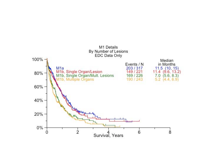
Classification of lung cancers with multiple lesions poses some problems, because the rules are sometimes ambiguous and their application may be interpreted differently in each situation. Therefore, a special subcommittee of the IASLC Staging and Prognostic Factors Committee studied the problem and made some recommendations regarding the uniform use of the classification rules depending on the pattern of disease. The subcommittee established four disease patterns: second primary tumors, lung cancers with separate tumor nodules of the same histopathological type, multiple tumors with predominant ground-glass features on CT and a lepidic pattern on pathological examination, and , finally, diffuse pneumonic-type lung cancer.16 Three in-depth articles expand the rationale for applying the classification rules to each disease pattern.17-19

The recommendations for classification are as follows:

  • Secondary primary tumors: Two or more synchronous or metachronous primary tumors should be classified separately, with an individual TNM for each one, regardless of whether they are in the same lobe, same lung, or contralateral lung. This rule applies to tumors identified clinically or grossly and also to those identified microscopically on pathological examination.18
  • Separate tumor nodules of the same histopathologic type (intrapulmonary metastases): The classification of these tumors is based on their lobar location. If the separate tumor nodule(s) is(are) in the same lobe of the primary, the tumor is classified as T3. If the separate tumor nodule(s) is(are) in another ipsilateral lobe, the tumor is classified as T4. The tumor classification is M1a if the separate tumor nodule(s) is(are) in the contralateral lung. This classification applies to separate tumor nodules identified clinically or grossly and also to those identified microscopically on pathological examination.17
  • Multifocal lung adenocarcinomas with ground-glass/lepidic features: These tumors should be classified by the T category of the lesion, with the highest T followed by the number of lesions (#) or simply (m) for multiple lesions indicated in parentheses, and an N and M category that applies to all of the multiple tumor foci collectively. The tumor size should be determined by the largest diameter of the solid component (by CT) or the invasive component on pathological examination. The T(#/m) multifocal classification should be applied equally whether the lesions are in the same lobe or in different ipsilateral or contralateral lobes. Furthermore, this classification should be applied to grossly recognizable lesions as well as to lesions discovered only on pathological examination.19
  • Diffuse pneumonic-type adenocarcinoma: In the case of a single tumor area, a stand ard TNM classification based on tumor size, nodal disease, and metastasis should be applied. In cases of multiple tumor areas, the T and M categories should be based on the location of the involved areas: T3 if the disease is confined to one lobe, T4 if it involves other ipsilateral lobes, and M1a if it involves the contralateral lung. If an area of involvement extends to the adjacent lobe, a T4 category should be assigned to recognize the extension into another ipsilateral lobe. If the tumor is confined to one lobe but its size is difficult to measure, a T3 category should be assigned. The N category is selected to apply to all pulmonary sites of the primary tumor collectively. Pleural/pericardial tumor nodules or distant metastases will lead to an M1a, M1b, or M1c designation. The classification should be applied to grossly recognizable lesions as well as to lesions discovered only on histologic examination. Miliary forms of adenocarcinoma should also be classified in this way. If the miliary involvement is limited to one lobe, because measurement of tumor size often is difficult, a T3 category should be assigned.19

The aforementioned recommendations for classifying the different patterns of disease are the result of a multidisciplinary and international consensus as well as a thorough literature review and statistical analysis of data from the IASLC database regarding separate tumor nodules. These suggestions are meant to minimize ambiguity and to serve as a guide in classifying these tumors uniformly.

Tables 36.3 36.4 36.5 36.6describe the clinical criteria used to define the different disease patterns in which lung cancers with multiple lesions may present.

36.3 Clinical criteria to distinguish second primary versus related tumors18*

Tumors may be considered second primary tumors if:

They clearly are of a different histologic type (e.g., squamous carcinoma and adenocarcinoma) on biopsy.

Tumors may be considered to be arising from a single tumor source if:

Exactly matching breakpoints are identified by comparative genomic hybridization.

Relative arguments that favor separate tumors:

Different radiographic appearance or metabolic uptake

Different biomarker pattern (driver gene mutations)

Different rates of growth (if previous imaging is available)

Absence of nodal or systemic metastases

Relative arguments that favor a single tumor source:

The same radiographic appearance

Similar growth patterns (if previous imaging is available)

Significant nodal or systemic metastases

The same biomarker pattern (and same histotype)

*A comprehensive histologic assessment is not included in clinical staging, as it requires the entire specimen to have been resected.

36.4 Clinical criteria to categorize a lesion as a separate tumor nodule (intrapulmonary metastasis)17

Tumors should be considered to have a separate tumor nodule(s) if:

There is a solid lung cancer and a separate tumor nodule(s) with a similar solid appearance and with (presumed) matching histologic appearance.

  • This applies regardless of whether or not the lesions have been biopsied, provided there is strong suspicion that the lesions are histologically identical.
  • This applies regardless of whether or not there are sites of extrathoracic metastases.

and provided that:

The lesions are NOT judged to be synchronous primary lung cancers.

The lesions are NOT multifocal GG/L lung cancer (multiple nodules with ground-glass/lepidic features) or pneumonic-type lung cancer.

GG/L, ground-glass/lepidic

A radiographically solid appearance and the specific histologic subtype of solid adenocarcinoma denote different things.

36.5 Clinical criteria to categorize a tumor as multifocal GG/L adenocarcinoma19

Tumors should be considered multifocal GG/L lung adenocarcinoma if:

There are multiple subsolid nodules (either pure ground glass or part solid), at least one of which is suspected (or proven) to be cancer.

  • This applies regardless of whether or not the nodules have been biopsied.
  • This applies if the other nodules(s) are suspected to be AIS, MIA, or LPA.
  • This applies if a nodule has become greater than 50% solid but is judged to have arisen from a GGN, provided there are other subsolid nodules.
  • GGN lesions less than 5 mm or lesions suspected to be AAH are not counted for TNM classification.

AAH, atypical adenomatous hyperplasia, AIS, adenocarcinoma in situ; GG/L, ground-glass/lepidic; GGN, ground-glass nodule; LPA, lepidic-predominant adenocarcinoma; MIA, minimally invasive adenocarcinoma

A radiographically solid appearance and the specific histologic subtype of solid adenocarcinoma denote different things.

36.6 Clinical criteria to categorize a tumor as pneumonic-type adenocarcinoma19

Tumors should be considered pneumonic-type adenocarcinoma if:

The cancer manifests in a regional distribution, similar to a pneumonic infiltrate or consolidation.

  • This applies whether there is one confluent area or multiple regions of disease. The region(s) may be confined to one lobe or in multiple lobes or bilateral, but should involve a regional pattern of distribution.
  • The appearance of involved areas may be ground glass, solid consolidation, or a combination thereof.
  • This can be applied when there is compelling suspicion of malignancy, whether or not a biopsy has been performed of the area(s).
  • This should not be applied to discrete nodules (i.e., GG/L nodules).
  • This should not be applied to tumors causing bronchial obstruction resulting in obstructive pneumonia or atelectasis.

GG/L, ground-glass/lepidic.

A radiographically solid appearance and the specific histologic subtype of solid adenocarcinoma denote different things.

Imaging

Medical history (e.g., family history of lung cancer or of any cancer, smoking history, exposure to asbestos or radon, history of passive smoking, presence of respiratory symptoms or chest pain) and physical examination findings (e.g., peripheral adenopathy, abnormal breath sounds, superior vena cava syndrome, hemoptysis, hepatomegaly) will prompt the request for a series of explorations to confirm or rule out lung cancer. There are many imaging techniques and invasive procedures that may be used to diagnose and stage lung cancer. Whenever possible, they should be performed sequentially and with an increasing degree of invasiveness.

The IASLC recommends a three-step protocol to rationalize the use of staging procedures. Step I includes medical history and physical examination, as well as plain radiographs of the chest and blood tests (hemoglobin, leukocytes, platelets, alanine aminotransferase, aspartate aminotransferase, lactate dehydrogenase, calcium, and albumin). Step II includes more complex investigations, such as contrast-enhanced CT scan of the chest and upper abdomen, bone scan, PET scan, brain CT, and bronchoscopy. Step III includes the more invasive procedures, including surgical exploration of the mediastinum (mediastinoscopy, extended cervical mediastinoscopy, mediastinotomy, video-assisted mediastinal lymphadenectomy, transcervical extended mediastinal lymphadenectomy), the pleural space (thoracentesis, percutaneous needle biopsy, thoracoscopy, video-assisted thoracoscopic surgery), or the pericardium (pericardiocentesis, pericardioscopy).20

Posteroanterior and lateral chest radiography usually is the first imaging technique requested for a patient with suspected lung cancer. The minimal information to be extracted from chest X-rays is:

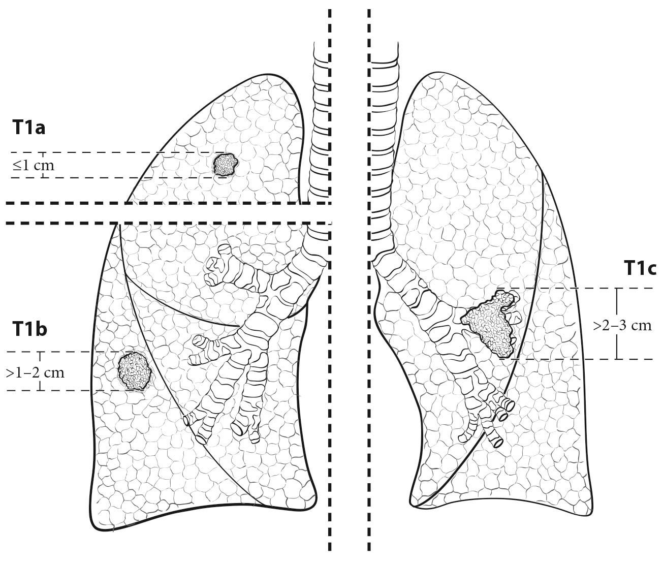
  • Tumor size (to assign a T category based on size)
  • Lobar and segmental location of the tumor
  • Presence of atelectasis and its extent (lobar or complete; cT2)
  • Presence of separate tumor nodules (cT3, cT4, or cM1a)
  • Evidence of lymphangitic carcinomatosis (cLy0: no radiologic evidence of lymphangitic carcinomatosis; cLy1: radiologic evidence of lymphangitic carcinomatosis confined to the area of the primary tumor; cLy2: lymphangitic carcinomatosis at a distance from the primary tumor but confined to the same lobe; cLy3: presence of lymphangitis in other ipsilateral lobes; cLy4: lymphangitis affecting the contralateral lung)
  • Relation of the primary tumor to the chest wall (contact or bone destruction [cT3]) or the mediastinum (elevated diaphragm may indicate invasion of the phrenic nerve [cT3])
  • Nodal spread: enlarged hili and abnormal mediastinum may indicate cN1, cN2, or cN3 disease
  • Intrathoracic spread: the presence of pleural or pericardial effusions (cM1a)
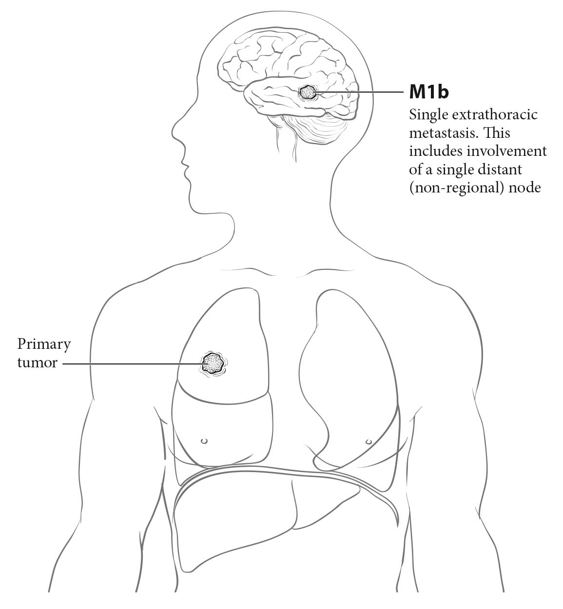
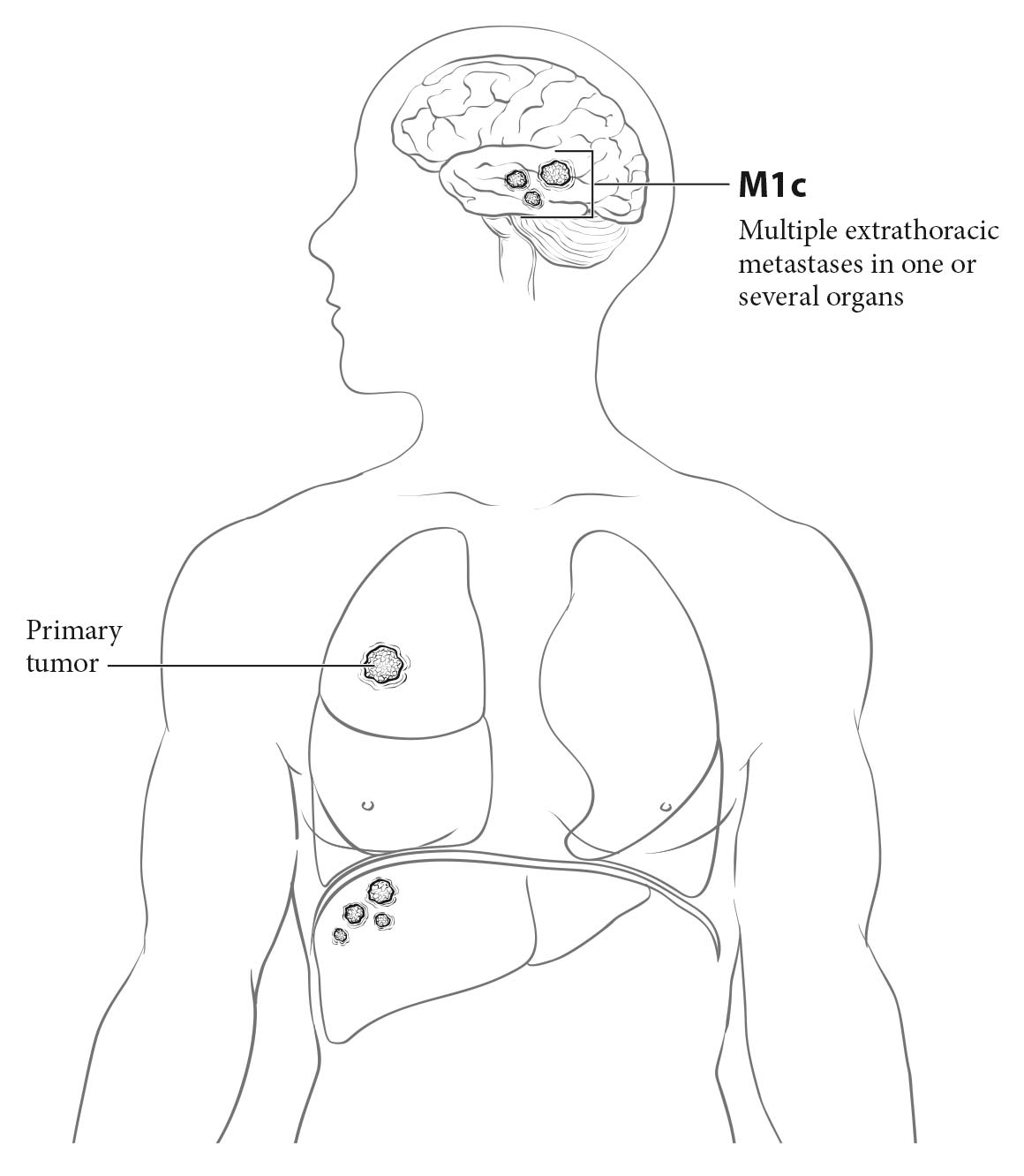
  • Extrathoracic spread: the integrity or involvement of the bones visible on chest X-rays—the ribs, the sternum, both scapulae, the vertebral column, the shoulder joint, and most of the length of both humeri; masses in the soft tissues of the chest wall (cM1b or cM1c)

Contrast CT of the chest and upper abdomen to include the liver and both adrenal gland s is recommended for patients who are cand idates for radical treatment, whether it be surgery, primarily or after induction, or definitive chemoradiation.21 CT should confirm and refine the information obtained from the chest X-rays:

  • Tumor size in its greatest dimension, which may be assessed by axial, coronal, or sagittal measurement; therefore, if technically possible, all the different projections should be studied to determine tumor size. Lung CT window display settings should be used when assessing images for tumor size.
  • Lobar and segmental location
  • Presence of atelectasis (partial or total; cT2) and endobronchial lesions
  • Presence of separate solid tumor nodules (cT3, cT4, or cM1a) and presence of part-solid lesions
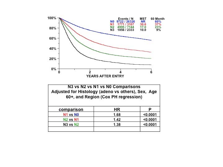
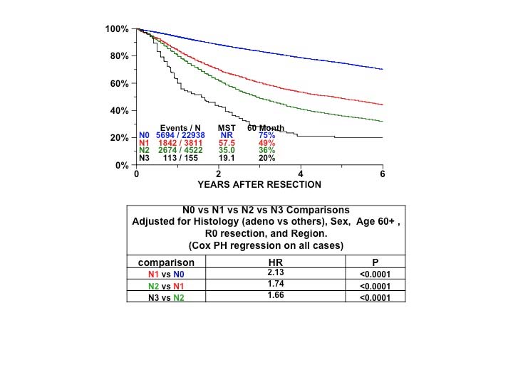
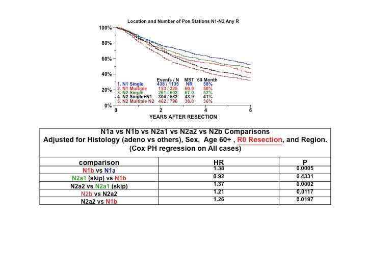
  • Nodal spread: hilar enlargement suggests cN1 disease if it is ipsilateral to the primary tumor or cN3 disease if it is contralateral. Nodes should be measured in their short axis; those larger than 1 cm in the short axis are considered abnormal and suggest metastatic involvement. Enlarged mediastinal lymph nodes may indicate cN2 disease if they are ipsilateral or subcarinal or cN3 disease if they are contralateral or supraclavicular. The number of nodal stations involved should be determined; bulky disease should be identified if present. Mediastinal CT window display settings should be used when assessing images for mediastinal structures, pleural or pericardial effusions, etc.
  • Evidence of lymphangitic carcinomatosis (cLy0, cLy1, cLy2, cLy3, and cLy4 as defined earlier)
  • Intrathoracic spread: pleural and pericardial effusion or nodules (cM1a)
  • Extrathoracic spread: bone lesions, soft tissue masses, adrenal masses, and liver nodules may indicate cM1b disease if single or cM1c disease if multiple

CT is important for assessing the size and location of any enlarged mediastinal lymph nodes present, because in the absence of metastasis, they are the strongest indicators of prognosis. In a review of 7,368 patients with a median prevalence of mediastinal nodal disease of 30%, staging values of chest CT were as follows: sensitivity, 0.55; specificity, 0.81; positive predictive value, 0.58; and negative predictive value, 0.83.21

PET is indicated in patients with no clinical abnormalities and no signs of metastatic spread on CT who are cand idates for treatment with curative intent. It is useful for evaluating metastatic spread, except that occurring in the brain. PET is not required in patients with ground-glass opacities or clinical stage IA tumors with no other abnormality on chest CT. Regarding mediastinal staging, in a review of 4,105 patients with a median prevalence of nodal disease of 28%, staging values were as follows: sensitivity, 0.8; specificity, 0.88; positive predictive value, 0.75; and negative predictive value, 0.91.21 PET should provide the following information:

  • Presence of normal or abnormal uptake in the primary tumor and quantification by maximum stand ardized uptake value (SUVmax)
  • Presence of normal or abnormal uptake in hilar and mediastinal nodes and quantification by SUVmax
  • Presence of normal or abnormal uptake in other parts of the lungs or in the rest of the body

Although SUVmax is subject to many intra- and interinstitutional variations, it is important to record it at initial staging to assess metabolic tumor response after treatment, especially after induction treatment to evaluate the possibility of tumor resection. SUVmax also has shown prognostic value, at least for Stage I-III squamous cell carcinomas and adenocarcinomas.22

Because PET has a poor anatomic resolution, the superimposition of PET with CT (e.g., with hybrid PET/CT scanners) may help the clinician locate the lesions with abnormal uptake. However, the mean staging values of combined PET/CT are similar to those of PET alone. In a review of 2,014 patients with a median prevalence of mediastinal nodal disease of 22%, the staging values for combined PET/CT were as follows: sensitivity, 0.62; specificity, 0.9; positive predictive value, 0.63; and negative predictive value, 0.9.21

The positive predictive value of PET is relatively low; therefore, histopathological confirmation of the lesions is recommended if this will affect therapy. Inflammations, granulomas, and infections may have high SUVmax, and if the correct histology remains unconfirmed, the patient may be excluded from radical treatment. If PET is not available, bone scanning and abdominal CT should be done to rule out metastatic spread.21

Magnetic resonance (MR) imaging has very specific indications in lung cancer staging. MR imaging of the brain currently is indicated in patients with Stage III and IV tumors, even if they have a negative clinical evaluation.21 It also is indicated in patients with brain metastasis identified on CT, as MR imaging may identify more lesions.23 It also may help define the involved anatomic structures in patients with apical (Pancoast) tumors or tumors invading the chest wall and mediastinum. MR imaging of the adrenals with in- and out-of-phase imaging may help exclude adrenal metastases in cases with indeterminate adrenal lesions on PET/CT.

The order in which the aforementioned anatomic and metabolic imaging tests are performed usually is chest X-rays first, followed by CT scan of the chest and upper abdomen, PET scan or PET/CT, and MR imaging in indicated cases.

The anatomic and metabolic imaging techniques described here provide a thorough description of the primary lesion and its local and distant spread, but do not provide its diagnosis. The TNM classification requires microscopic confirmation of malignancy24,25 and specification of histopathological type.14 The type of procedure used to obtain pathological confirmation of lung cancer differs depending on the location and spread of the tumor.

Sputum cytology may provide the diagnosis of lung cancer with high specificity. In a review of 29,145 patients, the diagnostic values of sputum cytology were as follows: sensitivity, 0.66; specificity, 0.99; false positive rate, 8%; and false negative rate, 10%.26 In certain patients with evident metastatic disease, this may be the only diagnostic test needed. However, molecular profiling of tumors is best performed on cell blocks; if these are not available in the sputum specimen, then larger samples may be needed.

Fiberoptic bronchoscopy is both a diagnostic and a staging procedure. As a diagnostic procedure including bronchial biopsy, brushings, washings, and endobronchial and transbronchial needle aspiration, its sensitivity is 0.88 and 0.78 for central and peripheral tumors, respectively.26 As a staging procedure, it shows the endobronchial location of the tumor: T2 if the main brochus is involved, regardless of its distance to the carina, and T4 if the carina is involved. It may suggest nodal involvement if there is extrinsic compression of the bronchi. The lymph nodes may be punctured with fine needles, either blindly—the classic transbronchial needle aspiration procedure—or with the assistance of EBUS and fine-needle aspiration (EBUS-FNA) or EUS and FNA (EUS-FNA). Peripheral tumors that remain undiagnosed by fiberoptic bronchoscopy may be diagnosed by transthoracic needle aspiration or biopsy, with a sensitivity of 0.9, a specificity of 0.97, a false positive rate of 1%, and a false negative rate of 22%.26

Thoracentesis and cytopathologic study of the pleural fluid may be enough in patients with malignant pleural effusion. It provides a diagnosis in 72% of patients.26 If cytology is negative, further pleural explorations with closed pleural biopsy and thoracoscopy should follow. Sensitivity and negative predictive values are both around 80% for closed pleural biopsy and greater than 80% and approximately 100%, respectively, for thoracoscopic biopsy.26 A malignant pleural effusion or tumor nodules on the pleural surface (parietal or visceral) classify the tumor as M1a. Thoracoscopy has the advantage of allowing exploration of the pleural cavity, lung surface, and mediastinum. Video-assisted thoracoscopic surgery also allows resection of peripheral nodules and assists in their diagnosis and staging. Ipsilateral hilar and mediastinal nodes may be biopsied as well.

The American College of Chest Physicians (ACCP) and the European Society of Thoracic Surgeons (ESTS) published guidelines on the preoperative staging of mediastinal lymph nodes.21,27 The 2013 ACCP Evidence-based Clinical Practice Guidelines favor invasive staging by needle aspiration techniques (EBUS-FNA, EUS-FNA) as the first procedures, but recommend confirmation with surgical biopsies (mediastinoscopy) if needle techniques are negative. In the absence of metastatic disease, the indications for invasive staging are as follows21:

  • Discrete mediastinal lymph node enlargement with or without PET uptake in mediastinal lymph nodes
  • PET activity in mediastinal lymph nodes and abnormal lymph nodes on CT
  • High suspicion of N2 or N3 disease, either by lymph node enlargement on CT or PET uptake
  • Intermediate suspicion of N2 or N3 disease by CT and PET and a central tumor or N1 disease

Invasive staging is not indicated for patients with extensive mediastinal infiltration or stage IA tumors with no suspicion of mediastinal lymph node involvement on CT or PET.21

The ESTS guidelines also recommend performing EBUS-FNA and EUS-FNA as the initial exploration in the following situations27:

  • Positive mediastinal nodes on CT and /or PET or PET/CT
  • Cases in which there is no evidence of N2-N3, but there is suspicion of N1 disease; central tumors larger than 3 cm; and adenocarcinomas with high PET uptake

Invasive staging may be avoided in patients with no evidence of mediastinal disease on CT and PET and tumors less than 3 cm in greatest dimension located peripherally, that is, in the outer third of the lung.

If needle techniques produce negative results, video-assisted mediastinoscopy is recommended to confirm the results or to identify mediastinal disease. In general, the negative predictive values of EBUS-FNA and EUS-FNA are too low, both in patients with normal and those with abnormal mediastinal lymph nodes, to make therapeutic decisions without proper confirmation by a surgical technique. In a recent article on the staging value of EBUS-FNA in patients with no mediastinal abnormalities, the sensitivity and negative predictive values for EBUS-FNA were 0.38 and 0.81, respectively, whereas they were 0.73 and 0.91 for mediastinoscopy.28 This article clearly highlights the importance of confirming negative results of EBUS-FNA and EUS-FNS with mediastinoscopy.

Additionally, the ESTS guidelines recommend exploration of the aortopulmonary window for left lung cancers and establish minimum requirements for mediastinoscopy in clinical practice: at least the inferior right and left paratracheal lymph nodes and the subcarinal lymph nodesshould be biopsied or removed; the superior right and left paratracheal lymph nodes and the hilar lymph nodes should be explored if there is evidence of involvement on CT or PET.27

Other invasive procedures should be performed as required, including pericardiocentesis or pericardioscopy, either transpleural or subxiphoid, for pericardial effusion; needle biopsies of liver and adrenal lesions; endoscopies of the gastrointestinal tract in cases of digestive symptoms or bleeding; and biopsy or excision of skin lesions.

These procedures should be performed sequentially from the least to most invasive: first, to rule out metastatic disease if imaging suggests metastatic spread, as this will avoid more invasive procedures; next, to rule out supraclavicular nodal disease (N3) if there is anatomic or metabolic suspicion; and finally, to explore the mediastinum as indicated by the aforementioned guidelines.

Pathological Classification

Pathological classification or postsurgical histopathological classification, designated pTNM, is used to guide adjuvant therapy and provides useful information to estimate prognosis and calculate end results. It is based on evidence acquired before treatment, supplemented or modified by additional evidence acquired from surgery and from pathological examination of the resected specimens.

The pathological assessment of the primary tumor (pT) entails resection of the primary tumor or a biopsy specimen adequate to evaluate the highest T category. The pathological assessment of the regional lymph nodes (pN) entails removal of enough nodes to validate the absence of regional lymph node metastasis (pN0) or to evaluate the highest pN category. The pathological assessment of distant metastasis (pM) entails microscopic examination. For pathological staging, cM0 or cM1 categories also are valid.

Visceral pleural invasion is defined as invasion beyond the elastic layer or to the surface of the visceral pleura. A tumor that falls short of completely traversing the elastic layer is defined as PL0. A tumor that extends through the elastic layer is defined as PL1, and one that extends to the surface of the visceral pleura as PL2. Elastic stains should be performed in cases in which there is any uncertainty based on review of hematoxylin and eosin sections. Either PL1 or PL2 status allows classification of the primary tumor as T2. Extension of the tumor to the parietal pleura or chest wall is defined as PL3 and categorizes the primary tumor as T3. Direct tumor invasion into an adjacent ipsilateral lobe (i.e., invasion across a fissure) is classified as T2a, unless tumor size indicates a higher T category. Figure 36.9 shows the graphic representation of visceral and parietal pleura invasion.29

36.9 Visceral pleura invasion. See text for definitions (From Travis et al.29 Copyright © 2008 Aletta Ann Frazier, MD).

For proper pathological lymph node staging and to fulfill the requirements for complete resection and pathological N0, the IASLC recommends performing a systematic nodal dissection or a lobe-specific systematic nodal dissection. Systematic nodal dissection is the en bloc removal of the mediastinal fatty tissue, including the lymph nodes, which should be followed by hilar and intrapulmonary nodal dissection.30 Lobe-specific systematic nodal dissection consists of the removal of certain mediastinal lymph nodes, depending on the lobar location of the primary tumor.31 The mediastinal nodal stations that should be biopsied or removed, according to the location of the primary tumor, are as follows (based on the IASLC lymph node map in Figure 36.1):

  • Right upper and middle lobes: 7, 2R, and 4R
  • Right lower lobe: 7, 4R, and 8 or 9
  • Left upper lobe: 7, 5, and 6
  • Light lower lobe: 7, 8, and 9

In any case, at least six lymph nodes/stations (six lymph nodes from six lymph node stations) should be removed or sampled. Three of these nodes/stations should be mediastinal, including the subcarinal nodes (nodal station #7), and three should be hilar-intrapulmonary lymph nodes/stations. The Union for International Cancer Control (UICC) and the AJCC accept that if all resected/sampled lymph nodes are negative, but the number recommended is not met, the classification is pN0. If resection has been performed, and otherwise fulfills the requirement for complete resection, the classification is R0.14,24 However, the ACCP recommends the classificiation of pN0(un), un for uncertain, if the number of recommended lymph nodes removed /sampled is not met. The suffix (un) also should be added to pN1, and pN2 if fewer than six lymph nodes are evaluated.32 This suffix is not added to the N categories, as it is an opinion of the ACCP that has not been discussed sufficiently at the international level or approved by the AJCC or UICC. Nevertheless, it makes sense to use it in cases in which the required stand ards of intraoperative lymph node examination are not met, especially when it has been proven that failure to perform a proper lymphadenectomy has had deleterious prognostic implications.33

Regarding tumors that underwent an attempt at resection but were not removed completely, the UICC and AJCC differ on when to consider pathological classification. According to the UICC, the following criteria should be satisfied14:

  • Biopsy confirmed a pT category, and there is microscopic confirmation of nodal disease at any level (pN1-3)
  • There is microscopic confirmation of the highest N category (pN3) or
  • There is microscopic confirmation of pM1

However, the AJCC requires24:

  • Microscopic confirmation of highest T and N categories or
  • Microscopic confirmation of M1

In view of the discrepancy and to avoid any confusion,34,35 the ACCP recommends the use of the p prefix for resected tumors, as well as for the rare cases in which a tumor was not resected, but extensive biopsy samples were taken during the resection attempt. In the clinical staging context, even if there is pathological confirmation of tumor extent, the c prefix should be used. It seems reasonable to use the ACCP recommendation and to discontinue the use of different criteria to assign pathological classification to tumors that have not been resected.32

Pleural lavage cytology is an easy and inexpensive method to further study tumor extent at the time of resection. In this procedure, performed before lung manipulation, a specified amount of saline is introduced into the pleural space and then retrieved for pathological study. If the fluid is positive for malignant cells, the prognosis is invariably worse than in the case of negative cytology, regardless of the T category of the primary tumor.36,37 Positive pleural lavage cytology is a descriptor of incomplete resection and is coded as R1(cy+).14

Since the 7th Edition TNM stage classification, WHO has defined new entities of adenocarcinoma in situ and minimally invasive adenocarcinoma. It also has histologically classified nonmucinous adenocarcinomas based on an estimate of percentages of lepidic and invasive (acinar, papillary, solid, and micropapillary) patterns. In general, the lepidic-versus-invasive patterns by histology correspond to ground-glass versus solid components by CT.

Adenocarcinoma in situ (AIS) is added to the category of Tis, which previously consisted only of squamous cell carcinoma in situ (SCIS). Because the histologic type of in situ carcinoma does not always match that of the associated primary lung carcinoma, it is important to specify Tis (AIS) versus Tis (SCIS).AIS is a localized small (less than or equal to 3-cm) adenocarcinoma with growth restricted to neoplastic cells along pre-existing alveolar structures (lepidic growth) and lacking stromal, vascular, alveolar space, or pleural invasion. Most AISs are nonmucinous, but rarely, they may be mucinous. Most AISs show a pure ground-glass nodule by CT, unless there are benign areas such as fibrous scar, inflammation, or organizing pneumonia contributing to a solid component.6,7

Minimally invasive adenocarcinoma (MIA) is defined as a lepidic-predominant adenocarcinoma measuring up to 3 cm with an invasive component measuring up to 0.5 cm. Most MIAs are nonmucinous, but rarely, they may be mucinous. In some nonmucinous MIA cases, there is a single, discrete focus of invasion or a solid component on CT. However, if the invasive/solid component assessed by histology/CT, respectively, consists of multiple foci, it is proposed that the percentage area of the invasive/solid be estimated and then multiplied by the total size. For example, a 2.0-cm total size with a 20% invasive or solid component on histology or CT, respectively, would have an estimated size of 0.4 cm.2,6,7

To measure tumor size in part-solid, nonmucinous adenocarcinomas, the recommendation is to follow the TNM rule to consider only the size of the invasive component in assigning a T category. This recommendation does not apply to other histologic types of lung cancer or to mucinous lung adenocarcinomas. Although this rule has been in place since 2001, until now it has not been applied in lung adenocarcinoma.2,14 Therefore, a lesion consisting of a 15-mm part-solid opacity with a 7-mm solid component would be classified as a cT1a lesion, because its solid component, excluding the ground-glass component, is less than 10 mm in greatest dimension. If the lesion is resected and proves to be an adenocarcinoma with lepidic and invasive components, the measurement of the invasive component at pathological examination will be used for the pathological classification. This recommendation is based on the increasing number of studies in small lung adenocarcinomas reporting that in part-solid adenocarcinomas, it is the invasive component that correlates with prognosis.38-40 Similar to MIA, in cases in which multiple invasive/solid areas, rather than a single, discrete focus, are observed on histology/CT, it is proposed that the percentage invasive/solid area be multiplied by the total tumor size to estimate the size of invasion.2 It is recommended that both total size and invasive/solid size continue to be documented in radiology and pathology reports.

In special situations, tumor size is determined after induction therapy. If no viable tumor cells remain after induction therapy, the tumor is classified as ypT0. However, no rules have been established to measure tumor size in patients who have had a partial response, the degree of which has prognostic relevance. A practical way to estimate tumor size is to multiply the percentage of viable tumor cells by the size of the total mass. This formula may be applied in cases in which there is a single focus of disease or multiple foci of viable cells.2

Pathological classification of lung cancers with multiple lesions follows the same criteria recommended for clinical classification of the four different patterns of disease: separate primary tumors, separate tumor nodules (intrapulmonary metastasis), ground-glass/lepidid adenocarcinomas, and pneumonic-type adenocarcinomas.16

Tables 36.7 36.8 36.9 36.10 36.11describe the pathological criteria for defining the different disease patterns in which lung cancers with multiple lesions may present.

36.7 Pathological criteria (i.e., after resection) for separate versus related pulmonary tumors18

Tumors may be considered second primary tumors if:

They clearly are a different histologic type (e.g., squamous carcinoma and adenocarcinoma).

They clearly are different based on a comprehensive histologic assessment.

They are squamous carcinomas that have arisen from carcinoma in situ.

Tumors may be considered to be arising from a single tumor source if:

Exactly matching breakpoints are identified by comparative genomic hybridization.

Relative arguments that favor separate tumors (to be considered together with clinical factors):

Different biomarker pattern

Absence of nodal or systemic metastases

Relative arguments that favor a single tumor source (to be considered together with clinical factors):

Matching appearance on comprehensive histologic assessment

The same biomarker pattern

Significant nodal or systemic metastases

36.8 Pathological criteria to categorize a lesion as a separate tumor nodule (intrapulmonary metastasis)17

Tumors should be considered to have a separate tumor nodule (intrapulmonary metastasis) if:

There is (are) a separate tumor nodule(s) of cancer in the lung with a similar histologic appearance to a primary lung cancer.

and provided that:

The lesions are NOT judged to be synchronous primary lung cancers.

The lesions are NOT multiple foci of LPA, MIA, or AIS.

AIS, adenocarcinoma in situ; LPA, lepidic-predominant adenocarcinoma; MIA, minimally invasive adenocarcinoma

A radiographically solid appearance and the specific histologic subtype of solid adenocarcinoma denote different things.

36.9 Pathological criteria identifying multifocal ground-glass/lepidic lung adenocarcinoma19

Tumors should be considered multifocal GG/L lung adenocarcinoma if:

There are multiple foci of LPA, MIA, or AIS.

  • This applies to whether a detailed histological assessment (i.e., proportion of subtypes, etc.) shows a matching or different appearance.
  • This applies if one lesion(s) is (are) LPA, MIA, or AIS and there are other subsolid nodules that have not been biopsied.
  • This applies whether the nodule(s) is (are) identified preoperatively or only on pathological examination.
  • Foci of AAH are not counted for TNM classification.

AAH, atypical adenomatous hyperplasia; AIS, adenocarcinoma in situ; GG/L, ground-glass/lepidic; LPA, lepidic-predominant adenocarcinoma; MIA, minimally invasive adenocarcinoma

A radiographically solid appearance and the specific histologic subtype of solid adenocarcinoma denote different things.

Tumors should be considered pneumonic-type adenocarcinoma if:

There is diffuse distribution of adenocarcinoma throughout a region(s) of the lung, as opposed to a single well-demarcated mass or multiple discrete well-demarcated nodules.

  • This typically involves an invasive mucinous adenocarcinoma, although a mixed mucinous and nonmucinous pattern may occur
  • The tumor may show a heterogeneous mixture of acinar, papillary, and micropapillary growth patterns, although it usually is lepidic perdominant.

A radiographically solid appearance and the specific histologic subtype of solid adenocarcinoma denote different things.

36.11 Schematic summary of disease patterns and TNM classification of patients with lung cancer with multiple pulmonary sites of involvement16

Second primary lung cancerMultifocal GG/L nodulesPneumonic-type adenocarcinomaSeparate tumor nodule
Imaging featuresTwo or more distinct masses with imaging characteristic of lung cancer (e.g., spiculated)Multiple ground-glass or part-solid nodulesPatchy areas of ground glass and consolidationTypical lung cancer (e.g., solid, spiculated) with separate solid nodule
Pathological featuresDifferent histotype or different morphology based on comprehensive histologic assessmentAdenocarcinomas with prominent lepidic component (typically varying degrees of AIS, MIA, LPA)Same histology throughout (most often invasive mucinous adenocarcinoma)Distinct masses with the same morphologic features based on comprehensive histologic assessment
TNM classificationSeparate cTNM and pTNM for each cancerT based on highest T lesion, with (#/m) indicating multiplicity; single N and MT based on size or T3 if in single lobe, T4 or M1a if in different ipsilateral or contralateral lobes; single N and MLocation of separate nodule relative to primary site determines whether T3, T4, or M1a; single N and M
Conceptual viewUnrelated tumorsSeparate tumors, albeit with similaritiesSingle tumor, diffuse pulmonary involvementSingle tumor with intrapulmonary metastasis

AIS, adenocarcinoma in situ; GG/L, ground-glass/lepidic; LPA, lepidic-predominant adenocarcinoma; MIA, minimally invasive adenocarcinoma

Prognostic Factors

Prognostic Factors Required for Stage Grouping

Beyond the factors used to assign T, N, or M categories, no additional prognostic factors are required for stage grouping.

Additional Factors Recommended for Clinical Care

The analyses of the database used to revise the 6th Edition of the TNM classification of lung cancer showed that prognosis beyond that indicated by the classification of anatomic extent may be refined by adding nonanatomic prognostic factors, both at clinical and pathological staging. In addition to anatomic stage, performance status, age, and gender were important prognostic factors, and their combination separated groups of tumors of significantly different prognoses.41,42

The following lists of nonanatomic prognostic factors are based on Prognostic Factors in Cancer, 3rd edition.43

Resectable Non-Small Cell Lung Cancer in Inoperable Patients Treated with Radiotherapy

Patient related:

  • Symptoms (presence/absence)
  • Performance status
  • Hemoglobin

Surgically Resected Non-Small Cell Lung Cancer

Tumor related:

  • Histologic type
  • Differentiation grade
  • Vascular invasion
  • Lymphatic invasion
  • Perineural invasion
  • Type of visceral pleura invasion: PL1 versus PL2
  • Positive pleural lavage cytology
  • SUVmax of primary tumor
  • Molecular/biologic markers

Patient related:

  • Age
  • Gender
  • Weight loss
  • Performance status
  • Quality of life
  • Marital status

Environment related:

  • Resection margins
  • Adequacy of mediastinal dissection
  • Radiotherapy dose
  • Adjuvant radiation
  • Adjuvant chemotherapy

Locally Advanced or Metastatic Non-Small Cell Lung Cancer

Tumor related:

  • Hemoglobin
  • Lactate dehydrogenase (LDH)
  • Albumin

Patient related:

  • Gender
  • Symptom burden
  • Weight loss
  • Performance status
  • Quality of life
  • Marital status
  • Anxiety/depression

Environment related:

  • Chemoradiotherapy
  • Chemotherapy

Small Cell Lung Cancer

Tumor related:

  • LDH
  • Alkaline phosphatase
  • Cushing syndrome
  • White blood cells
  • Platelet count
  • Molecular/biologic markers

Patient related:

  • Age
  • Comorbidity
  • Performance status

Environment related:

  • Chemotherapy
  • Thoracic radiotherapy
  • Prophylactic cranial radiotherapy

A recent review on prognostic tools for non-small cell and small cell lung cancers shows that most of them lack internal and external validation. However, prognostic tools combining a series of variables may contribute to the development of personalized medicine.44

The authors have not identified emerging factors for clinical care at this time.

Risk Assessment

Risk Assesment Models

The AJCC recently established guidelines that will be used to evaluate published statistical prediction models for the purpose of granting endorsement for clinical use.45 Although this is a monumental step toward the goal of precision medicine, this work was published only very recently. Therefore, the existing models that have been published or may be in clinical use have not yet been evaluated for this cancer site by the Precision Medicine Core of the AJCC. In the future, the statistical prediction models for this cancer site will be evaluated, and those that meet all AJCC criteria will be endorsed.

Recommendations

Trials for adjuvant chemotherapy for completely resected T1 non-small cell lung cancer:

Trials for adjuvant therapy (chemotherapy, radiotherapy, or both) for resected non-small cell lung cancer with N2 disease:

Trials for therapy of nonmetastatic small cell lung cancer:

TNM Definitions

Clinical T (cT)

cT CategorycT Criteria
cTXPrimary tumor cannot be assessed, or tumor proven by the presence of malignant cells in sputum or bronchial washings but not visualized by imaging or bronchoscopy
cT0No evidence of primary tumor
cTisCarcinoma in situ; Squamous cell carcinoma in situ (SCIS); Adenocarcinoma in situ (AIS): adenocarcinoma with pure lepidic pattern, less than or equal to 3 cm in greatest dimension
cT1Tumor less than or equal to 3 cm in greatest dimension, surrounded by lung or visceral pleura, without bronchoscopic evidence of invasion more proximal than the lobar bronchus (i.e., not in the main bronchus)
cT1miMinimally invasive adenocarcinoma: adenocarcinoma (less than or equal to 3 cm in greatest dimension) with a predominantly lepidic pattern and less than or equal to 5 mm invasion in greatest dimension
cT1aTumor less than or equal to 1 cm in greatest dimension. A superficial, spreading tumor of any size whose invasive component is limited to the bronchial wall and may extend proximal to the main bronchus also is classified as T1a, but these tumors are uncommon.
cT1bTumor greater than 1 cm but less than or equal to 2 cm in greatest dimension
cT1cTumor greater than 2 cm but less than or equal to 3 cm in greatest dimension
cT2Tumor greater than 3 cm but less than or equal to 5 cm or having any of the following features: Involves the main bronchus regardless of distance to the carina, but without involvement of the carina; Invades visceral pleura (PL1 or PL2); Associated with atelectasis or obstructive pneumonitis that extends to the hilar region, involving part or all of the lung. T2 tumors with these features are classified as T2a if less than or equal to 4 cm or if the size cannot be determined and T2b if greater than 4 cm but less than or equal to 5 cm.
cT2aTumor greater than 3 cm but less than or equal to 4 cm in greatest dimension
cT2bTumor greater than 4 cm but less than or equal to 5 cm in greatest dimension
cT3Tumor greater than 5 cm but less than or equal to 7 cm in greatest dimension or directly invading any of the following: parietal pleura (PL3), chest wall (including superior sulcus tumors), phrenic nerve, parietal pericardium; or separate tumor nodule(s) in the same lobe as the primary
cT4Tumor greater than 7 cm or tumor of any size invading one or more of the following: diaphragm, mediastinum, heart, great vessels, trachea, recurrent laryngeal nerve, esophagus, vertebral body, or carina; separate tumor nodule(s) in an ipsilateral lobe different from that of the primary

Pathological T (pT)

pT CategorypT Criteria
pTXPrimary tumor cannot be assessed, or tumor proven by the presence of malignant cells in sputum or bronchial washings but not visualized by imaging or bronchoscopy
pT0No evidence of primary tumor
pTisCarcinoma in situ; Squamous cell carcinoma in situ (SCIS); Adenocarcinoma in situ (AIS): adenocarcinoma with pure lepidic pattern, less than or equal to 3 cm in greatest dimension
pT1Tumor less than or equal to 3 cm in greatest dimension, surrounded by lung or visceral pleura, without bronchoscopic evidence of invasion more proximal than the lobar bronchus (i.e., not in the main bronchus)
pT1miMinimally invasive adenocarcinoma: adenocarcinoma (less than or equal to 3 cm in greatest dimension) with a predominantly lepidic pattern and less than or equal to 5 mm invasion in greatest dimension
pT1aTumor less than or equal to 1 cm in greatest dimension. A superficial, spreading tumor of any size whose invasive component is limited to the bronchial wall and may extend proximal to the main bronchus also is classified as T1a, but these tumors are uncommon.
pT1bTumor greater than 1 cm but less than or equal to 2 cm in greatest dimension
pT1cTumor greater than 2 cm but less than or equal to 3 cm in greatest dimension
pT2Tumor greater than 3 cm but less than or equal to 5 cm or having any of the following features: Involves the main bronchus regardless of distance to the carina, but without involvement of the carina; Invades visceral pleura (PL1 or PL2); Associated with atelectasis or obstructive pneumonitis that extends to the hilar region, involving part or all of the lung. T2 tumors with these features are classified as T2a if less than or equal to 4 cm or if the size cannot be determined and T2b if greater than 4 cm but less than or equal to 5 cm.
pT2aTumor greater than 3 cm but less than or equal to 4 cm in greatest dimension
pT2bTumor greater than 4 cm but less than or equal to 5 cm in greatest dimension
pT3Tumor greater than 5 cm but less than or equal to 7 cm in greatest dimension or directly invading any of the following: parietal pleura (PL3), chest wall (including superior sulcus tumors), phrenic nerve, parietal pericardium; or separate tumor nodule(s) in the same lobe as the primary
pT4Tumor greater than 7 cm or tumor of any size invading one or more of the following: diaphragm, mediastinum, heart, great vessels, trachea, recurrent laryngeal nerve, esophagus, vertebral body, or carina; separate tumor nodule(s) in an ipsilateral lobe different from that of the primary
cTXPrimary tumor cannot be assessed, or tumor proven by the presence of malignant cells in sputum or bronchial washings but not visualized by imaging or bronchoscopy
cT0No evidence of primary tumor
cTisCarcinoma in situ; Squamous cell carcinoma in situ (SCIS); Adenocarcinoma in situ (AIS): adenocarcinoma with pure lepidic pattern, less than or equal to 3 cm in greatest dimension
cT1Tumor less than or equal to 3 cm in greatest dimension, surrounded by lung or visceral pleura, without bronchoscopic evidence of invasion more proximal than the lobar bronchus (i.e., not in the main bronchus)
cT1miMinimally invasive adenocarcinoma: adenocarcinoma (less than or equal to 3 cm in greatest dimension) with a predominantly lepidic pattern and less than or equal to 5 mm invasion in greatest dimension
cT1aTumor less than or equal to 1 cm in greatest dimension. A superficial, spreading tumor of any size whose invasive component is limited to the bronchial wall and may extend proximal to the main bronchus also is classified as T1a, but these tumors are uncommon.
cT1bTumor greater than 1 cm but less than or equal to 2 cm in greatest dimension
cT1cTumor greater than 2 cm but less than or equal to 3 cm in greatest dimension
cT2Tumor greater than 3 cm but less than or equal to 5 cm or having any of the following features: Involves the main bronchus regardless of distance to the carina, but without involvement of the carina; Invades visceral pleura (PL1 or PL2); Associated with atelectasis or obstructive pneumonitis that extends to the hilar region, involving part or all of the lung. T2 tumors with these features are classified as T2a if less than or equal to 4 cm or if the size cannot be determined and T2b if greater than 4 cm but less than or equal to 5 cm.
cT2aTumor greater than 3 cm but less than or equal to 4 cm in greatest dimension
cT2bTumor greater than 4 cm but less than or equal to 5 cm in greatest dimension
cT3Tumor greater than 5 cm but less than or equal to 7 cm in greatest dimension or directly invading any of the following: parietal pleura (PL3), chest wall (including superior sulcus tumors), phrenic nerve, parietal pericardium; or separate tumor nodule(s) in the same lobe as the primary
cT4Tumor greater than 7 cm or tumor of any size invading one or more of the following: diaphragm, mediastinum, heart, great vessels, trachea, recurrent laryngeal nerve, esophagus, vertebral body, or carina; separate tumor nodule(s) in an ipsilateral lobe different from that of the primary

Neoadjuvant Clinical T (yT)

ycT CategoryycT Criteria
ycTXPrimary tumor cannot be assessed, or tumor proven by the presence of malignant cells in sputum or bronchial washings but not visualized by imaging or bronchoscopy
ycT0No evidence of primary tumor
ycTisCarcinoma in situ; Squamous cell carcinoma in situ (SCIS); Adenocarcinoma in situ (AIS): adenocarcinoma with pure lepidic pattern, less than or equal to 3 cm in greatest dimension
ycT1Tumor less than or equal to 3 cm in greatest dimension, surrounded by lung or visceral pleura, without bronchoscopic evidence of invasion more proximal than the lobar bronchus (i.e., not in the main bronchus)
ycT1miMinimally invasive adenocarcinoma: adenocarcinoma (less than or equal to 3 cm in greatest dimension) with a predominantly lepidic pattern and less than or equal to 5 mm invasion in greatest dimension
ycT1aTumor less than or equal to 1 cm in greatest dimension. A superficial, spreading tumor of any size whose invasive component is limited to the bronchial wall and may extend proximal to the main bronchus also is classified as T1a, but these tumors are uncommon.
ycT1bTumor greater than 1 cm but less than or equal to 2 cm in greatest dimension
ycT1cTumor greater than 2 cm but less than or equal to 3 cm in greatest dimension
ycT2Tumor greater than 3 cm but less than or equal to 5 cm or having any of the following features: Involves the main bronchus regardless of distance to the carina, but without involvement of the carina; Invades visceral pleura (PL1 or PL2); Associated with atelectasis or obstructive pneumonitis that extends to the hilar region, involving part or all of the lung. T2 tumors with these features are classified as T2a if less than or equal to 4 cm or if the size cannot be determined and T2b if greater than 4 cm but less than or equal to 5 cm.
ycT2aTumor greater than 3 cm but less than or equal to 4 cm in greatest dimension
ycT2bTumor greater than 4 cm but less than or equal to 5 cm in greatest dimension
ycT3Tumor greater than 5 cm but less than or equal to 7 cm in greatest dimension or directly invading any of the following: parietal pleura (PL3), chest wall (including superior sulcus tumors), phrenic nerve, parietal pericardium; or separate tumor nodule(s) in the same lobe as the primary
ycT4Tumor greater than 7 cm or tumor of any size invading one or more of the following: diaphragm, mediastinum, heart, great vessels, trachea, recurrent laryngeal nerve, esophagus, vertebral body, or carina; separate tumor nodule(s) in an ipsilateral lobe different from that of the primary

Neoadjuvant Pathological T (yT)

ypT CategoryypT Criteria
ypTXPrimary tumor cannot be assessed, or tumor proven by the presence of malignant cells in sputum or bronchial washings but not visualized by imaging or bronchoscopy
ypT0No evidence of primary tumor
ypTisCarcinoma in situ; Squamous cell carcinoma in situ (SCIS); Adenocarcinoma in situ (AIS): adenocarcinoma with pure lepidic pattern, less than or equal to 3 cm in greatest dimension
ypT1Tumor less than or equal to 3 cm in greatest dimension, surrounded by lung or visceral pleura, without bronchoscopic evidence of invasion more proximal than the lobar bronchus (i.e., not in the main bronchus)
ypT1miMinimally invasive adenocarcinoma: adenocarcinoma (less than or equal to 3 cm in greatest dimension) with a predominantly lepidic pattern and less than or equal to 5 mm invasion in greatest dimension
ypT1aTumor less than or equal to 1 cm in greatest dimension. A superficial, spreading tumor of any size whose invasive component is limited to the bronchial wall and may extend proximal to the main bronchus also is classified as T1a, but these tumors are uncommon.
ypT1bTumor greater than 1 cm but less than or equal to 2 cm in greatest dimension
ypT1cTumor greater than 2 cm but less than or equal to 3 cm in greatest dimension
ypT2Tumor greater than 3 cm but less than or equal to 5 cm or having any of the following features: Involves the main bronchus regardless of distance to the carina, but without involvement of the carina; Invades visceral pleura (PL1 or PL2); Associated with atelectasis or obstructive pneumonitis that extends to the hilar region, involving part or all of the lung. T2 tumors with these features are classified as T2a if less than or equal to 4 cm or if the size cannot be determined and T2b if greater than 4 cm but less than or equal to 5 cm.
ypT2aTumor greater than 3 cm but less than or equal to 4 cm in greatest dimension
ypT2bTumor greater than 4 cm but less than or equal to 5 cm in greatest dimension
ypT3Tumor greater than 5 cm but less than or equal to 7 cm in greatest dimension or directly invading any of the following: parietal pleura (PL3), chest wall (including superior sulcus tumors), phrenic nerve, parietal pericardium; or separate tumor nodule(s) in the same lobe as the primary
ypT4Tumor greater than 7 cm or tumor of any size invading one or more of the following: diaphragm, mediastinum, heart, great vessels, trachea, recurrent laryngeal nerve, esophagus, vertebral body, or carina; separate tumor nodule(s) in an ipsilateral lobe different from that of the primary

Definition of Regional Lymph Node (N)

Clinical N (cN)
cN CategorycN Criteria
cNXRegional lymph nodes cannot be assessed
cN0No regional lymph node metastasis
cN1Metastasis in ipsilateral peribronchial and /or ipsilateral hilar lymph nodes and intrapulmonary nodes, including involvement by direct extension
cN2Metastasis in ipsilateral mediastinal and /or subcarinal lymph node(s)
cN3Metastasis in contralateral mediastinal, contralateral hilar, ipsilateral or contralateral scalene, or supraclavicular lymph node(s)
Pathological N (pN)
pN CategorypN Criteria
pNXRegional lymph nodes cannot be assessed
pN0No regional lymph node metastasis
pN1Metastasis in ipsilateral peribronchial and /or ipsilateral hilar lymph nodes and intrapulmonary nodes, including involvement by direct extension
pN2Metastasis in ipsilateral mediastinal and /or subcarinal lymph node(s)
pN3Metastasis in contralateral mediastinal, contralateral hilar, ipsilateral or contralateral scalene, or supraclavicular lymph node(s)
cNXRegional lymph nodes cannot be assessed
cN0No regional lymph node metastasis
cN1Metastasis in ipsilateral peribronchial and /or ipsilateral hilar lymph nodes and intrapulmonary nodes, including involvement by direct extension
cN2Metastasis in ipsilateral mediastinal and /or subcarinal lymph node(s)
cN3Metastasis in contralateral mediastinal, contralateral hilar, ipsilateral or contralateral scalene, or supraclavicular lymph node(s)
Neoadjuvant Clinical N (pY)
ycN CategoryycN Criteria
ycNXRegional lymph nodes cannot be assessed
ycN0No regional lymph node metastasis
ycN1Metastasis in ipsilateral peribronchial and /or ipsilateral hilar lymph nodes and intrapulmonary nodes, including involvement by direct extension
ycN2Metastasis in ipsilateral mediastinal and /or subcarinal lymph node(s)
ycN3Metastasis in contralateral mediastinal, contralateral hilar, ipsilateral or contralateral scalene, or supraclavicular lymph node(s)
Neoadjuvant Pathological N (pY)
ypN CategoryypN Criteria
ypNXRegional lymph nodes cannot be assessed
ypN0No regional lymph node metastasis
ypN1Metastasis in ipsilateral peribronchial and /or ipsilateral hilar lymph nodes and intrapulmonary nodes, including involvement by direct extension
ypN2Metastasis in ipsilateral mediastinal and /or subcarinal lymph node(s)
ypN3Metastasis in contralateral mediastinal, contralateral hilar, ipsilateral or contralateral scalene, or supraclavicular lymph node(s)

Definition of Distant Metastasis (M)- Clinical M (cN)

cM CategorycM Criteria
cM0No distant metastasis
cM1Distant metastasis
cM1aSeparate tumor nodule(s) in a contralateral lobe; tumor with pleural or pericardial nodules or malignant pleural or pericardial effusion. Most pleural (pericardial) effusions with lung cancer are a result of the tumor. In a few patients, however, multiple microscopic examinations of pleural (pericardial) fluid are negative for tumor, and the fluid is nonbloody and not an exudate. If these elements and clinical judgment dictate that the effusion is not related to the tumor, the effusion should be excluded as a staging descriptor.
cM1bSingle extrathoracic metastasis in a single organ (including involvement of a single nonregional node)
cM1cMultiple extrathoracic metastases in a single organ or in multiple organs
pM1Microscopic evidence of distant metastasis
pM1aMicroscopic evidence of separate tumor nodule(s) in a contralateral lobe; tumor with pleural or pericardial nodules or malignant pleural or pericardial effusion, microscopically confirmed. Most pleural (pericardial) effusions with lung cancer are a result of the tumor. In a few patients, however, multiple microscopic examinations of pleural (pericardial) fluid are negative for tumor, and the fluid is nonbloody and not an exudate. If these elements and clinical judgment dictate that the effusion is not related to the tumor, the effusion should be excluded as a staging descriptor.
pM1bMicroscopic evidence of single extrathoracic metastasis in a single organ (including involvement of a single nonregional node), microscopically confirmed
pM1cMicroscopic evidence of multiple extrathoracic metastases in a single organ or in multiple organs, microscopically confirmed

Definition of Distant Metastasis (M)- Pathological M (pN)

pM CategorypM Criteria
cM0No distant metastasis
cM1Distant metastasis
cM1aSeparate tumor nodule(s) in a contralateral lobe; tumor with pleural or pericardial nodules or malignant pleural or pericardial effusion. Most pleural (pericardial) effusions with lung cancer are a result of the tumor. In a few patients, however, multiple microscopic examinations of pleural (pericardial) fluid are negative for tumor, and the fluid is nonbloody and not an exudate. If these elements and clinical judgment dictate that the effusion is not related to the tumor, the effusion should be excluded as a staging descriptor.
cM1bSingle extrathoracic metastasis in a single organ (including involvement of a single nonregional node)
cM1cMultiple extrathoracic metastases in a single organ or in multiple organs
pM1Microscopic evidence of distant metastasis, microscopically confirmed
pM1aMicroscopic evidence of separate tumor nodule(s) in a contralateral lobe; tumor with pleural or pericardial nodules or malignant pleural or pericardial effusion, microscopically confirmed. Most pleural (pericardial) effusions with lung cancer are a result of the tumor. In a few patients, however, multiple microscopic examinations of pleural (pericardial) fluid are negative for tumor, and the fluid is nonbloody and not an exudate. If these elements and clinical judgment dictate that the effusion is not related to the tumor, the effusion should be excluded as a staging descriptor.
pM1bMicroscopic evidence of single extrathoracic metastasis in a single organ (including involvement of a single nonregional node), microscopically confirmed
pM1cMicroscopic evidence of multiple extrathoracic metastases in a single organ or in multiple organs, microscopically confirmed

Definition of Distant Metastasis (M)- Neoadjuvant Clinical M (pY)

ycM CategoryycM Criteria
cM0No distant metastasis
cM1Distant metastasis
cM1aSeparate tumor nodule(s) in a contralateral lobe; tumor with pleural or pericardial nodules or malignant pleural or pericardial effusion. Most pleural (pericardial) effusions with lung cancer are a result of the tumor. In a few patients, however, multiple microscopic examinations of pleural (pericardial) fluid are negative for tumor, and the fluid is nonbloody and not an exudate. If these elements and clinical judgment dictate that the effusion is not related to the tumor, the effusion should be excluded as a staging descriptor.
cM1bSingle extrathoracic metastasis in a single organ (including involvement of a single nonregional node)
cM1cMultiple extrathoracic metastases in a single organ or in multiple organs
pM1Microscopic evidence of distant metastasis, microscopically confirmed
pM1aMicroscopic evidence of separate tumor nodule(s) in a contralateral lobe; tumor with pleural or pericardial nodules or malignant pleural or pericardial effusion, microscopically confirmed. Most pleural (pericardial) effusions with lung cancer are a result of the tumor. In a few patients, however, multiple microscopic examinations of pleural (pericardial) fluid are negative for tumor, and the fluid is nonbloody and not an exudate. If these elements and clinical judgment dictate that the effusion is not related to the tumor, the effusion should be excluded as a staging descriptor.
pM1bMicroscopic evidence of single extrathoracic metastasis in a single organ (including involvement of a single nonregional node), microscopically confirmed
pM1cMicroscopic evidence of multiple extrathoracic metastases in a single organ or in multiple organs, microscopically confirmed

Definition of Distant Metastasis (M)- Neoadjuvant Pathological M (pY)

ypM CategoryypM Criteria
cM0No distant metastasis
cM1Distant metastasis
cM1aSeparate tumor nodule(s) in a contralateral lobe; tumor with pleural or pericardial nodules or malignant pleural or pericardial effusion. Most pleural (pericardial) effusions with lung cancer are a result of the tumor. In a few patients, however, multiple microscopic examinations of pleural (pericardial) fluid are negative for tumor, and the fluid is nonbloody and not an exudate. If these elements and clinical judgment dictate that the effusion is not related to the tumor, the effusion should be excluded as a staging descriptor.
cM1bSingle extrathoracic metastasis in a single organ (including involvement of a single nonregional node)
cM1cMultiple extrathoracic metastases in a single organ or in multiple organs
pM1Microscopic evidence of distant metastasis, microscopically confirmed
pM1aMicroscopic evidence of separate tumor nodule(s) in a contralateral lobe; tumor with pleural or pericardial nodules or malignant pleural or pericardial effusion, microscopically confirmed. Most pleural (pericardial) effusions with lung cancer are a result of the tumor. In a few patients, however, multiple microscopic examinations of pleural (pericardial) fluid are negative for tumor, and the fluid is nonbloody and not an exudate. If these elements and clinical judgment dictate that the effusion is not related to the tumor, the effusion should be excluded as a staging descriptor.
pM1bMicroscopic evidence of single extrathoracic metastasis in a single organ (including involvement of a single nonregional node), microscopically confirmed
pM1cMicroscopic evidence of multiple extrathoracic metastases in a single organ or in multiple organs, microscopically confirmed

36.12 Guide to uniform categorization of situations beyond the stand ard descriptors14

SituationCategory
Direct invasion of an adjacent lobe, across the fissure or directly if the fissure is incomplete, unless other criteria assign a higher TT2a
Invasion of phrenic nerveT3
Paralysis of the recurrent laryngeal nerve, superior vena caval obstruction, or compression of the trachea or esophagus related to direct extension of the primary tumorT4
Paralysis of the recurrent laryngeal nerve, superior vena caval obstruction, or compression of the trachea or esophagus related to lymph node involvementN2
Involvement of great vessels: aorta, superior vena cava, inferior vena cava, main pulmonary artery (pulmonary trunk), intrapericardial portions of the right and left pulmonary artery, intrapericardial portions of the superior and inferior right and left pulmonary veinsT4
Pancoast tumors with evidence of invasion of the vertebral body or spinal canal, encasement of the subclavian vessels, or unequivocal involvement of the superior branches of the brachial plexus (C8 or above)T4
Pancoast tumors without the criteria for T4 classificationT3
Direct extension to parietal pericardiumT3
Direct extension to visceral pericardiumT4
Tumor extending to ribT3
Invasion into hilar fat, unless other criteria assign a higher TT2a
Invasion into mediastinal fatT4
Discontinuous tumor nodules in the ipsilateral parietal or visceral pleuraM1a
Discontinuous tumor nodules outside the parietal pleura in the chest wall or in the diaphragmM1b or M1c

Stage Prognostic

Clinical

When T is…and N is…and M is…Then the Clinical Prognostic Stage Group is…
cTXcN0cM0OccultCarcinoma
cTiscN0cM00
cT1micN0cM0IA1
cT1acN0cM0IA1
cT1acN1cM0IIB
cT1acN2cM0IIIA
cT1acN3cM0IIIB
cT1bcN0cM0IA2
cT1bcN1cM0IIB
cT1bcN2cM0IIIA
cT1bcN3cM0IIIB
cT1ccN0cM0IA3
cT1ccN1cM0IIB
cT1ccN2cM0IIIA
cT1ccN3cM0IIIB
cT2acN0cM0IB
cT2cN1cM0IIB
cT2cN2cM0IIIA
cT2cN3cM0IIIB
cT2acN1cM0IIB
cT2acN2cM0IIIA
cT2acN3cM0IIIB
cT2bcN0cM0IIA
cT2bcN1cM0IIB
cT2bcN2cM0IIIA
cT2bcN3cM0IIIB
cT3cN0cM0IIB
cT3cN1cM0IIIA
cT3cN2cM0IIIB
cT3cN3cM0IIIC
cT4cN0cM0IIIA
cT4cN1cM0IIIA
cT4cN2cM0IIIB
cT4cN3cM0IIIC
cTXcNXcM1IV
cTXcN0cM1IV
cTXcN1cM1IV
cTXcN2cM1IV
cTXcN3cM1IV
cT0cNXcM1IV
cT0cN0cM1IV
cT0cN1cM1IV
cT0cN2cM1IV
cT0cN3cM1IV
cT1cNXcM1IV
cT1cN0cM1IV
cT1cN1cM1IV
cT1cN2cM1IV
cT1cN3cM1IV
cT1micNXcM1IV
cT1micN0cM1IV
cT1micN1cM1IV
cT1micN2cM1IV
cT1micN3cM1IV
cT1acNXcM1IV
cT1acN0cM1IV
cT1acN1cM1IV
cT1acN2cM1IV
cT1acN3cM1IV
cT1bcNXcM1IV
cT1bcN0cM1IV
cT1bcN1cM1IV
cT1bcN2cM1IV
cT1bcN3cM1IV
cT1ccNXcM1IV
cT1ccN0cM1IV
cT1ccN1cM1IV
cT1ccN2cM1IV
cT1ccN3cM1IV
cT2cNXcM1IV
cT2cN0cM1IV
cT2cN1cM1IV
cT2cN2cM1IV
cT2cN3cM1IV
cT2acNXcM1IV
cT2acN0cM1IV
cT2acN1cM1IV
cT2acN2cM1IV
cT2acN3cM1IV
cT2bcNXcM1IV
cT2bcN0cM1IV
cT2bcN1cM1IV
cT2bcN2cM1IV
cT2bcN3cM1IV
cT3cNXcM1IV
cT3cN0cM1IV
cT3cN1cM1IV
cT3cN2cM1IV
cT3cN3cM1IV
cT4cNXcM1IV
cT4cN0cM1IV
cT4cN1cM1IV
cT4cN2cM1IV
cT4cN3cM1IV
cTXcNXcM1aIVA
cTXcN0cM1aIVA
cTXcN1cM1aIVA
cTXcN2cM1aIVA
cTXcN3cM1aIVA
cT0cNXcM1aIVA
cT0cN0cM1aIVA
cT0cN1cM1aIVA
cT0cN2cM1aIVA
cT0cN3cM1aIVA
cT1cNXcM1aIVA
cT1cN0cM1aIVA
cT1cN1cM1aIVA
cT1cN2cM1aIVA
cT1cN3cM1aIVA
cT1micNXcM1aIVA
cT1micN0cM1aIVA
cT1micN1cM1aIVA
cT1micN2cM1aIVA
cT1micN3cM1aIVA
cT1acNXcM1aIVA
cT1acN0cM1aIVA
cT1acN1cM1aIVA
cT1acN2cM1aIVA
cT1acN3cM1aIVA
cT1bcNXcM1aIVA
cT1bcN0cM1aIVA
cT1bcN1cM1aIVA
cT1bcN2cM1aIVA
cT1bcN3cM1aIVA
cT1ccNXcM1aIVA
cT1ccN0cM1aIVA
cT1ccN1cM1aIVA
cT1ccN2cM1aIVA
cT1ccN3cM1aIVA
cT2cNXcM1aIVA
cT2cN0cM1aIVA
cT2cN1cM1aIVA
cT2cN2cM1aIVA
cT2cN3cM1aIVA
cT2acNXcM1aIVA
cT2acN0cM1aIVA
cT2acN1cM1aIVA
cT2acN2cM1aIVA
cT2acN3cM1aIVA
cT2bcNXcM1aIVA
cT2bcN0cM1aIVA
cT2bcN1cM1aIVA
cT2bcN2cM1aIVA
cT2bcN3cM1aIVA
cT3cNXcM1aIVA
cT3cN0cM1aIVA
cT3cN1cM1aIVA
cT3cN2cM1aIVA
cT3cN3cM1aIVA
cT4cNXcM1aIVA
cT4cN0cM1aIVA
cT4cN1cM1aIVA
cT4cN2cM1aIVA
cT4cN3cM1aIVA
cTXcNXcM1bIVA
cTXcN0cM1bIVA
cTXcN1cM1bIVA
cTXcN2cM1bIVA
cTXcN3cM1bIVA
cT0cNXcM1bIVA
cT0cN0cM1bIVA
cT0cN1cM1bIVA
cT0cN2cM1bIVA
cT0cN3cM1bIVA
cT1cNXcM1bIVA
cT1cN0cM1bIVA
cT1cN1cM1bIVA
cT1cN2cM1bIVA
cT1cN3cM1bIVA
cT1micNXcM1bIVA
cT1micN0cM1bIVA
cT1micN1cM1bIVA
cT1micN2cM1bIVA
cT1micN3cM1bIVA
cT1acNXcM1bIVA
cT1acN0cM1bIVA
cT1acN1cM1bIVA
cT1acN2cM1bIVA
cT1acN3cM1bIVA
cT1bcNXcM1bIVA
cT1bcN0cM1bIVA
cT1bcN1cM1bIVA
cT1bcN2cM1bIVA
cT1bcN3cM1bIVA
cT1ccNXcM1bIVA
cT1ccN0cM1bIVA
cT1ccN1cM1bIVA
cT1ccN2cM1bIVA
cT1ccN3cM1bIVA
cT2cNXcM1bIVA
cT2cN0cM1bIVA
cT2cN1cM1bIVA
cT2cN2cM1bIVA
cT2cN3cM1bIVA
cT2acNXcM1bIVA
cT2acN0cM1bIVA
cT2acN1cM1bIVA
cT2acN2cM1bIVA
cT2acN3cM1bIVA
cT2bcNXcM1bIVA
cT2bcN0cM1bIVA
cT2bcN1cM1bIVA
cT2bcN2cM1bIVA
cT2bcN3cM1bIVA
cT3cNXcM1bIVA
cT3cN0cM1bIVA
cT3cN1cM1bIVA
cT3cN2cM1bIVA
cT3cN3cM1bIVA
cT4cNXcM1bIVA
cT4cN0cM1bIVA
cT4cN1cM1bIVA
cT4cN2cM1bIVA
cT4cN3cM1bIVA
cTXcNXcM1cIVB
cTXcN0cM1cIVB
cTXcN1cM1cIVB
cTXcN2cM1cIVB
cTXcN3cM1cIVB
cT0cNXcM1cIVB
cT0cN0cM1cIVB
cT0cN1cM1cIVB
cT0cN2cM1cIVB
cT0cN3cM1cIVB
cT1cNXcM1cIVB
cT1cN0cM1cIVB
cT1cN1cM1cIVB
cT1cN2cM1cIVB
cT1cN3cM1cIVB
cT1micNXcM1cIVB
cT1micN0cM1cIVB
cT1micN1cM1cIVB
cT1micN2cM1cIVB
cT1micN3cM1cIVB
cT1acNXcM1cIVB
cT1acN0cM1cIVB
cT1acN1cM1cIVB
cT1acN2cM1cIVB
cT1acN3cM1cIVB
cT1bcNXcM1cIVB
cT1bcN0cM1cIVB
cT1bcN1cM1cIVB
cT1bcN2cM1cIVB
cT1bcN3cM1cIVB
cT1ccNXcM1cIVB
cT1ccN0cM1cIVB
cT1ccN1cM1cIVB
cT1ccN2cM1cIVB
cT1ccN3cM1cIVB
cT2cNXcM1cIVB
cT2cN0cM1cIVB
cT2cN1cM1cIVB
cT2cN2cM1cIVB
cT2cN3cM1cIVB
cT2acNXcM1cIVB
cT2acN0cM1cIVB
cT2acN1cM1cIVB
cT2acN2cM1cIVB
cT2acN3cM1cIVB
cT2bcNXcM1cIVB
cT2bcN0cM1cIVB
cT2bcN1cM1cIVB
cT2bcN2cM1cIVB
cT2bcN3cM1cIVB
cT3cNXcM1cIVB
cT3cN0cM1cIVB
cT3cN1cM1cIVB
cT3cN2cM1cIVB
cT3cN3cM1cIVB
cT4cNXcM1cIVB
cT4cN0cM1cIVB
cT4cN1cM1cIVB
cT4cN2cM1cIVB
cT4cN3cM1cIVB
cTXcNXpM1IV
cTXcN0pM1IV
cTXcN1pM1IV
cTXcN2pM1IV
cTXcN3pM1IV
cT0cNXpM1IV
cT0cN0pM1IV
cT0cN1pM1IV
cT0cN2pM1IV
cT0cN3pM1IV
cT1cNXpM1IV
cT1cN0pM1IV
cT1cN1pM1IV
cT1cN2pM1IV
cT1cN3pM1IV
cT1micNXpM1IV
cT1micN0pM1IV
cT1micN1pM1IV
cT1micN2pM1IV
cT1micN3pM1IV
cT1acNXpM1IV
cT1acN0pM1IV
cT1acN1pM1IV
cT1acN2pM1IV
cT1acN3pM1IV
cT1bcNXpM1IV
cT1bcN0pM1IV
cT1bcN1pM1IV
cT1bcN2pM1IV
cT1bcN3pM1IV
cT1ccNXpM1IV
cT1ccN0pM1IV
cT1ccN1pM1IV
cT1ccN2pM1IV
cT1ccN3pM1IV
cT2cNXpM1IV
cT2cN0pM1IV
cT2cN1pM1IV
cT2cN2pM1IV
cT2cN3pM1IV
cT2acNXpM1IV
cT2acN0pM1IV
cT2acN1pM1IV
cT2acN2pM1IV
cT2acN3pM1IV
cT2bcNXpM1IV
cT2bcN0pM1IV
cT2bcN1pM1IV
cT2bcN2pM1IV
cT2bcN3pM1IV
cT3cNXpM1IV
cT3cN0pM1IV
cT3cN1pM1IV
cT3cN2pM1IV
cT3cN3pM1IV
cT4cNXpM1IV
cT4cN0pM1IV
cT4cN1pM1IV
cT4cN2pM1IV
cT4cN3pM1IV
cTXcNXpM1aIVA
cTXcN0pM1aIVA
cTXcN1pM1aIVA
cTXcN2pM1aIVA
cTXcN3pM1aIVA
cT0cNXpM1aIVA
cT0cN0pM1aIVA
cT0cN1pM1aIVA
cT0cN2pM1aIVA
cT0cN3pM1aIVA
cT1cNXpM1aIVA
cT1cN0pM1aIVA
cT1cN1pM1aIVA
cT1cN2pM1aIVA
cT1cN3pM1aIVA
cT1micNXpM1aIVA
cT1micN0pM1aIVA
cT1micN1pM1aIVA
cT1micN2pM1aIVA
cT1micN3pM1aIVA
cT1acNXpM1aIVA
cT1acN0pM1aIVA
cT1acN1pM1aIVA
cT1acN2pM1aIVA
cT1acN3pM1aIVA
cT1bcNXpM1aIVA
cT1bcN0pM1aIVA
cT1bcN1pM1aIVA
cT1bcN2pM1aIVA
cT1bcN3pM1aIVA
cT1ccNXpM1aIVA
cT1ccN0pM1aIVA
cT1ccN1pM1aIVA
cT1ccN2pM1aIVA
cT1ccN3pM1aIVA
cT2cNXpM1aIVA
cT2cN0pM1aIVA
cT2cN1pM1aIVA
cT2cN2pM1aIVA
cT2cN3pM1aIVA
cT2acNXpM1aIVA
cT2acN0pM1aIVA
cT2acN1pM1aIVA
cT2acN2pM1aIVA
cT2acN3pM1aIVA
cT2bcNXpM1aIVA
cT2bcN0pM1aIVA
cT2bcN1pM1aIVA
cT2bcN2pM1aIVA
cT2bcN3pM1aIVA
cT3cNXpM1aIVA
cT3cN0pM1aIVA
cT3cN1pM1aIVA
cT3cN2pM1aIVA
cT3cN3pM1aIVA
cT4cNXpM1aIVA
cT4cN0pM1aIVA
cT4cN1pM1aIVA
cT4cN2pM1aIVA
cT4cN3pM1aIVA
cTXcNXpM1bIVA
cTXcN0pM1bIVA
cTXcN1pM1bIVA
cTXcN2pM1bIVA
cTXcN3pM1bIVA
cT0cNXpM1bIVA
cT0cN0pM1bIVA
cT0cN1pM1bIVA
cT0cN2pM1bIVA
cT0cN3pM1bIVA
cT1cNXpM1bIVA
cT1cN0pM1bIVA
cT1cN1pM1bIVA
cT1cN2pM1bIVA
cT1cN3pM1bIVA
cT1micNXpM1bIVA
cT1micN0pM1bIVA
cT1micN1pM1bIVA
cT1micN2pM1bIVA
cT1micN3pM1bIVA
cT1acNXpM1bIVA
cT1acN0pM1bIVA
cT1acN1pM1bIVA
cT1acN2pM1bIVA
cT1acN3pM1bIVA
cT1bcNXpM1bIVA
cT1bcN0pM1bIVA
cT1bcN1pM1bIVA
cT1bcN2pM1bIVA
cT1bcN3pM1bIVA
cT1ccNXpM1bIVA
cT1ccN0pM1bIVA
cT1ccN1pM1bIVA
cT1ccN2pM1bIVA
cT1ccN3pM1bIVA
cT2cNXpM1bIVA
cT2cN0pM1bIVA
cT2cN1pM1bIVA
cT2cN2pM1bIVA
cT2cN3pM1bIVA
cT2acNXpM1bIVA
cT2acN0pM1bIVA
cT2acN1pM1bIVA
cT2acN2pM1bIVA
cT2acN3pM1bIVA
cT2bcNXpM1bIVA
cT2bcN0pM1bIVA
cT2bcN1pM1bIVA
cT2bcN2pM1bIVA
cT2bcN3pM1bIVA
cT3cNXpM1bIVA
cT3cN0pM1bIVA
cT3cN1pM1bIVA
cT3cN2pM1bIVA
cT3cN3pM1bIVA
cT4cNXpM1bIVA
cT4cN0pM1bIVA
cT4cN1pM1bIVA
cT4cN2pM1bIVA
cT4cN3pM1bIVA
cTXcNXpM1cIVB
cTXcN0pM1cIVB
cTXcN1pM1cIVB
cTXcN2pM1cIVB
cTXcN3pM1cIVB
cT0cNXpM1cIVB
cT0cN0pM1cIVB
cT0cN1pM1cIVB
cT0cN2pM1cIVB
cT0cN3pM1cIVB
cT1cNXpM1cIVB
cT1cN0pM1cIVB
cT1cN1pM1cIVB
cT1cN2pM1cIVB
cT1cN3pM1cIVB
cT1micNXpM1cIVB
cT1micN0pM1cIVB
cT1micN1pM1cIVB
cT1micN2pM1cIVB
cT1micN3pM1cIVB
cT1acNXpM1cIVB
cT1acN0pM1cIVB
cT1acN1pM1cIVB
cT1acN2pM1cIVB
cT1acN3pM1cIVB
cT1bcNXpM1cIVB
cT1bcN0pM1cIVB
cT1bcN1pM1cIVB
cT1bcN2pM1cIVB
cT1bcN3pM1cIVB
cT1ccNXpM1cIVB
cT1ccN0pM1cIVB
cT1ccN1pM1cIVB
cT1ccN2pM1cIVB
cT1ccN3pM1cIVB
cT2cNXpM1cIVB
cT2cN0pM1cIVB
cT2cN1pM1cIVB
cT2cN2pM1cIVB
cT2cN3pM1cIVB
cT2acNXpM1cIVB
cT2acN0pM1cIVB
cT2acN1pM1cIVB
cT2acN2pM1cIVB
cT2acN3pM1cIVB
cT2bcNXpM1cIVB
cT2bcN0pM1cIVB
cT2bcN1pM1cIVB
cT2bcN2pM1cIVB
cT2bcN3pM1cIVB
cT3cNXpM1cIVB
cT3cN0pM1cIVB
cT3cN1pM1cIVB
cT3cN2pM1cIVB
cT3cN3pM1cIVB
cT4cNXpM1cIVB
cT4cN0pM1cIVB
cT4cN1pM1cIVB
cT4cN2pM1cIVB
cT4cN3pM1cIVB

Pathological

When T is…and N is…and M is…Then the Pathological Prognostic Stage Group is…
pTXpN0cM0OccultCarcinoma
pTispN0cM00
pTiscN0cM00
pT1mipN0cM0IA1
pT1apN0cM0IA1
pT1apN1cM0IIB
pT1apN2cM0IIIA
pT1apN3cM0IIIB
pT1bpN0cM0IA2
pT1bpN1cM0IIB
pT1bpN2cM0IIIA
pT1bpN3cM0IIIB
pT1cpN0cM0IA3
pT1cpN1cM0IIB
pT1cpN2cM0IIIA
pT1cpN3cM0IIIB
pT2apN0cM0IB
pT2pN1cM0IIB
pT2pN2cM0IIIA
pT2pN3cM0IIIB
pT2apN1cM0IIB
pT2apN2cM0IIIA
pT2apN3cM0IIIB
pT2bpN0cM0IIA
pT2bpN1cM0IIB
pT2bpN2cM0IIIA
pT2bpN3cM0IIIB
pT3pN0cM0IIB
pT3pN1cM0IIIA
pT3pN2cM0IIIB
pT3pN3cM0IIIC
pT4pN0cM0IIIA
pT4pN1cM0IIIA
pT4pN2cM0IIIB
pT4pN3cM0IIIC
pTXpNXpM1IV
pTXpN0pM1IV
pTXpN1pM1IV
pTXpN2pM1IV
pTXpN3pM1IV
pT0pNXpM1IV
pT0pN0pM1IV
pT0pN1pM1IV
pT0pN2pM1IV
pT0pN3pM1IV
pT1pNXpM1IV
pT1pN0pM1IV
pT1pN1pM1IV
pT1pN2pM1IV
pT1pN3pM1IV
pT1mipNXpM1IV
pT1mipN0pM1IV
pT1mipN1pM1IV
pT1mipN2pM1IV
pT1mipN3pM1IV
pT1apNXpM1IV
pT1apN0pM1IV
pT1apN1pM1IV
pT1apN2pM1IV
pT1apN3pM1IV
pT1bpNXpM1IV
pT1bpN0pM1IV
pT1bpN1pM1IV
pT1bpN2pM1IV
pT1bpN3pM1IV
pT1cpNXpM1IV
pT1cpN0pM1IV
pT1cpN1pM1IV
pT1cpN2pM1IV
pT1cpN3pM1IV
pT2pNXpM1IV
pT2pN0pM1IV
pT2pN1pM1IV
pT2pN2pM1IV
pT2pN3pM1IV
pT2apNXpM1IV
pT2apN0pM1IV
pT2apN1pM1IV
pT2apN2pM1IV
pT2apN3pM1IV
pT2bpNXpM1IV
pT2bpN0pM1IV
pT2bpN1pM1IV
pT2bpN2pM1IV
pT2bpN3pM1IV
pT3pNXpM1IV
pT3pN0pM1IV
pT3pN1pM1IV
pT3pN2pM1IV
pT3pN3pM1IV
pT4pNXpM1IV
pT4pN0pM1IV
pT4pN1pM1IV
pT4pN2pM1IV
pT4pN3pM1IV
pTXpNXpM1aIVA
pTXpN0pM1aIVA
pTXpN1pM1aIVA
pTXpN2pM1aIVA
pTXpN3pM1aIVA
pT0pNXpM1aIVA
pT0pN0pM1aIVA
pT0pN1pM1aIVA
pT0pN2pM1aIVA
pT0pN3pM1aIVA
pT1pNXpM1aIVA
pT1pN0pM1aIVA
pT1pN1pM1aIVA
pT1pN2pM1aIVA
pT1pN3pM1aIVA
pT1mipNXpM1aIVA
pT1mipN0pM1aIVA
pT1mipN1pM1aIVA
pT1mipN2pM1aIVA
pT1mipN3pM1aIVA
pT1apNXpM1aIVA
pT1apN0pM1aIVA
pT1apN1pM1aIVA
pT1apN2pM1aIVA
pT1apN3pM1aIVA
pT1bpNXpM1aIVA
pT1bpN0pM1aIVA
pT1bpN1pM1aIVA
pT1bpN2pM1aIVA
pT1bpN3pM1aIVA
pT1cpNXpM1aIVA
pT1cpN0pM1aIVA
pT1cpN1pM1aIVA
pT1cpN2pM1aIVA
pT1cpN3pM1aIVA
pT2pNXpM1aIVA
pT2pN0pM1aIVA
pT2pN1pM1aIVA
pT2pN2pM1aIVA
pT2pN3pM1aIVA
pT2apNXpM1aIVA
pT2apN0pM1aIVA
pT2apN1pM1aIVA
pT2apN2pM1aIVA
pT2apN3pM1aIVA
pT2bpNXpM1aIVA
pT2bpN0pM1aIVA
pT2bpN1pM1aIVA
pT2bpN2pM1aIVA
pT2bpN3pM1aIVA
pT3pNXpM1aIVA
pT3pN0pM1aIVA
pT3pN1pM1aIVA
pT3pN2pM1aIVA
pT3pN3pM1aIVA
pT4pNXpM1aIVA
pT4pN0pM1aIVA
pT4pN1pM1aIVA
pT4pN2pM1aIVA
pT4pN3pM1aIVA
pTXpNXpM1bIVA
pTXpN0pM1bIVA
pTXpN1pM1bIVA
pTXpN2pM1bIVA
pTXpN3pM1bIVA
pT0pNXpM1bIVA
pT0pN0pM1bIVA
pT0pN1pM1bIVA
pT0pN2pM1bIVA
pT0pN3pM1bIVA
pT1pNXpM1bIVA
pT1pN0pM1bIVA
pT1pN1pM1bIVA
pT1pN2pM1bIVA
pT1pN3pM1bIVA
pT1mipNXpM1bIVA
pT1mipN0pM1bIVA
pT1mipN1pM1bIVA
pT1mipN2pM1bIVA
pT1mipN3pM1bIVA
pT1apNXpM1bIVA
pT1apN0pM1bIVA
pT1apN1pM1bIVA
pT1apN2pM1bIVA
pT1apN3pM1bIVA
pT1bpNXpM1bIVA
pT1bpN0pM1bIVA
pT1bpN1pM1bIVA
pT1bpN2pM1bIVA
pT1bpN3pM1bIVA
pT1cpNXpM1bIVA
pT1cpN0pM1bIVA
pT1cpN1pM1bIVA
pT1cpN2pM1bIVA
pT1cpN3pM1bIVA
pT2pNXpM1bIVA
pT2pN0pM1bIVA
pT2pN1pM1bIVA
pT2pN2pM1bIVA
pT2pN3pM1bIVA
pT2apNXpM1bIVA
pT2apN0pM1bIVA
pT2apN1pM1bIVA
pT2apN2pM1bIVA
pT2apN3pM1bIVA
pT2bpNXpM1bIVA
pT2bpN0pM1bIVA
pT2bpN1pM1bIVA
pT2bpN2pM1bIVA
pT2bpN3pM1bIVA
pT3pNXpM1bIVA
pT3pN0pM1bIVA
pT3pN1pM1bIVA
pT3pN2pM1bIVA
pT3pN3pM1bIVA
pT4pNXpM1bIVA
pT4pN0pM1bIVA
pT4pN1pM1bIVA
pT4pN2pM1bIVA
pT4pN3pM1bIVA
pTXpNXpM1cIVB
pTXpN0pM1cIVB
pTXpN1pM1cIVB
pTXpN2pM1cIVB
pTXpN3pM1cIVB
pT0pNXpM1cIVB
pT0pN0pM1cIVB
pT0pN1pM1cIVB
pT0pN2pM1cIVB
pT0pN3pM1cIVB
pT1pNXpM1cIVB
pT1pN0pM1cIVB
pT1pN1pM1cIVB
pT1pN2pM1cIVB
pT1pN3pM1cIVB
pT1mipNXpM1cIVB
pT1mipN0pM1cIVB
pT1mipN1pM1cIVB
pT1mipN2pM1cIVB
pT1mipN3pM1cIVB
pT1apNXpM1cIVB
pT1apN0pM1cIVB
pT1apN1pM1cIVB
pT1apN2pM1cIVB
pT1apN3pM1cIVB
pT1bpNXpM1cIVB
pT1bpN0pM1cIVB
pT1bpN1pM1cIVB
pT1bpN2pM1cIVB
pT1bpN3pM1cIVB
pT1cpNXpM1cIVB
pT1cpN0pM1cIVB
pT1cpN1pM1cIVB
pT1cpN2pM1cIVB
pT1cpN3pM1cIVB
pT2pNXpM1cIVB
pT2pN0pM1cIVB
pT2pN1pM1cIVB
pT2pN2pM1cIVB
pT2pN3pM1cIVB
pT2apNXpM1cIVB
pT2apN0pM1cIVB
pT2apN1pM1cIVB
pT2apN2pM1cIVB
pT2apN3pM1cIVB
pT2bpNXpM1cIVB
pT2bpN0pM1cIVB
pT2bpN1pM1cIVB
pT2bpN2pM1cIVB
pT2bpN3pM1cIVB
pT3pNXpM1cIVB
pT3pN0pM1cIVB
pT3pN1pM1cIVB
pT3pN2pM1cIVB
pT3pN3pM1cIVB
pT4pNXpM1cIVB
pT4pN0pM1cIVB
pT4pN1pM1cIVB
pT4pN2pM1cIVB
pT4pN3pM1cIVB
pTXpNXcM1IV
pTXpN0cM1IV
pTXpN1cM1IV
pTXpN2cM1IV
pTXpN3cM1IV
pT0pNXcM1IV
pT0pN0cM1IV
pT0pN1cM1IV
pT0pN2cM1IV
pT0pN3cM1IV
pT1pNXcM1IV
pT1pN0cM1IV
pT1pN1cM1IV
pT1pN2cM1IV
pT1pN3cM1IV
pT1mipNXcM1IV
pT1mipN0cM1IV
pT1mipN1cM1IV
pT1mipN2cM1IV
pT1mipN3cM1IV
pT1apNXcM1IV
pT1apN0cM1IV
pT1apN1cM1IV
pT1apN2cM1IV
pT1apN3cM1IV
pT1bpNXcM1IV
pT1bpN0cM1IV
pT1bpN1cM1IV
pT1bpN2cM1IV
pT1bpN3cM1IV
pT1cpNXcM1IV
pT1cpN0cM1IV
pT1cpN1cM1IV
pT1cpN2cM1IV
pT1cpN3cM1IV
pT2pNXcM1IV
pT2pN0cM1IV
pT2pN1cM1IV
pT2pN2cM1IV
pT2pN3cM1IV
pT2apNXcM1IV
pT2apN0cM1IV
pT2apN1cM1IV
pT2apN2cM1IV
pT2apN3cM1IV
pT2bpNXcM1IV
pT2bpN0cM1IV
pT2bpN1cM1IV
pT2bpN2cM1IV
pT2bpN3cM1IV
pT3pNXcM1IV
pT3pN0cM1IV
pT3pN1cM1IV
pT3pN2cM1IV
pT3pN3cM1IV
pT4pNXcM1IV
pT4pN0cM1IV
pT4pN1cM1IV
pT4pN2cM1IV
pT4pN3cM1IV
pTXpNXcM1aIVA
pTXpN0cM1aIVA
pTXpN1cM1aIVA
pTXpN2cM1aIVA
pTXpN3cM1aIVA
pT0pNXcM1aIVA
pT0pN0cM1aIVA
pT0pN1cM1aIVA
pT0pN2cM1aIVA
pT0pN3cM1aIVA
pT1pNXcM1aIVA
pT1pN0cM1aIVA
pT1pN1cM1aIVA
pT1pN2cM1aIVA
pT1pN3cM1aIVA
pT1mipNXcM1aIVA
pT1mipN0cM1aIVA
pT1mipN1cM1aIVA
pT1mipN2cM1aIVA
pT1mipN3cM1aIVA
pT1apNXcM1aIVA
pT1apN0cM1aIVA
pT1apN1cM1aIVA
pT1apN2cM1aIVA
pT1apN3cM1aIVA
pT1bpNXcM1aIVA
pT1bpN0cM1aIVA
pT1bpN1cM1aIVA
pT1bpN2cM1aIVA
pT1bpN3cM1aIVA
pT1cpNXcM1aIVA
pT1cpN0cM1aIVA
pT1cpN1cM1aIVA
pT1cpN2cM1aIVA
pT1cpN3cM1aIVA
pT2pNXcM1aIVA
pT2pN0cM1aIVA
pT2pN1cM1aIVA
pT2pN2cM1aIVA
pT2pN3cM1aIVA
pT2apNXcM1aIVA
pT2apN0cM1aIVA
pT2apN1cM1aIVA
pT2apN2cM1aIVA
pT2apN3cM1aIVA
pT2bpNXcM1aIVA
pT2bpN0cM1aIVA
pT2bpN1cM1aIVA
pT2bpN2cM1aIVA
pT2bpN3cM1aIVA
pT3pNXcM1aIVA
pT3pN0cM1aIVA
pT3pN1cM1aIVA
pT3pN2cM1aIVA
pT3pN3cM1aIVA
pT4pNXcM1aIVA
pT4pN0cM1aIVA
pT4pN1cM1aIVA
pT4pN2cM1aIVA
pT4pN3cM1aIVA
pTXpNXcM1bIVA
pTXpN0cM1bIVA
pTXpN1cM1bIVA
pTXpN2cM1bIVA
pTXpN3cM1bIVA
pT0pNXcM1bIVA
pT0pN0cM1bIVA
pT0pN1cM1bIVA
pT0pN2cM1bIVA
pT0pN3cM1bIVA
pT1pNXcM1bIVA
pT1pN0cM1bIVA
pT1pN1cM1bIVA
pT1pN2cM1bIVA
pT1pN3cM1bIVA
pT1mipNXcM1bIVA
pT1mipN0cM1bIVA
pT1mipN1cM1bIVA
pT1mipN2cM1bIVA
pT1mipN3cM1bIVA
pT1apNXcM1bIVA
pT1apN0cM1bIVA
pT1apN1cM1bIVA
pT1apN2cM1bIVA
pT1apN3cM1bIVA
pT1bpNXcM1bIVA
pT1bpN0cM1bIVA
pT1bpN1cM1bIVA
pT1bpN2cM1bIVA
pT1bpN3cM1bIVA
pT1cpNXcM1bIVA
pT1cpN0cM1bIVA
pT1cpN1cM1bIVA
pT1cpN2cM1bIVA
pT1cpN3cM1bIVA
pT2pNXcM1bIVA
pT2pN0cM1bIVA
pT2pN1cM1bIVA
pT2pN2cM1bIVA
pT2pN3cM1bIVA
pT2apNXcM1bIVA
pT2apN0cM1bIVA
pT2apN1cM1bIVA
pT2apN2cM1bIVA
pT2apN3cM1bIVA
pT2bpNXcM1bIVA
pT2bpN0cM1bIVA
pT2bpN1cM1bIVA
pT2bpN2cM1bIVA
pT2bpN3cM1bIVA
pT3pNXcM1bIVA
pT3pN0cM1bIVA
pT3pN1cM1bIVA
pT3pN2cM1bIVA
pT3pN3cM1bIVA
pT4pNXcM1bIVA
pT4pN0cM1bIVA
pT4pN1cM1bIVA
pT4pN2cM1bIVA
pT4pN3cM1bIVA
pTXpNXcM1cIVB
pTXpN0cM1cIVB
pTXpN1cM1cIVB
pTXpN2cM1cIVB
pTXpN3cM1cIVB
pT0pNXcM1cIVB
pT0pN0cM1cIVB
pT0pN1cM1cIVB
pT0pN2cM1cIVB
pT0pN3cM1cIVB
pT1pNXcM1cIVB
pT1pN0cM1cIVB
pT1pN1cM1cIVB
pT1pN2cM1cIVB
pT1pN3cM1cIVB
pT1mipNXcM1cIVB
pT1mipN0cM1cIVB
pT1mipN1cM1cIVB
pT1mipN2cM1cIVB
pT1mipN3cM1cIVB
pT1apNXcM1cIVB
pT1apN0cM1cIVB
pT1apN1cM1cIVB
pT1apN2cM1cIVB
pT1apN3cM1cIVB
pT1bpNXcM1cIVB
pT1bpN0cM1cIVB
pT1bpN1cM1cIVB
pT1bpN2cM1cIVB
pT1bpN3cM1cIVB
pT1cpNXcM1cIVB
pT1cpN0cM1cIVB
pT1cpN1cM1cIVB
pT1cpN2cM1cIVB
pT1cpN3cM1cIVB
pT2pNXcM1cIVB
pT2pN0cM1cIVB
pT2pN1cM1cIVB
pT2pN2cM1cIVB
pT2pN3cM1cIVB
pT2apNXcM1cIVB
pT2apN0cM1cIVB
pT2apN1cM1cIVB
pT2apN2cM1cIVB
pT2apN3cM1cIVB
pT2bpNXcM1cIVB
pT2bpN0cM1cIVB
pT2bpN1cM1cIVB
pT2bpN2cM1cIVB
pT2bpN3cM1cIVB
pT3pNXcM1cIVB
pT3pN0cM1cIVB
pT3pN1cM1cIVB
pT3pN2cM1cIVB
pT3pN3cM1cIVB
pT4pNXcM1cIVB
pT4pN0cM1cIVB
pT4pN1cM1cIVB
pT4pN2cM1cIVB
pT4pN3cM1cIVB
cTXcNXpM1IV
cTXcN0pM1IV
cTXcN1pM1IV
cTXcN2pM1IV
cTXcN3pM1IV
cT0cNXpM1IV
cT0cN0pM1IV
cT0cN1pM1IV
cT0cN2pM1IV
cT0cN3pM1IV
cT1cNXpM1IV
cT1cN0pM1IV
cT1cN1pM1IV
cT1cN2pM1IV
cT1cN3pM1IV
cT1micNXpM1IV
cT1micN0pM1IV
cT1micN1pM1IV
cT1micN2pM1IV
cT1micN3pM1IV
cT1acNXpM1IV
cT1acN0pM1IV
cT1acN1pM1IV
cT1acN2pM1IV
cT1acN3pM1IV
cT1bcNXpM1IV
cT1bcN0pM1IV
cT1bcN1pM1IV
cT1bcN2pM1IV
cT1bcN3pM1IV
cT1ccNXpM1IV
cT1ccN0pM1IV
cT1ccN1pM1IV
cT1ccN2pM1IV
cT1ccN3pM1IV
cT2cNXpM1IV
cT2cN0pM1IV
cT2cN1pM1IV
cT2cN2pM1IV
cT2cN3pM1IV
cT2acNXpM1IV
cT2acN0pM1IV
cT2acN1pM1IV
cT2acN2pM1IV
cT2acN3pM1IV
cT2bcNXpM1IV
cT2bcN0pM1IV
cT2bcN1pM1IV
cT2bcN2pM1IV
cT2bcN3pM1IV
cT3cNXpM1IV
cT3cN0pM1IV
cT3cN1pM1IV
cT3cN2pM1IV
cT3cN3pM1IV
cT4cNXpM1IV
cT4cN0pM1IV
cT4cN1pM1IV
cT4cN2pM1IV
cT4cN3pM1IV
cTXcNXpM1aIVA
cTXcN0pM1aIVA
cTXcN1pM1aIVA
cTXcN2pM1aIVA
cTXcN3pM1aIVA
cT0cNXpM1aIVA
cT0cN0pM1aIVA
cT0cN1pM1aIVA
cT0cN2pM1aIVA
cT0cN3pM1aIVA
cT1cNXpM1aIVA
cT1cN0pM1aIVA
cT1cN1pM1aIVA
cT1cN2pM1aIVA
cT1cN3pM1aIVA
cT1micNXpM1aIVA
cT1micN0pM1aIVA
cT1micN1pM1aIVA
cT1micN2pM1aIVA
cT1micN3pM1aIVA
cT1acNXpM1aIVA
cT1acN0pM1aIVA
cT1acN1pM1aIVA
cT1acN2pM1aIVA
cT1acN3pM1aIVA
cT1bcNXpM1aIVA
cT1bcN0pM1aIVA
cT1bcN1pM1aIVA
cT1bcN2pM1aIVA
cT1bcN3pM1aIVA
cT1ccNXpM1aIVA
cT1ccN0pM1aIVA
cT1ccN1pM1aIVA
cT1ccN2pM1aIVA
cT1ccN3pM1aIVA
cT2cNXpM1aIVA
cT2cN0pM1aIVA
cT2cN1pM1aIVA
cT2cN2pM1aIVA
cT2cN3pM1aIVA
cT2acNXpM1aIVA
cT2acN0pM1aIVA
cT2acN1pM1aIVA
cT2acN2pM1aIVA
cT2acN3pM1aIVA
cT2bcNXpM1aIVA
cT2bcN0pM1aIVA
cT2bcN1pM1aIVA
cT2bcN2pM1aIVA
cT2bcN3pM1aIVA
cT3cNXpM1aIVA
cT3cN0pM1aIVA
cT3cN1pM1aIVA
cT3cN2pM1aIVA
cT3cN3pM1aIVA
cT4cNXpM1aIVA
cT4cN0pM1aIVA
cT4cN1pM1aIVA
cT4cN2pM1aIVA
cT4cN3pM1aIVA
cTXcNXpM1bIVA
cTXcN0pM1bIVA
cTXcN1pM1bIVA
cTXcN2pM1bIVA
cTXcN3pM1bIVA
cT0cNXpM1bIVA
cT0cN0pM1bIVA
cT0cN1pM1bIVA
cT0cN2pM1bIVA
cT0cN3pM1bIVA
cT1cNXpM1bIVA
cT1cN0pM1bIVA
cT1cN1pM1bIVA
cT1cN2pM1bIVA
cT1cN3pM1bIVA
cT1micNXpM1bIVA
cT1micN0pM1bIVA
cT1micN1pM1bIVA
cT1micN2pM1bIVA
cT1micN3pM1bIVA
cT1acNXpM1bIVA
cT1acN0pM1bIVA
cT1acN1pM1bIVA
cT1acN2pM1bIVA
cT1acN3pM1bIVA
cT1bcNXpM1bIVA
cT1bcN0pM1bIVA
cT1bcN1pM1bIVA
cT1bcN2pM1bIVA
cT1bcN3pM1bIVA
cT1ccNXpM1bIVA
cT1ccN0pM1bIVA
cT1ccN1pM1bIVA
cT1ccN2pM1bIVA
cT1ccN3pM1bIVA
cT2cNXpM1bIVA
cT2cN0pM1bIVA
cT2cN1pM1bIVA
cT2cN2pM1bIVA
cT2cN3pM1bIVA
cT2acNXpM1bIVA
cT2acN0pM1bIVA
cT2acN1pM1bIVA
cT2acN2pM1bIVA
cT2acN3pM1bIVA
cT2bcNXpM1bIVA
cT2bcN0pM1bIVA
cT2bcN1pM1bIVA
cT2bcN2pM1bIVA
cT2bcN3pM1bIVA
cT3cNXpM1bIVA
cT3cN0pM1bIVA
cT3cN1pM1bIVA
cT3cN2pM1bIVA
cT3cN3pM1bIVA
cT4cNXpM1bIVA
cT4cN0pM1bIVA
cT4cN1pM1bIVA
cT4cN2pM1bIVA
cT4cN3pM1bIVA
cTXcNXpM1cIVB
cTXcN0pM1cIVB
cTXcN1pM1cIVB
cTXcN2pM1cIVB
cTXcN3pM1cIVB
cT0cNXpM1cIVB
cT0cN0pM1cIVB
cT0cN1pM1cIVB
cT0cN2pM1cIVB
cT0cN3pM1cIVB
cT1cNXpM1cIVB
cT1cN0pM1cIVB
cT1cN1pM1cIVB
cT1cN2pM1cIVB
cT1cN3pM1cIVB
cT1micNXpM1cIVB
cT1micN0pM1cIVB
cT1micN1pM1cIVB
cT1micN2pM1cIVB
cT1micN3pM1cIVB
cT1acNXpM1cIVB
cT1acN0pM1cIVB
cT1acN1pM1cIVB
cT1acN2pM1cIVB
cT1acN3pM1cIVB
cT1bcNXpM1cIVB
cT1bcN0pM1cIVB
cT1bcN1pM1cIVB
cT1bcN2pM1cIVB
cT1bcN3pM1cIVB
cT1ccNXpM1cIVB
cT1ccN0pM1cIVB
cT1ccN1pM1cIVB
cT1ccN2pM1cIVB
cT1ccN3pM1cIVB
cT2cNXpM1cIVB
cT2cN0pM1cIVB
cT2cN1pM1cIVB
cT2cN2pM1cIVB
cT2cN3pM1cIVB
cT2acNXpM1cIVB
cT2acN0pM1cIVB
cT2acN1pM1cIVB
cT2acN2pM1cIVB
cT2acN3pM1cIVB
cT2bcNXpM1cIVB
cT2bcN0pM1cIVB
cT2bcN1pM1cIVB
cT2bcN2pM1cIVB
cT2bcN3pM1cIVB
cT3cNXpM1cIVB
cT3cN0pM1cIVB
cT3cN1pM1cIVB
cT3cN2pM1cIVB
cT3cN3pM1cIVB
cT4cNXpM1cIVB
cT4cN0pM1cIVB
cT4cN1pM1cIVB
cT4cN2pM1cIVB
cT4cN3pM1cIVB

Neoadjuvant Clinical

There is no Postneoadjuvant Clinical Therapy (ycTNM) stage group available at this time.

Neoadjuvant Pathological

When T is…and N is…and M is…Then the Postneoadjuvant Pathological Stage Group is…
ypTXypN0cM0OccultCarcinoma
ypTisypN0cM00
ypT1miypN0cM0IA1
ypT1aypN0cM0IA1
ypT1aypN1cM0IIB
ypT1aypN2cM0IIIA
ypT1aypN3cM0IIIB
ypT1bypN0cM0IA2
ypT1bypN1cM0IIB
ypT1bypN2cM0IIIA
ypT1bypN3cM0IIIB
ypT1cypN0cM0IA3
ypT1cypN1cM0IIB
ypT1cypN2cM0IIIA
ypT1cypN3cM0IIIB
ypT2aypN0cM0IB
ypT2ypN1cM0IIB
ypT2ypN2cM0IIIA
ypT2ypN3cM0IIIB
ypT2aypN1cM0IIB
ypT2aypN2cM0IIIA
ypT2aypN3cM0IIIB
ypT2bypN0cM0IIA
ypT2bypN1cM0IIB
ypT2bypN2cM0IIIA
ypT2bypN3cM0IIIB
ypT3ypN0cM0IIB
ypT3ypN1cM0IIIA
ypT3ypN2cM0IIIB
ypT3ypN3cM0IIIC
ypT4ypN0cM0IIIA
ypT4ypN1cM0IIIA
ypT4ypN2cM0IIIB
ypT4ypN3cM0IIIC
ypTXypNXpM1IV
ypTXypN0pM1IV
ypTXypN1pM1IV
ypTXypN2pM1IV
ypTXypN3pM1IV
ypT0ypNXpM1IV
ypT0ypN0pM1IV
ypT0ypN1pM1IV
ypT0ypN2pM1IV
ypT0ypN3pM1IV
ypT1ypNXpM1IV
ypT1ypN0pM1IV
ypT1ypN1pM1IV
ypT1ypN2pM1IV
ypT1ypN3pM1IV
ypT1miypNXpM1IV
ypT1miypN0pM1IV
ypT1miypN1pM1IV
ypT1miypN2pM1IV
ypT1miypN3pM1IV
ypT1aypNXpM1IV
ypT1aypN0pM1IV
ypT1aypN1pM1IV
ypT1aypN2pM1IV
ypT1aypN3pM1IV
ypT1bypNXpM1IV
ypT1bypN0pM1IV
ypT1bypN1pM1IV
ypT1bypN2pM1IV
ypT1bypN3pM1IV
ypT1cypNXpM1IV
ypT1cypN0pM1IV
ypT1cypN1pM1IV
ypT1cypN2pM1IV
ypT1cypN3pM1IV
ypT2ypNXpM1IV
ypT2ypN0pM1IV
ypT2ypN1pM1IV
ypT2ypN2pM1IV
ypT2ypN3pM1IV
ypT2aypNXpM1IV
ypT2aypN0pM1IV
ypT2aypN1pM1IV
ypT2aypN2pM1IV
ypT2aypN3pM1IV
ypT2bypNXpM1IV
ypT2bypN0pM1IV
ypT2bypN1pM1IV
ypT2bypN2pM1IV
ypT2bypN3pM1IV
ypT3ypNXpM1IV
ypT3ypN0pM1IV
ypT3ypN1pM1IV
ypT3ypN2pM1IV
ypT3ypN3pM1IV
ypT4ypNXpM1IV
ypT4ypN0pM1IV
ypT4ypN1pM1IV
ypT4ypN2pM1IV
ypT4ypN3pM1IV
ypTXypNXpM1aIVA
ypTXypN0pM1aIVA
ypTXypN1pM1aIVA
ypTXypN2pM1aIVA
ypTXypN3pM1aIVA
ypT0ypNXpM1aIVA
ypT0ypN0pM1aIVA
ypT0ypN1pM1aIVA
ypT0ypN2pM1aIVA
ypT0ypN3pM1aIVA
ypT1ypNXpM1aIVA
ypT1ypN0pM1aIVA
ypT1ypN1pM1aIVA
ypT1ypN2pM1aIVA
ypT1ypN3pM1aIVA
ypT1miypNXpM1aIVA
ypT1miypN0pM1aIVA
ypT1miypN1pM1aIVA
ypT1miypN2pM1aIVA
ypT1miypN3pM1aIVA
ypT1aypNXpM1aIVA
ypT1aypN0pM1aIVA
ypT1aypN1pM1aIVA
ypT1aypN2pM1aIVA
ypT1aypN3pM1aIVA
ypT1bypNXpM1aIVA
ypT1bypN0pM1aIVA
ypT1bypN1pM1aIVA
ypT1bypN2pM1aIVA
ypT1bypN3pM1aIVA
ypT1cypNXpM1aIVA
ypT1cypN0pM1aIVA
ypT1cypN1pM1aIVA
ypT1cypN2pM1aIVA
ypT1cypN3pM1aIVA
ypT2ypNXpM1aIVA
ypT2ypN0pM1aIVA
ypT2ypN1pM1aIVA
ypT2ypN2pM1aIVA
ypT2ypN3pM1aIVA
ypT2aypNXpM1aIVA
ypT2aypN0pM1aIVA
ypT2aypN1pM1aIVA
ypT2aypN2pM1aIVA
ypT2aypN3pM1aIVA
ypT2bypNXpM1aIVA
ypT2bypN0pM1aIVA
ypT2bypN1pM1aIVA
ypT2bypN2pM1aIVA
ypT2bypN3pM1aIVA
ypT3ypNXpM1aIVA
ypT3ypN0pM1aIVA
ypT3ypN1pM1aIVA
ypT3ypN2pM1aIVA
ypT3ypN3pM1aIVA
ypT4ypNXpM1aIVA
ypT4ypN0pM1aIVA
ypT4ypN1pM1aIVA
ypT4ypN2pM1aIVA
ypT4ypN3pM1aIVA
ypTXypNXpM1bIVA
ypTXypN0pM1bIVA
ypTXypN1pM1bIVA
ypTXypN2pM1bIVA
ypTXypN3pM1bIVA
ypT0ypNXpM1bIVA
ypT0ypN0pM1bIVA
ypT0ypN1pM1bIVA
ypT0ypN2pM1bIVA
ypT0ypN3pM1bIVA
ypT1ypNXpM1bIVA
ypT1ypN0pM1bIVA
ypT1ypN1pM1bIVA
ypT1ypN2pM1bIVA
ypT1ypN3pM1bIVA
ypT1miypNXpM1bIVA
ypT1miypN0pM1bIVA
ypT1miypN1pM1bIVA
ypT1miypN2pM1bIVA
ypT1miypN3pM1bIVA
ypT1aypNXpM1bIVA
ypT1aypN0pM1bIVA
ypT1aypN1pM1bIVA
ypT1aypN2pM1bIVA
ypT1aypN3pM1bIVA
ypT1bypNXpM1bIVA
ypT1bypN0pM1bIVA
ypT1bypN1pM1bIVA
ypT1bypN2pM1bIVA
ypT1bypN3pM1bIVA
ypT1cypNXpM1bIVA
ypT1cypN0pM1bIVA
ypT1cypN1pM1bIVA
ypT1cypN2pM1bIVA
ypT1cypN3pM1bIVA
ypT2ypNXpM1bIVA
ypT2ypN0pM1bIVA
ypT2ypN1pM1bIVA
ypT2ypN2pM1bIVA
ypT2ypN3pM1bIVA
ypT2aypNXpM1bIVA
ypT2aypN0pM1bIVA
ypT2aypN1pM1bIVA
ypT2aypN2pM1bIVA
ypT2aypN3pM1bIVA
ypT2bypNXpM1bIVA
ypT2bypN0pM1bIVA
ypT2bypN1pM1bIVA
ypT2bypN2pM1bIVA
ypT2bypN3pM1bIVA
ypT3ypNXpM1bIVA
ypT3ypN0pM1bIVA
ypT3ypN1pM1bIVA
ypT3ypN2pM1bIVA
ypT3ypN3pM1bIVA
ypT4ypNXpM1bIVA
ypT4ypN0pM1bIVA
ypT4ypN1pM1bIVA
ypT4ypN2pM1bIVA
ypT4ypN3pM1bIVA
ypTXypNXpM1cIVB
ypTXypN0pM1cIVB
ypTXypN1pM1cIVB
ypTXypN2pM1cIVB
ypTXypN3pM1cIVB
ypT0ypNXpM1cIVB
ypT0ypN0pM1cIVB
ypT0ypN1pM1cIVB
ypT0ypN2pM1cIVB
ypT0ypN3pM1cIVB
ypT1ypNXpM1cIVB
ypT1ypN0pM1cIVB
ypT1ypN1pM1cIVB
ypT1ypN2pM1cIVB
ypT1ypN3pM1cIVB
ypT1miypNXpM1cIVB
ypT1miypN0pM1cIVB
ypT1miypN1pM1cIVB
ypT1miypN2pM1cIVB
ypT1miypN3pM1cIVB
ypT1aypNXpM1cIVB
ypT1aypN0pM1cIVB
ypT1aypN1pM1cIVB
ypT1aypN2pM1cIVB
ypT1aypN3pM1cIVB
ypT1bypNXpM1cIVB
ypT1bypN0pM1cIVB
ypT1bypN1pM1cIVB
ypT1bypN2pM1cIVB
ypT1bypN3pM1cIVB
ypT1cypNXpM1cIVB
ypT1cypN0pM1cIVB
ypT1cypN1pM1cIVB
ypT1cypN2pM1cIVB
ypT1cypN3pM1cIVB
ypT2ypNXpM1cIVB
ypT2ypN0pM1cIVB
ypT2ypN1pM1cIVB
ypT2ypN2pM1cIVB
ypT2ypN3pM1cIVB
ypT2aypNXpM1cIVB
ypT2aypN0pM1cIVB
ypT2aypN1pM1cIVB
ypT2aypN2pM1cIVB
ypT2aypN3pM1cIVB
ypT2bypNXpM1cIVB
ypT2bypN0pM1cIVB
ypT2bypN1pM1cIVB
ypT2bypN2pM1cIVB
ypT2bypN3pM1cIVB
ypT3ypNXpM1cIVB
ypT3ypN0pM1cIVB
ypT3ypN1pM1cIVB
ypT3ypN2pM1cIVB
ypT3ypN3pM1cIVB
ypT4ypNXpM1cIVB
ypT4ypN0pM1cIVB
ypT4ypN1pM1cIVB
ypT4ypN2pM1cIVB
ypT4ypN3pM1cIVB
ypTXypNXcM1IV
ypTXypN0cM1IV
ypTXypN1cM1IV
ypTXypN2cM1IV
ypTXypN3cM1IV
ypT0ypNXcM1IV
ypT0ypN0cM1IV
ypT0ypN1cM1IV
ypT0ypN2cM1IV
ypT0ypN3cM1IV
ypT1ypNXcM1IV
ypT1ypN0cM1IV
ypT1ypN1cM1IV
ypT1ypN2cM1IV
ypT1ypN3cM1IV
ypT1miypNXcM1IV
ypT1miypN0cM1IV
ypT1miypN1cM1IV
ypT1miypN2cM1IV
ypT1miypN3cM1IV
ypT1aypNXcM1IV
ypT1aypN0cM1IV
ypT1aypN1cM1IV
ypT1aypN2cM1IV
ypT1aypN3cM1IV
ypT1bypNXcM1IV
ypT1bypN0cM1IV
ypT1bypN1cM1IV
ypT1bypN2cM1IV
ypT1bypN3cM1IV
ypT1cypNXcM1IV
ypT1cypN0cM1IV
ypT1cypN1cM1IV
ypT1cypN2cM1IV
ypT1cypN3cM1IV
ypT2ypNXcM1IV
ypT2ypN0cM1IV
ypT2ypN1cM1IV
ypT2ypN2cM1IV
ypT2ypN3cM1IV
ypT2aypNXcM1IV
ypT2aypN0cM1IV
ypT2aypN1cM1IV
ypT2aypN2cM1IV
ypT2aypN3cM1IV
ypT2bypNXcM1IV
ypT2bypN0cM1IV
ypT2bypN1cM1IV
ypT2bypN2cM1IV
ypT2bypN3cM1IV
ypT3ypNXcM1IV
ypT3ypN0cM1IV
ypT3ypN1cM1IV
ypT3ypN2cM1IV
ypT3ypN3cM1IV
ypT4ypNXcM1IV
ypT4ypN0cM1IV
ypT4ypN1cM1IV
ypT4ypN2cM1IV
ypT4ypN3cM1IV
ypTXypNXcM1aIVA
ypTXypN0cM1aIVA
ypTXypN1cM1aIVA
ypTXypN2cM1aIVA
ypTXypN3cM1aIVA
ypT0ypNXcM1aIVA
ypT0ypN0cM1aIVA
ypT0ypN1cM1aIVA
ypT0ypN2cM1aIVA
ypT0ypN3cM1aIVA
ypT1ypNXcM1aIVA
ypT1ypN0cM1aIVA
ypT1ypN1cM1aIVA
ypT1ypN2cM1aIVA
ypT1ypN3cM1aIVA
ypT1miypNXcM1aIVA
ypT1miypN0cM1aIVA
ypT1miypN1cM1aIVA
ypT1miypN2cM1aIVA
ypT1miypN3cM1aIVA
ypT1aypNXcM1aIVA
ypT1aypN0cM1aIVA
ypT1aypN1cM1aIVA
ypT1aypN2cM1aIVA
ypT1aypN3cM1aIVA
ypT1bypNXcM1aIVA
ypT1bypN0cM1aIVA
ypT1bypN1cM1aIVA
ypT1bypN2cM1aIVA
ypT1bypN3cM1aIVA
ypT1cypNXcM1aIVA
ypT1cypN0cM1aIVA
ypT1cypN1cM1aIVA
ypT1cypN2cM1aIVA
ypT1cypN3cM1aIVA
ypT2ypNXcM1aIVA
ypT2ypN0cM1aIVA
ypT2ypN1cM1aIVA
ypT2ypN2cM1aIVA
ypT2ypN3cM1aIVA
ypT2aypNXcM1aIVA
ypT2aypN0cM1aIVA
ypT2aypN1cM1aIVA
ypT2aypN2cM1aIVA
ypT2aypN3cM1aIVA
ypT2bypNXcM1aIVA
ypT2bypN0cM1aIVA
ypT2bypN1cM1aIVA
ypT2bypN2cM1aIVA
ypT2bypN3cM1aIVA
ypT3ypNXcM1aIVA
ypT3ypN0cM1aIVA
ypT3ypN1cM1aIVA
ypT3ypN2cM1aIVA
ypT3ypN3cM1aIVA
ypT4ypNXcM1aIVA
ypT4ypN0cM1aIVA
ypT4ypN1cM1aIVA
ypT4ypN2cM1aIVA
ypT4ypN3cM1aIVA
ypTXypNXcM1bIVA
ypTXypN0cM1bIVA
ypTXypN1cM1bIVA
ypTXypN2cM1bIVA
ypTXypN3cM1bIVA
ypT0ypNXcM1bIVA
ypT0ypN0cM1bIVA
ypT0ypN1cM1bIVA
ypT0ypN2cM1bIVA
ypT0ypN3cM1bIVA
ypT1ypNXcM1bIVA
ypT1ypN0cM1bIVA
ypT1ypN1cM1bIVA
ypT1ypN2cM1bIVA
ypT1ypN3cM1bIVA
ypT1miypNXcM1bIVA
ypT1miypN0cM1bIVA
ypT1miypN1cM1bIVA
ypT1miypN2cM1bIVA
ypT1miypN3cM1bIVA
ypT1aypNXcM1bIVA
ypT1aypN0cM1bIVA
ypT1aypN1cM1bIVA
ypT1aypN2cM1bIVA
ypT1aypN3cM1bIVA
ypT1bypNXcM1bIVA
ypT1bypN0cM1bIVA
ypT1bypN1cM1bIVA
ypT1bypN2cM1bIVA
ypT1bypN3cM1bIVA
ypT1cypNXcM1bIVA
ypT1cypN0cM1bIVA
ypT1cypN1cM1bIVA
ypT1cypN2cM1bIVA
ypT1cypN3cM1bIVA
ypT2ypNXcM1bIVA
ypT2ypN0cM1bIVA
ypT2ypN1cM1bIVA
ypT2ypN2cM1bIVA
ypT2ypN3cM1bIVA
ypT2aypNXcM1bIVA
ypT2aypN0cM1bIVA
ypT2aypN1cM1bIVA
ypT2aypN2cM1bIVA
ypT2aypN3cM1bIVA
ypT2bypNXcM1bIVA
ypT2bypN0cM1bIVA
ypT2bypN1cM1bIVA
ypT2bypN2cM1bIVA
ypT2bypN3cM1bIVA
ypT3ypNXcM1bIVA
ypT3ypN0cM1bIVA
ypT3ypN1cM1bIVA
ypT3ypN2cM1bIVA
ypT3ypN3cM1bIVA
ypT4ypNXcM1bIVA
ypT4ypN0cM1bIVA
ypT4ypN1cM1bIVA
ypT4ypN2cM1bIVA
ypT4ypN3cM1bIVA
ypTXypNXcM1cIVB
ypTXypN0cM1cIVB
ypTXypN1cM1cIVB
ypTXypN2cM1cIVB
ypTXypN3cM1cIVB
ypT0ypNXcM1cIVB
ypT0ypN0cM1cIVB
ypT0ypN1cM1cIVB
ypT0ypN2cM1cIVB
ypT0ypN3cM1cIVB
ypT1ypNXcM1cIVB
ypT1ypN0cM1cIVB
ypT1ypN1cM1cIVB
ypT1ypN2cM1cIVB
ypT1ypN3cM1cIVB
ypT1miypNXcM1cIVB
ypT1miypN0cM1cIVB
ypT1miypN1cM1cIVB
ypT1miypN2cM1cIVB
ypT1miypN3cM1cIVB
ypT1aypNXcM1cIVB
ypT1aypN0cM1cIVB
ypT1aypN1cM1cIVB
ypT1aypN2cM1cIVB
ypT1aypN3cM1cIVB
ypT1bypNXcM1cIVB
ypT1bypN0cM1cIVB
ypT1bypN1cM1cIVB
ypT1bypN2cM1cIVB
ypT1bypN3cM1cIVB
ypT1cypNXcM1cIVB
ypT1cypN0cM1cIVB
ypT1cypN1cM1cIVB
ypT1cypN2cM1cIVB
ypT1cypN3cM1cIVB
ypT2ypNXcM1cIVB
ypT2ypN0cM1cIVB
ypT2ypN1cM1cIVB
ypT2ypN2cM1cIVB
ypT2ypN3cM1cIVB
ypT2aypNXcM1cIVB
ypT2aypN0cM1cIVB
ypT2aypN1cM1cIVB
ypT2aypN2cM1cIVB
ypT2aypN3cM1cIVB
ypT2bypNXcM1cIVB
ypT2bypN0cM1cIVB
ypT2bypN1cM1cIVB
ypT2bypN2cM1cIVB
ypT2bypN3cM1cIVB
ypT3ypNXcM1cIVB
ypT3ypN0cM1cIVB
ypT3ypN1cM1cIVB
ypT3ypN2cM1cIVB
ypT3ypN3cM1cIVB
ypT4ypNXcM1cIVB
ypT4ypN0cM1cIVB
ypT4ypN1cM1cIVB
ypT4ypN2cM1cIVB
ypT4ypN3cM1cIVB

Registry Data

Registry Data Collection Variables

For data collection, all T, N, and M descriptors and at least the prognostic factors considered essential and additional in Additional Factors Recommended for Clinical Care should be collected.43

For surgically resected non-small cell lung cancer

Patient related:

Environment related:

For advanced non-small cell lung cancer

Tumor related:

Patient related:

Environment related:

For small cell lung cancer

Patient related:

Environment related:

Histopathologic type

This classification applies to carcinomas of the lung, including non-small cell and small cell carcinomas, and bronchopulmonary carcinoid tumors.

Histologic grade

HISTOLOGIC GRADE (G)

GG Definition
GXGrade of differentiation cannot be assessed
G1Well differentiated
G2Moderately differentiated
G3Poorly differentiated
G4Undifferentiated

Survival

Figures 36.10 and 36.11 show the survival graphs and 2- and 5-year survival rates for 8th Edition clinical and pathological stages.

36.10 Overall survival graph and 2- and 5-year overall survival rates for 8th Edition clinical stages (From Goldstraw P et al.5).

36.11 Overall survival graph and 2- and 5-year overall survival rates for 8th Edition pathological stages (From Goldstraw P et al.5).

Illustrations

36.12 T1 is defined as a tumor less than or equal to 3 cm in greatest dimension, surrounded by lung or visceral pleura, without bronchoscopic evidence of invasion more proximal than the lobar bronchus (i.e., not in the main bronchus). T1a is defined as tumor less than or equal to 1 cm in greatest dimension. A superficial, spreading tumor of any size whose invasive component is limited to the bronchial wall and may extend proximal to the main bronchus also is classified as T1a, but these tumors are uncommon. T1b is defined as tumor greater than 1 cm but less than or equal to 2 cm in greatest dimension. T1c is defined as tumor greater than 2 cm but less than ore equal to 3 cm in greatest dimension.

36.13 T2 is defined as tumor greater than 3 cm but less than or equal to 5 cm or having any of the following features: involves the main bronchus regardless of distance to the carina, but without involvement of the carina; invades visceral pleura (PL1 or PL2); associated with atelectasis or obstructive pneumonitis that extends to the hilar region, involving part or all of the lung. T2 tumors with these features are classified as T2a if less than or equal to 4 cm or if the size cannot be determined and T2b if greater than 4 cm but less than or equal to 5 cm. T2a is defined as tumor greater than 3 cm but less than or equal to 4 cm in greatest dimension. T2b is defined as tumor greater than 4 cm but less than or equal to 5 cm in greatest dimension.

36.14 T3 is defined as tumor greater than 5 cm but less than or equal to 7 cm in greatest dimension or directly invading any of the following: parietal pleura (PL3), chest wall (including superior sulcus tumors), phrenic nerve, parietal pericardium; or separate tumor nodule(s) in the same lobe as the primary.

36.15 T4 is defined as tumor greater than 7 cm or tumor of any size invading one or more of the following: diaphragm, mediastinum, heart, great vessels, trachea, recurrent laryngeal nerve, esophagus, vertebral body, or carina; separate tumor nodule(s) in an ipsilateral lobe different from that of the primary.

36.16 T3 includes separate tumor nodule(s) in the same lobe as the primary. T4 includes separate tumor nodule(s) in an ipsilateral lobe different from that of the primary. M1a includes separate tumor nodule(s) in a contralateral lobe; tumor with pleural or pericardial nodules or malignant pleural or pericardial effusion.

36.17 M1b is defined as single extrathoracic metastasis in a single organ (including involvement of a single nonregional node)

36.18 M1c includes multiple extrathoracic metastases in a single organ or in multiple organs.

Bibliography

  1. Rami-Porta R, Bolejack V, Giroux DJ, et al. The IASLC lung cancer staging project: the new database to inform the eighth edition of the TNM classification of lung cancer. J Thorac Oncol. Nov 2014;9(11):1618-1624.
  2. Travis WD AH, Bankier A et al. The IASLC lung cancer staging project: proposals for coding T categories for subsolid nodules and assessment of tumor size in part-solid tumors in the forthcoming eighth edition of the TNM classification of lung cancer. J Thorac Oncol 2015; in press. 2015.
  3. Rami-Porta R, Bolejack V, Crowley J, et al. The IASLC Lung Cancer Staging Project: Proposals for the Revisions of the T Descriptors in the Forthcoming Eighth Edition of the TNM Classification for Lung Cancer. J Thorac Oncol. Jul 2015;10(7):990-1003.
  4. Eberhardt WE, Mitchell A, Crowley J, et al. The IASLC Lung Cancer Staging Project: Proposals for the Revision of the M Descriptors in the Forthcoming Eighth Edition of the TNM Classification of Lung Cancer. J Thorac Oncol. Nov 2015;10(11):1515-1522.
  5. Goldstraw P, Chansky K, Crowley J, et al. The IASLC Lung Cancer Staging Project: Proposals for Revision of the TNM Stage Groupings in the Forthcoming (Eighth) Edition of the TNM Classification for Lung Cancer. J Thorac Oncol. Jan 2016;11(1):39-51.
  6. Travis W, Brambilla E, Burke A, Marx A, AG N. WHO Classification of Tumours of the Lung, Pleura, Thymus and Heart. Fourth ed. Lyon: IARC; 2015.
  7. Travis WD, Brambilla E, Nicholson AG, et al. The 2015 World Health Organization Classification of Lung Tumors: Impact of Genetic, Clinical and Radiologic Advances Since the 2004 Classification. J Thorac Oncol. Sep 2015;10(9):1243-1260.
  8. Globocan 2012. Estimated cancer incidence, mortality and relevance worldwide in 2012. International Agency for Research on Cancer. World Health Organization. 2015.
  9. Asamura H, Chansky K, Crowley J, et al. The International Association for the Study of Lung Cancer Lung Cancer Staging Project: Proposals for the Revision of the N Descriptors in the Forthcoming 8th Edition of the TNM Classification for Lung Cancer. J Thorac Oncol. Dec 2015;10(12):1675-1684.
  10. Nicholson AG CK, Crowley J et al. The IASLC lung cancer staging project: proposals for the revision of the clinical and pathologic staging of small cell lung cancer in the forthcoming eighth edition of the TNM classification for lung cancer. J Thorac Oncol 2015; in press. 2015.
  11. Shepherd FA, Crowley J, Van Houtte P, et al. The International Association for the Study of Lung Cancer lung cancer staging project: proposals regarding the clinical staging of small cell lung cancer in the forthcoming (seventh) edition of the tumor, node, metastasis classification for lung cancer. J Thorac Oncol. Dec 2007;2(12):1067-1077.
  12. Vallieres E, Shepherd FA, Crowley J, et al. The IASLC Lung Cancer Staging Project: proposals regarding the relevance of TNM in the pathologic staging of small cell lung cancer in the forthcoming (seventh) edition of the TNM classification for lung cancer. J Thorac Oncol. Sep 2009;4(9):1049-1059.
  13. Rami-Porta R, Asamura H, Goldstraw P. Predicting the prognosis of lung cancer: the evolution of tumor, node and metastasis in the molecular age-challenges and opportunities. Transl Lung Cancer Res. Aug 2015;4(4):415-423.
  14. Wittekind C. TNM supplement: a commentary on uniform use. John Wiley & Sons; 2012.
  15. Rusch VW, Asamura H, Watanabe H, et al. The IASLC lung cancer staging project: a proposal for a new international lymph node map in the forthcoming seventh edition of the TNM classification for lung cancer. J Thorac Oncol. May 2009;4(5):568-577.
  16. Detterbeck FC, Nicholson AG, Franklin WA, et al. The IASLC Lung Cancer Staging Project: Summary of Proposals for Revisions of the Classification of Lung Cancers with Multiple Pulmonary Sites of Involvement in the Forthcoming Eighth Edition of the TNM Classification. J Thorac Oncol. Feb 29 2016.
  17. Detterbeck FC, Bolejack V, Arenberg DA, et al. The IASLC Lung Cancer Staging Project: Background Data and Proposals for the Classification of Lung Cancer with Separate Tumor Nodules in the Forthcoming Eighth Edition of the TNM Classification for Lung Cancer. J Thorac Oncol. Feb 29 2016.
  18. Detterbeck FC, Franklin WA, Nicholson AG, et al. The IASLC Lung Cancer Staging Project: Background Data and Proposed Criteria to Distinguish Separate Primary Lung Cancers from Metastatic Foci in Patients with Two Lung Tumors in the Forthcoming Eighth Edition of the TNM Classification for Lung Cancer. J Thorac Oncol. Mar 1 2016.
  19. Detterbeck FC, Marom EM, Arenberg DA, et al. The IASLC Lung Cancer Staging Project: Background Data and Proposals for the Application of TNM Staging Rules to Lung Cancer Presenting as Multiple Nodules with Ground Glass or Lepidic Features or a Pneumonic-Type of Involvement in the Forthcoming Eighth Edition of the TNM Classification. J Thorac Oncol. Feb 29 2016.
  20. Postmus PE, Rocmans P, Asamura H, et al. Consensus report IASLC workshop Bruges, September 2002: pretreatment minimal staging for non-small cell lung cancer. Lung cancer. Dec 2003;42 Suppl 1(2):S3-6.
  21. Silvestri GA, Gonzalez AV, Jantz MA, et al. Methods for staging non-small cell lung cancer: Diagnosis and management of lung cancer, 3rd ed: American College of Chest Physicians evidence-based clinical practice guidelines. Chest. May 2013;143(5 Suppl):e211S-250S.
  22. Paesmans M, Garcia C, Wong CY, et al. Primary tumour stand ardised uptake value is prognostic in nonsmall cell lung cancer: a multivariate pooled analysis of individual data. Eur Respir J. Dec 2015;46(6):1751-1761.
  23. Davis PC, Hudgins PA, Peterman SB, Hoffman JC, Jr. Diagnosis of cerebral metastases: double-dose delayed CT vs contrast-enhanced MR imaging. AJNR. American journal of neuroradiology. Mar-Apr 1991;12(2):293-300.
  24. Edge SB, Compton CC. The AJCC Cancer Staging Manual. 7th ed: Springer; 2009.
  25. Sobin LH, Gospodarowicz MK, Wittekind C. TNM classification of malignant tumours. John Wiley & Sons; 2011.
  26. Rivera MP, Mehta AC, Wahidi MM. Establishing the diagnosis of lung cancer: Diagnosis and management of lung cancer, 3rd ed: American College of Chest Physicians evidence-based clinical practice guidelines. Chest. May 2013;143(5 Suppl):e142S-165S.
  27. De Leyn P, Dooms C, Kuzdzal J, et al. Revised ESTS guidelines for preoperative mediastinal lymph node staging for non-small-cell lung cancer. European journal of cardio-thoracic surgery : official journal of the European Association for Cardio-thoracic Surgery. May 2014;45(5):787-798.
  28. Dooms C, Tournoy KG, Schuurbiers O, et al. Endosonography for mediastinal nodal staging of clinical N1 non-small cell lung cancer: a prospective multicenter study. Chest. Jan 2015;147(1):209-215.
  29. Travis WD, Brambilla E, Rami-Porta R, et al. Visceral pleural invasion: pathologic criteria and use of elastic stains: proposal for the 7th edition of the TNM classification for lung cancer. J Thorac Oncol. Dec 2008;3(12):1384-1390.
  30. Goldstraw P. Report on the international workshop on intrathoracic staging. London, October 1996. Lung cancer. 1997;18(1):107-111.
  31. Rami-Porta R, Wittekind C, Goldstraw P, International Association for the Study of Lung Cancer Staging C. Complete resection in lung cancer surgery: proposed definition. Lung cancer. Jul 2005;49(1):25-33.
  32. Detterbeck FC, Postmus PE, Tanoue LT. The stage classification of lung cancer: Diagnosis and management of lung cancer, 3rd ed: American College of Chest Physicians evidence-based clinical practice guidelines. Chest. May 2013;143(5 Suppl):e191S-210S.
  33. Osarogiagbon RU, Allen JW, Farooq A, Berry A, Spencer D, O'Brien T. Outcome of surgical resection for pathologic N0 and Nx non-small cell lung cancer. J Thorac Oncol. Feb 2010;5(2):191-196.
  34. Rami-Porta R, Lopez-Encuentra A, Duque-Medina JL. Caution! The latest AJCC's rules for lung cancer classification differ from the latest UICC's. Lung cancer. Mar 2004;43(3):361-362.
  35. Lopez-Encuentra A, Duque-Medina JL, Rami-Porta R. Persistent confusion on the clinical and pathologic nodal staging in lung cancer. J Thorac Oncol. Feb 2010;5(2):285-286; discussion 286-287.
  36. Lim E, Clough R, Goldstraw P, et al. Impact of positive pleural lavage cytology on survival in patients having lung resection for non-small-cell lung cancer: An international individual patient data meta-analysis. J Thorac Cardiovasc Surg. Jun 2010;139(6):1441-1446.
  37. Kameyama K, Okumura N, Miyaoka E, et al. Prognostic value of intraoperative pleural lavage cytology for non-small cell lung cancer: the influence of positive pleural lavage cytology results on T classification. J Thorac Cardiovasc Surg. Dec 2014;148(6):2659-2664.
  38. Tsutani Y, Miyata Y, Nakayama H, et al. Prognostic significance of using solid versus whole tumor size on high-resolution computed tomography for predicting pathologic malignant grade of tumors in clinical stage IA lung adenocarcinoma: a multicenter study. J Thorac Cardiovasc Surg. Mar 2012;143(3):607-612.
  39. Yoshizawa A, Motoi N, Riely GJ, et al. Impact of proposed IASLC/ATS/ERS classification of lung adenocarcinoma: prognostic subgroups and implications for further revision of staging based on analysis of 514 stage I cases. Modern pathology : an official journal of the United States and Canadian Academy of Pathology, Inc. May 2011;24(5):653-664.
  40. Maeyashiki T, Suzuki K, Hattori A, Matsunaga T, Takamochi K, Oh S. The size of consolidation on thin-section computed tomography is a better predictor of survival than the maximum tumour dimension in resectable lung cancer. European journal of cardio-thoracic surgery : official journal of the European Association for Cardio-thoracic Surgery. May 2013;43(5):915-918.
  41. Sculier JP, Chansky K, Crowley JJ, et al. The impact of additional prognostic factors on survival and their relationship with the anatomical extent of disease expressed by the 6th Edition of the TNM Classification of Malignant Tumors and the proposals for the 7th Edition. J Thorac Oncol. May 2008;3(5):457-466.
  42. Chansky K, Sculier JP, Crowley JJ, et al. The International Association for the Study of Lung Cancer Staging Project: prognostic factors and pathologic TNM stage in surgically managed non-small cell lung cancer. J Thorac Oncol. Jul 2009;4(7):792-801.
  43. Brundage MD MW. Lung cancer. In: Gospodarowicz MK OSB, Sobin LH, ed. International Union Against Cancer Prognostic Factors in Cancer, Third Edition. Hoboken, NJ: Wiley-Liss; 2006.
  44. Mahar AL, Compton C, McShane LM, et al. Refining Prognosis in Lung Cancer: A Report on the Quality and Relevance of Clinical Prognostic Tools. J Thorac Oncol. Nov 2015;10(11):1576-1589.
  45. Kattan MW, Hess KR, Amin MB, et al. American Joint Committee on Cancer acceptance criteria for inclusion of risk models for individualized prognosis in the practice of precision medicine. CA: a cancer journal for clinicians. Jan 19 2016.