section name header

Disease Prologue

Summary

Cancers Staged Using This Staging System

This staging system applies to all carcinomas arising in the anal canal, including carcinomas that arise within anorectal fistulas and those arising in the perianal area. High-grade neuroendocrine carcinomas (small cell neuroendocrine carcinoma and large cell neuroendocrine carcinoma) are staged using this system.

Cancers Not Staged Using This Staging System

These histopathologic types of cancer…Are staged according to the classification for…and can be found in chapter…
Mucosal melanoma of the anusNo AJCC staging systemN/A
Well-differentiated neuroendocrine tumorsNo AJCC staging systemN/A

Summary of Changes

ChangeDetails of ChangeLevel of Evidence
Anatomy—Primary Site(s)Land marks that define anal and perianal tumors have been clarified and made consistent with terminology in the colon and rectum chapter.IV
Anatomy—Primary Site(s)Land marks that define vulvar and perianal areas are discussed, and a classification that differentiates the two is proposed to allow collection of data for subsequent review.IV
Anatomy—Regional Lymph NodesNew terminology referring to regional lymph nodes draining the region has been made consistent with terminology used in the colon and rectum chapter.IV
Definition of Regional Lymph Node (N)N2 and N3 categories were removed, and new categories of N1a, N1b, and N1c are defined.II
AJCC Prognostic Stage GroupsStage groups were revised to accommodate the new N categories.II

ICD-O-3 Topography Codes

CodeDescription
C21.0Anus, NOS
C21.1Anal canal
C21.8Overlapping lesion of rectum, anus, and anal canal

WHO Classification of Tumors

This list includes histology codes and preferred terms from the WHO Classification of Tumors and the International Classification of Diseases for Oncology (ICD-O). Most of the terms in this list represent malignant behavior. For cancer reporting purposes, behavior codes /3 (denoting malignant neoplasms), /2 (denoting in situ neoplasms), and in some cases /1 (denoting neoplasms with uncertain and unknown behavior) may be appended to the 4-digit histology codes to create a complete morphology code.

CodeDescription
8000Neoplasm, malignant
8010Carcinoma in situ, NOS
8010Carcinoma, NOS
8013Large cell neuroendocrine carcinoma (NEC)
8020Undifferentiated carcinoma
8041Small cell neuroendocrine carcinoma (NEC)
8051Verrucous carcinoma
8070Squamous cell carcinoma
8071Squamous cell carcinoma, keratinizing, NOS
8072Squamous cell carcinoma, large cell, non-keratinizing, NOS
8077Anal intraepithelial neoplasia, high grade
8083Basaloid squamous cell carcinoma
8090Basal cell carcinoma, NOS
8123Basaloid carcinoma
8124Cloacogenic carcinoma
8140Adenocarcinoma
8244Mixed adenoneuroendocrine carcinoma
8246Neuroendocrine carcinoma (NEC)
8480Mucinous adenocarcinoma
8542Paget disease, extramammary

Histology is not ideal for clinical use in patient care, as it describes an unspecified or outdated diagnosis. Data collectors may use this code only if there is not enough information in the medical record to document a more specific diagnosis.

Bosman FT, Carneiro F, Hruban RH, Theise ND, eds. World Health Organization Classification of Tumours of the Digestive System. Lyon: IARC; 2010. Used with permission.

International Agency for Research on Cancer, World Health Organization. International Classification of Diseases for Oncology. ICD-O-3-Online.http://codes.iarc.fr/home. Accessed September 29, 2017. Used with permission.

Introduction

This classification applies to carcinomas of the anal canal and perianus (formerly anal margin). The land marks that define the anal canal and perianus are discussed and illustrated in the anatomy section. This chapter also covers the pathological classification and staging of carcinomas of the perianal region.

Anal cancer is rare, representing only 0.4% of all new cancer cases in the United States. The incidence is higher in women than men, 2.0 versus 1.5 per 100,000, respectively, with an overall rate of 1.8 per 100,000 persons. The incidence of anal cancer in both men and women has been rising steadily in the United States over the past decade, increasing roughly 2.2% each year.1 In 2014 alone, 7,210 new anal cancer cases were estimated in the United States, with 950 deaths.1,2 In comparison, only approximately 27,000 cases of anal cancer were diagnosed worldwide in 2008.

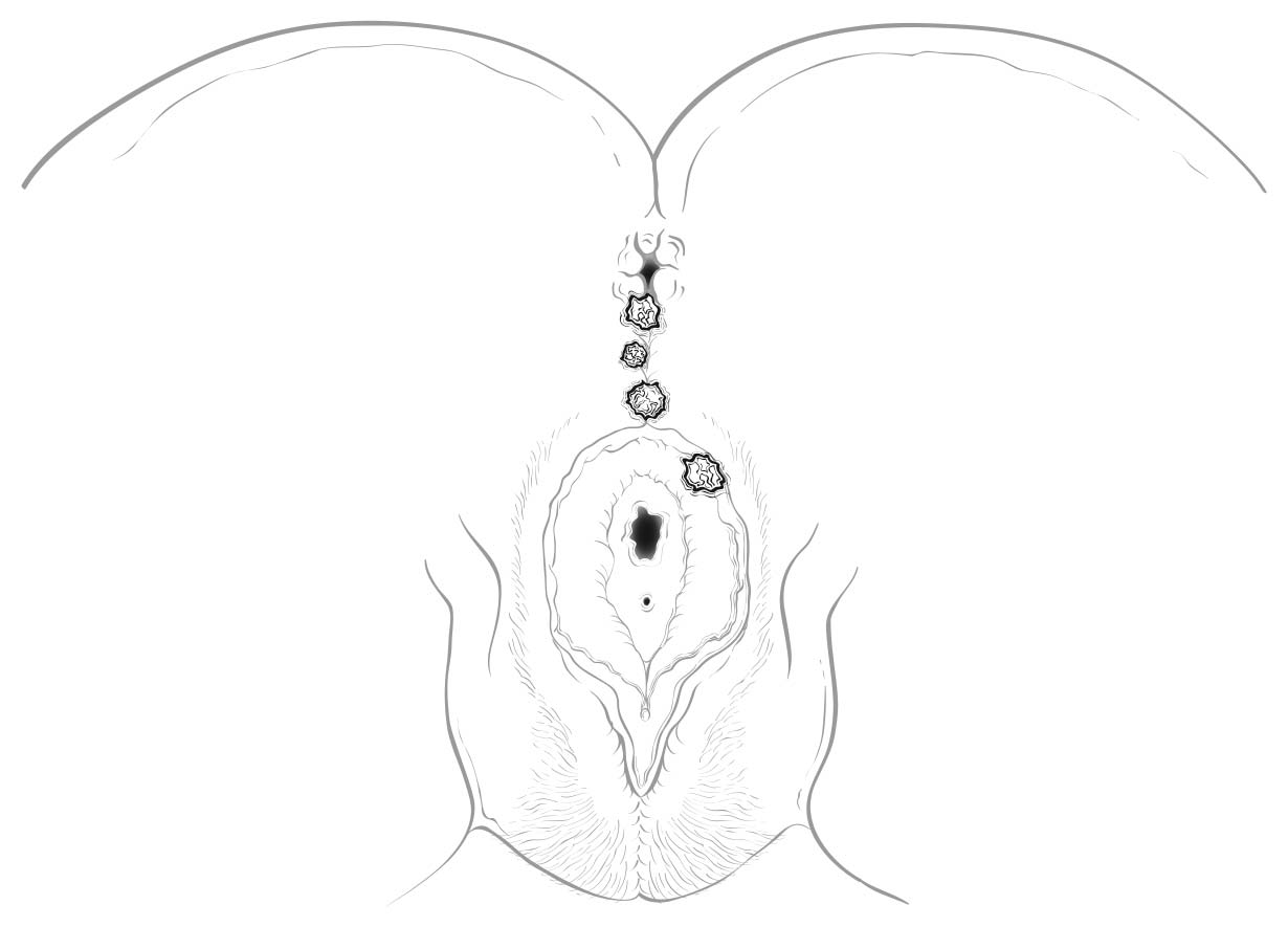
A focus of this chapter is to further refine the TNM staging system to clarify what constitutes anal canal versus perianal cancer and to specify additional considerations that may be of value for cancers in the region of the perianus and vulva. For instance, squamous cell carcinomas (SCCs) overlying the perineal body may be classified as perianal or vulvar, and the treatment plans may be quite dissimilar. For this reason, we recommend the following: lesions that clearly arise from the vulva and extend onto the perineum and potentially involve the anus should be classified as vulvar. Similarly, lesions that clearly arise from the distal anal mucosa and extend onto the perineum should be classified as perianal. Lesions localized to the perineum that are not clearly arising from either the vulva or the anus should be categorized based on the clinician's clinical impression. Thus, we recommend the following terminology: perineum favor vulva and perineum favor perianus. We also recommend consulting with colleagues in gynecologic oncology, colorectal or general surgery, or surgical oncology, because classification has a significant impact on treatment.

Most carcinomas of the anal canal and perianal region are SCCs. The terms transitional cell and cloacogenic carcinoma have been aband oned, because these tumors are now recognized as nonkeratinizing types of SCC.

Squamous Cell Carcinoma

The predominant histologic type of malignancy arising in the anal canal is SCC. Estimates suggest that 95% of these cancers are caused by oncogenic human papillomavirus (HPV) types, with HPV 16 associated with 89% of cases.3-6 Higher-risk populations are men who have sex with men and people who are immunosuppressed. Mortality rates are rising as well, with an average increase of 1.7% each year from 2001 to 2010. Only 65.5% of patients survive 5 years or more.1 Nonsquamous anal cancers include adenocarcinomas, basal cell carcinomas (BCCs), and melanomas (not discussed here).

Carcinomas of the anal canal typically are staged clinically according to the size and extent of the untreated primary tumor. Patients with cancer of the anal canal typically are staged at the time of presentation with inspection, palpation, and biopsy of the mass; palpation (and biopsy as needed) of regional lymph nodes; and radiologic imaging of the chest, abdomen, and pelvis.

High-grade squamous intraepithelial lesion (HSIL) is not a malignancy and should not be coded as such. Direct evidence supports the progression of HSIL to SCC, but the impact of treatment on the progression to carcinoma is being studied in the ANCHOR trial, a multi-institutional rand omized prospective trial comparing observation with excision/destruction.7

The primary management of SCCs of the anal canal is nonoperative, involving combined chemotherapy and radiation therapy. Fortunately, localized SCC of the anal canal has been associated with excellent outcomes with combined-modality therapy, with a 5-year overall survival rate of 78% in the positive arm of a US phase III rand omized trial, Radiation Therapy Oncology Group (RTOG) 98-11.8

In contrast, the management of perianal carcinomas remains mixed, with both operative and nonoperative treatments used selectively, based on involvement of adjacent structures, tumor size, and histology of the primary lesion. Complete pathological staging is possible for a perianal primary tumor treated surgically. The remainder of the staging of regional lymph nodes and distant disease in perianal tumors is as described for anal cancers.

Other Histologic Types

Verrucous Carcinoma

Verrucous carcinoma historically has been distinguished from ordinary condyloma acuminatum by its apparent combination of exophytic and endophytic growth.9-11 However, the appearance of endophytic growth may, in fact, represent growth along preexisting cryptogland ular fistulous disease rather than actual endophytic invasion, as convincing evidence of actual invasion is rare. Some verrucous carcinomas contain HPV, the most common types being HPV 6 and HPV 11. This is another distinction from SCCs, which demonstrate predominantly HPV 16. Treatment typically focuses on local control in the absence of invasion. However, any histologic evidence of invasive disease or metastasis should lead to the diagnosis of SCC and appropriate therapy.

Basal Cell Carcinoma

BCC of the perianus is an uncommon entity, with a reported incidence of about 0.1% of all BCCs and less than 1% of all anorectal neoplasms.12-14 Differentiating basaloid SCC from BCC may be challenging, as they have similar histologic features. However, SCCs arise from a known precursor lesion (anal squamous intraepithelial neoplasia), whereas BCCs do not have a well-defined precursor. SCCs may be distinguished further from BCCs in that they may arise from the anal mucosa or the perianal skin, whereas BCCs usually arise in the perianal skin. Rarely, BCCs may extend from the perianal skin into the anal canal. In general, BCCs are associated with a low recurrence rate (0-29%), and wide local excision with negative margins remains the stand ard of care, especially for smaller lesions. Electrodesiccation and curettage, Mohs micrographic surgery, and external beam irradiation also have been reported with successful results.15 Radiation and /or abdominal-perineal resection may be required for large lesions and recurrent tumors.

Adenocarcinoma

Adenocarcinomas of the anus and perianus are rare and generally fall into three categories: those that extend down from above the dentate line, those that originate from the underlying anorectal gland s or longstand ing fistulous disease, and those arising primarily from the anal mucosa or perianal skin.16-18 Lesions in the perianal skin may be amenable to wide local excision in select cases. Most perianal, anal, and distal rectal lesions extending into the anal mucosa are treated with abdominal perineal excision with or without neoadjuvant chemoradiation. The outcomes for all other histologic types, including adenocarcinomas, are poorer, stage for stage, than for SCC (Table 21.1).

The presumed precursor lesion for perianal adenocarcinoma, extramammary Paget disease, is adenocarcinoma in situ. Anal Paget disease may be divided into two classes. About half the cases are associated with synchronous or metachronous colorectal malignancies; the other subset does not appear to be associated with internal malignancies but is associated with a high local recurrence rate and is more likely to progress to invasive cancer.19 Perianal Paget disease may be treated with wide local excision.20,21
Anatomy

Primary Site(s)

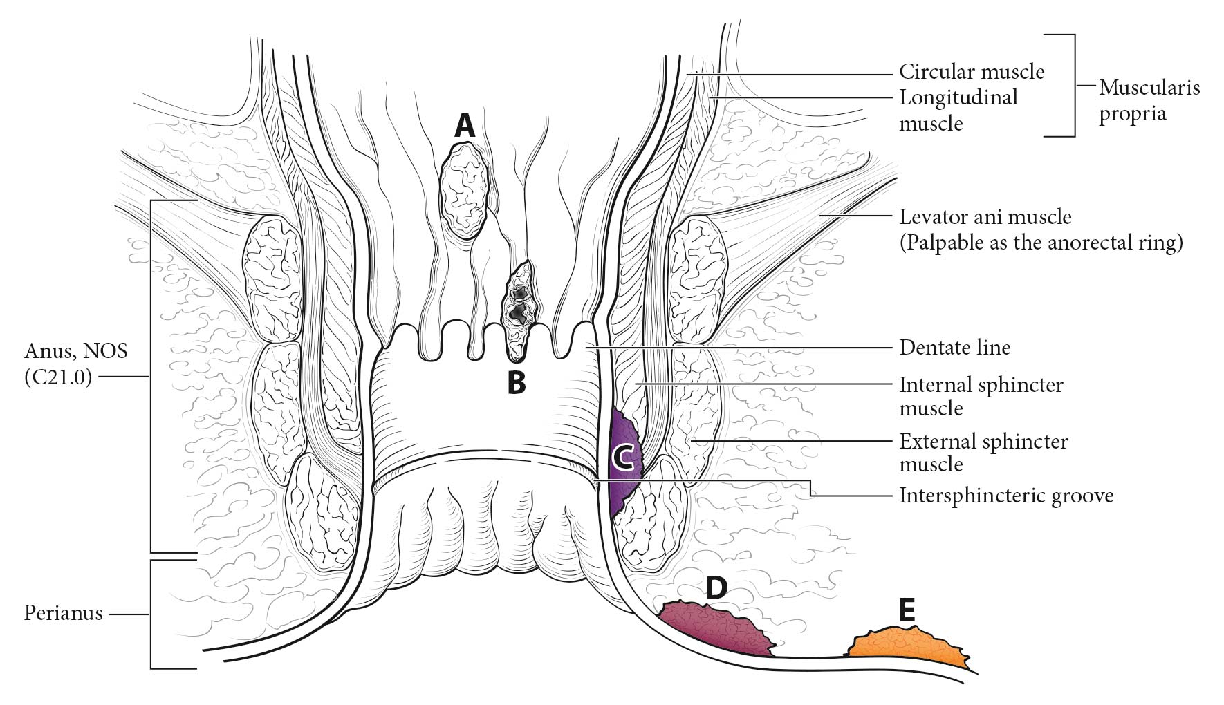
The anatomic subsites of the anal canal are illustrated in Figures 21.1_A and 21.1_B. The anal canal begins where the rectum enters the puborectalis sling at the apex of the anal sphincter complex (palpable as the anorectal ring on digital rectal examination and approximately 1 to 2 cm proximal to the dentate line) and ends with the squamous mucosa blending with the perianal skin, which coincides roughly with the palpable intersphincteric groove or the outermost boundary of the internal sphincter muscle, easily visualized on endoanal ultrasound. The anus encompasses true mucosa of three different histologic types: gland ular, transitional, and squamous (proximal to distal, respectively). The most proximal aspect of the anal canal is lined by colorectal mucosa in which squamous metaplasia may occur. When involved by metaplasia, this zone also may be referred to as the transformation zone. Immediately proximal to the macroscopically visible dentate line, a narrow zone of multilayered transitional mucosa is variably present. In the region of the dentate line, anal gland s are subjacent to the mucosa, often penetrating through the internal sphincter into the intersphincteric plane. The distal zone of the anal canal extends from the dentate line to the mucocutaneous junction with the perianal skin and is lined by a nonkeratinizing squamous epithelium devoid of epidermal appendages (hair follicles, apocrine gland s, and sweat gland s).

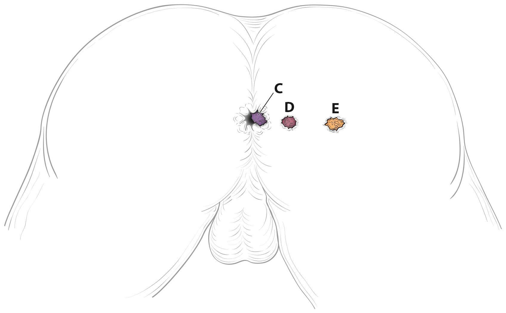
Tumors that develop from mucosa (of any of the three types) that cannot be visualized in their entirety while gentle traction is placed on the buttocks are termed anal cancers, whereas those that arise within the skin at or distal to the squamous mucocutaneous junction, can be seen in their entirety with gentle traction placed on the buttocks, and are within 5 cm of the anus are termed perianal cancers.

21.1_A Anal cancer (A-C), perianal cancer (D), and skin cancer (E) as visualized with gentle traction placed on the buttocks.

21.1_B Anal cancer (A-C), perianal cancer (D), and skin cancer (E) as visualized with gentle traction placed on the buttocks.

21.2 Perineal and vulvar lesions, from top to bottom. The lesion at the top is perianal, the middle two lesions are perineal, and the lesion at the bottom is considered vulvar.

Regional Lymph Nodes

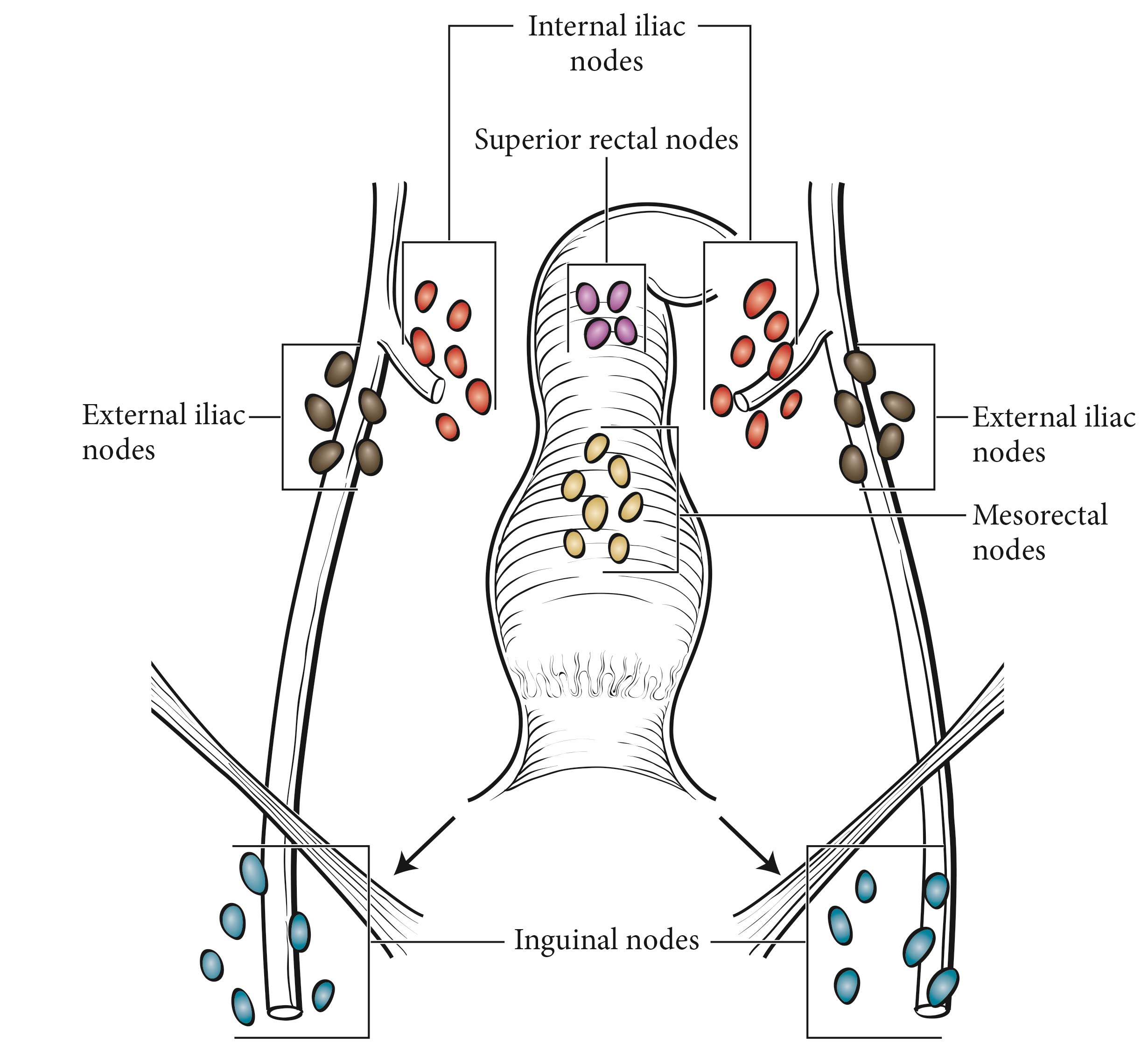
Lymphatic drainage and nodal involvement of anal cancers often depend on the location of the primary tumor. Tumors above the dentate line spread primarily to the mesorectal and internal iliac nodes, whereas tumors below the dentate line also may spread to the superficial inguinal and external iliac (deep inguinal) nodes.

The regional lymph nodes are as follows (Figure 21.3):

  • Mesocrectal
  • Inguinal: superficial, deep
  • Superior rectal (hemorrhoidal)
  • External iliac
  • Internal iliac (hypogastric)

All other nodal groups represent sites of distant metastasis.

21.3 Regional lymph nodes of the anal canal.

Metastatic Sites

Cancers of the anus may metastasize to any organ, but the liver and lungs are the distal organs involved most frequently.

Classification Rules

Clinical Classification

Clinical assessment is based on medical history, physical examination, radiology, and endoscopy, with biopsy for histologic confirmation of malignancy. HIV testing should be performed in patients with established risk factors. Given patterns of spread, a thorough examination of lymph node areas, including inguinofemoral regions, should be performed. If possible, suspicious inguinal lymphadenopathy should be biopsied, generally through fine-needle aspiration, to further facilitate diagnosis and tumor staging, particularly in the HIV-positive population, in whom reactive nodes are common. In female patients, vaginal examination should be performed to rule out posterior vaginal invasion and fistula, and the cervix should be evaluated to rule out gynecologic malignancy.

Imaging

Radiologic examinations may include chest radiographic films, computed tomography (CT, of the abdomen, pelvis, and chest), magnetic resonance imaging, and positron emission tomography (PET)/CT scans. These are obtained simultanteously during the initial evaluation. Of note, HIV-positive patients may have falsely positive nodes on PET imaging.

Imaging is the primary modality for diagnosing nonpalpable nodal disease and distant metastatic disease. Nonregional nodes or involvement of extrapelvic sites on imaging studies are considered M1 disease.

Pathological Classification

Because the primary treatment for anal canal SCC usually is combined chemoradiation, a surgical resection specimen is not commonly available for complete evaluation by the pathologist (pTNM), unless there is persistent or recurrent local tumor after chemoradiation. For the latter group of patients who were assigned a clinical stage (cTNM) before beginning primary chemoradiation, a modified pathological stage is determined after surgical resection (usually combined abdominoperineal resection) and annotated by the y prefix (ypTNM). For tumors treated by surgery and without neoadjuvant chemoradiation, and for those not clinically staged before surgery, pTN is assigned on pathological examination of the resection specimen, in consultation with the surgeon, who may comment on residual disease status and assign the c/pM.

Prognostic Factors

Prognostic Factors Required for Stage Grouping

Regarding SCC of the anal canal, based on data from the National Cancer Data Base (NCDB) for 2003 to 2006, the 5-year observed survival rates for each of the stage groups are as follows: Stage I (n = 1,516), 76.9%; Stage II (n = 3,214), 66.7%; Stage IIIA (n = 735), 57.7%; Stage IIIB (n = 1,117), 50.7%; Stage IV (n = 364), 15.2%. Stage-related survival is shown in Table 21.1. These figures reflect in-patient data; therefore, because most early tumors are not treated in the in-patient setting, the reported data might be skewed.

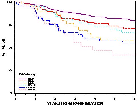
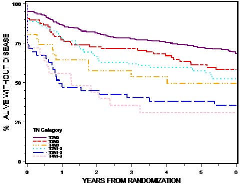
The TN category of disease is shown to have an impact on overall and disease-free survival (OS and DFS), the need for colostomy, and disease relapse (locoregional failure [LRF] and distant metastasis [DM]) in patients with SCC treated with primary chemoradiation.22 In the most recent analysis of the US GI Intergroup phase III trial RTOG 98-11, 620 of 682 rand omly assigned patients were analyzable for outcomes by TN category, and six TN categories were compared (T2N0, T3N0, T4N0, T2N1-3, T3N1-3, and T4N1-3). All end points showed statistically significant differences among the TN categories of disease (Table 21.2 and Figures 21.4_Aand 21.4_B). The best OS, DFS, and LRF outcomes were found in patients with T2N0 and T3N0 categories and the poorest outcomes in those with T4N0 and T3-4N+ disease. Survival and LRF outcomes for T2N+ patients were intermediate between T3N0 and T4N0 patients, as observed in previous TN outcome analyses in patients with colon and rectal cancer. 22 The need for colostomy was lowest for T2N0 and T2N+ disease (both 11%) and worst for T4N0, T3N+, and T4N+ (26%, 27%, and 24%, respectively). In summary, the prognosis of T2N+ category tumors is more akin to T2-3N0 tumors, and that of T3N+ tumors to T4N0 and T4N+.

The most important risk factor for squamous cell anal cancer is HPV infection, primarily types 16 and 18. Viral proteins in these high-risk HPV types mediate oncogenic transformation of the anal squamous epithelia.23,24 Recent evidence indicates that virtually every anal SCC is related to HPV, although uncertainty continues to exist regarding the impact of various subclassifications of the virus.6 A recent meta-analysis suggests that HPV 16 is found more frequently (75%) and HPV 18 less frequently (10%) in anal carcinomas than in cervical carcinomas.25 In contrast to squamous carcinomas of the head and neck, there do not appear to be two major categories with entirely different biologies and prognoses based on the HPV status.

Other known risk factors for anal SCC include a history of sexually transmitted disease, a history of multiple sexual partners, and anal intercourse, all of which may be associated with a higher incidence of HPV infection.26,27 SCCs of the anus are more common in women; however, men have a worse prognosis.2,27 The poorer prognosis in males also was confirmed in three phase III chemoradiation trials (US GI Intergroup 98-118,28, European Organisation for Research and Treatment of Cancer [EORTC] 22861,29 and United Kingdom ACT I30). Chronic immunosuppression, which may be related to HIV-positive status or a history of organ transplantation, and tobacco use also are important risk factors for anal cancer.26,31-33

Nonsquamous histologies of anal canal carcinomas, including adenocarcinoma, mucinous adenocarcinoma, high-grade neuroendocrine carcinoma, and undifferentiated carcinoma, are associated with worse 5-year survival (OS) rates than SCCs of the anal canal. At each stage, survival rates for patients with SCCs are better than those for patients with nonsquamous tumors, as shown in Table 21.1. However, historically recognized histologic SCC variants, such as large cell keratinizing, large cell nonkeratinizing, and basaloid subtypes, have no associated prognostic differences. Therefore, the World Health Organization recommends that the generic term squamous cell carcinoma be used for all squamous tumors of the anal canal. As previously stated, BCCs of the perianal region tend to have a better prognosis, with lower risk of relapse.

Beyond the factors used to assign T, N, or M categories, no additional prognostic factors are required for stage grouping.

Additional Factors Recommended for Clinical Care

Tumor Location

Tumor location defines nodal drainage fields at risk and treatment approach. For anal canal lesions, external beam irradiation fields differ from those for perianal lesions. Perianal cancers would be treated as skin cancers elsewhere in the body with focused irradiation fields without inclusion of regional nodes, unless there was deep invasion of the tumor, necessitating inguinal node coverage. For cancers of the anal canal, irradiation fields commonly include the primary tumor plus all regional node groups felt to be at risk (inguinal, mesorectal/superior rectal, internal iliac, external iliac). Tumor location is reported as anal, perianal, or perineal and left/right/anterior/posterior/lateral, and it is documented in and abstracted from the medical record.

AJCC Level of Evidence: I

HIV Status

The impact of HIV status on prognosis remains incompletely defined. The literature remains controversial regarding the perception that HIV-positive patients tolerate chemoradiation therapy less well and have poorer outcomes compared with HIV-negative patients. Patients whose HIV is well controlled with highly active antiretroviral therapy appear to do as well as HIV-negative patients. Better documentation of HIV status is needed.34-36 HIV status is reported as positive or negative, and it is documented in and abstracted from the medical record.

AJCC Level of Evidence: I

Gender
Male gender has a negative impact on prognosis. Although SCCs of the anus are more common in women, men have a worse prognosis.2,27 The poorer prognosis in males also was confirmed in three phase III chemoradiation trials (US GI Intergroup 98-118,28, EORTC 22861,29 and United Kingdom ACT I30). Gender is reported as male or female, and it is documented in and abstracted from the medical record.
AJCC Level of Evidence: I

Grade

High-grade (poorly differentiated) squamous carcinomas or adenocarcinomas of the anus have a worse prognosis than low-grade tumors. Grade is reported as well- (G1), moderately (G2), or poorly differentiated (G3), or undifferentiated (G4) carcinoma, and it is documented in and abstracted from the medical record.

AJCC Level of Evidence: I

HPV Status and p16 or p18 Expression

The most important risk factor for squamous cell anal cancer is infection of the anus, cervix, or vulva with HPV, primarily types 16 and 18. Recent evidence indicates that virtually every anal SCC is related to HPV, although uncertainty continues to exist regarding the impact of various subclassifications of the virus.6 A recent meta-analysis suggests that HPV 16 is found more frequently (75%) and HPV 18 less frequently (10%) in anal carcinomas than in cervical carcinomas.25 This factor is reported as HPV type, and it is documented in and abstracted from the medical record.

AJCC Level of Evidence: I

The authors have not noted any emerging factors for clinical care.

Risk Assessment

Risk Assesment Models

The AJCC recently established guidelines that will be used to evaluate published statistical prediction models for the purpose of granting endorsement for clinical use.37 Although this is a monumental step toward the goal of precision medicine, this work was published only very recently. Therefore, the existing models that have been published or may be in clinical use have not yet been evaluated for this cancer site by the Precision Medicine Core of the AJCC. In the future, the statistical prediction models for this cancer site will be evaluated, and those that meet all AJCC criteria will be endorsed.

Recommendations

Trials are few, and therefore investigators are very familiar with stratification issues. However, we suggest a focus on male gender, specific tumor location (anal vs. perianal vs. perineal, as we may be overtreating many patients with perianal and perineal lesions), and HIV status, including measures of disease control, such as CD4 counts and viral load.

TNM Definitions

Clinical T (cT)

cT CategorycT Criteria
cTXPrimary tumor not assessed
cT0No evidence of primary tumor
cTisHigh-grade squamous intraepithelial lesion (previously termed carcinoma in situ, Bowen disease, anal intraepithelial neoplasia II-III, high-grade anal intraepithelial neoplasia)
cT1Tumor less than or equal to 2 cm
cT2Tumor greater than 2 cm but less than or equal to 5 cm
cT3Tumor greater than 5 cm
cT4Tumor of any size invading adjacent organ(s), such as the vagina, urethra, or bladder

Pathological T (pT)

pT CategorypT Criteria
pTXPrimary tumor not assessed
pT0No evidence of primary tumor
pTisHigh-grade squamous intraepithelial lesion (previously termed carcinoma in situ, Bowen disease, anal intraepithelial neoplasia II-III, high-grade anal intraepithelial neoplasia)
pT1Tumor less than or equal to 2 cm
pT2Tumor greater than 2 cm but less than or equal to 5 cm
pT3Tumor greater than 5 cm
pT4Tumor of any size invading adjacent organ(s), such as the vagina, urethra, or bladder
cTXPrimary tumor not assessed
cT0No evidence of primary tumor
cTisHigh-grade squamous intraepithelial lesion (previously termed carcinoma in situ, Bowen disease, anal intraepithelial neoplasia II-III, high-grade anal intraepithelial neoplasia)
cT1Tumor less than or equal to 2 cm
cT2Tumor greater than 2 cm but less than or equal to 5 cm
cT3Tumor greater than 5 cm
cT4Tumor of any size invading adjacent organ(s), such as the vagina, urethra, or bladder

Neoadjuvant Clinical T (yT)

ycT CategoryycT Criteria
ycTXPrimary tumor not assessed
ycT0No evidence of primary tumor
ycTisHigh-grade squamous intraepithelial lesion (previously termed carcinoma in situ, Bowen disease, anal intraepithelial neoplasia II-III, high-grade anal intraepithelial neoplasia)
ycT1Tumor less than or equal to 2 cm
ycT2Tumor greater than 2 cm but less than or equal to 5 cm
ycT3Tumor greater than 5 cm
ycT4Tumor of any size invading adjacent organ(s), such as the vagina, urethra, or bladder

Neoadjuvant Pathological T (yT)

ypT CategoryypT Criteria
ypTXPrimary tumor not assessed
ypT0No evidence of primary tumor
ypTisHigh-grade squamous intraepithelial lesion (previously termed carcinoma in situ, Bowen disease, anal intraepithelial neoplasia II-III, high-grade anal intraepithelial neoplasia)
ypT1Tumor less than or equal to 2 cm
ypT2Tumor greater than 2 cm but less than or equal to 5 cm
ypT3Tumor greater than 5 cm
ypT4Tumor of any size invading adjacent organ(s), such as the vagina, urethra, or bladder

Definition of Regional Lymph Node (N)

Clinical N (cN)
cN CategorycN Criteria
cNXRegional lymph nodes cannot be assessed
cN0No regional lymph node metastasis
cN1Metastasis in inguinal, mesorectal, internal iliac, or external iliac nodes
cN1aMetastasis in inguinal, mesorectal, or internal iliac lymph nodes
cN1bMetastasis in external iliac lymph nodes
cN1cMetastasis in external iliac with any N1a nodes
Pathological N (pN)
pN CategorypN Criteria
pNXRegional lymph nodes cannot be assessed
pN0No regional lymph node metastasis
pN1Metastasis in inguinal, mesorectal, internal iliac, or external iliac nodes
pN1aMetastasis in inguinal, mesorectal, or internal iliac lymph nodes
pN1bMetastasis in external iliac lymph nodes
pN1cMetastasis in external iliac with any N1a nodes
cNXRegional lymph nodes cannot be assessed
cN0No regional lymph node metastasis
cN1Metastasis in inguinal, mesorectal, internal iliac, or external iliac nodes
cN1aMetastasis in inguinal, mesorectal, or internal iliac lymph nodes
cN1bMetastasis in external iliac lymph nodes
cN1cMetastasis in external iliac with any N1a nodes
Neoadjuvant Clinical N (pY)
ycN CategoryycN Criteria
ycNXRegional lymph nodes cannot be assessed
ycN0No regional lymph node metastasis
ycN1Metastasis in inguinal, mesorectal, internal iliac, or external iliac nodes
ycN1aMetastasis in inguinal, mesorectal, or internal iliac lymph nodes
ycN1bMetastasis in external iliac lymph nodes
ycN1cMetastasis in external iliac with any N1a nodes
Neoadjuvant Pathological N (pY)
ypN CategoryypN Criteria
ypNXRegional lymph nodes cannot be assessed
ypN0No regional lymph node metastasis
ypN1Metastasis in inguinal, mesorectal, internal iliac, or external iliac nodes
ypN1aMetastasis in inguinal, mesorectal, or internal iliac lymph nodes
ypN1bMetastasis in external iliac lymph nodes
ypN1cMetastasis in external iliac with any N1a nodes

Definition of Distant Metastasis (M)- Clinical M (cN)

cM CategorycM Criteria
cM0No distant metastasis
cM1Distant metastasis
pM1Microscopic evidence of distant metastasis

Definition of Distant Metastasis (M)- Pathological M (pN)

pM CategorypM Criteria
cM0No distant metastasis
cM1Distant metastasis
pM1Microscopic evidence of distant metastasis

Definition of Distant Metastasis (M)- Neoadjuvant Clinical M (pY)

ycM CategoryycM Criteria
cM0No distant metastasis
cM1Distant metastasis
pM1Microscopic evidence of distant metastasis

Definition of Distant Metastasis (M)- Neoadjuvant Pathological M (pY)

ypM CategoryypM Criteria
cM0No distant metastasis
cM1Distant metastasis
pM1Microscopic evidence of distant metastasis

Stage Prognostic

Clinical

When T is…and N is…and M is…Then the Clinical Prognostic Stage Group is…
cTiscN0cM00
cT1cN0cM0I
cT1cN1cM0IIIA
cT1cN1acM0IIIA
cT1cN1bcM0IIIA
cT1cN1ccM0IIIA
cT2cN0cM0IIA
cT2cN1cM0IIIA
cT2cN1acM0IIIA
cT2cN1bcM0IIIA
cT2cN1ccM0IIIA
cT3cN0cM0IIB
cT3cN1cM0IIIC
cT3cN1acM0IIIC
cT3cN1bcM0IIIC
cT3cN1ccM0IIIC
cT4cN0cM0IIIB
cT4cN1cM0IIIC
cT4cN1acM0IIIC
cT4cN1bcM0IIIC
cT4cN1ccM0IIIC
cTXcNXcM1IV
cTXcN0cM1IV
cTXcN1cM1IV
cTXcN1acM1IV
cTXcN1bcM1IV
cTXcN1ccM1IV
cT0cNXcM1IV
cT0cN0cM1IV
cT0cN1cM1IV
cT0cN1acM1IV
cT0cN1bcM1IV
cT0cN1ccM1IV
cT1cNXcM1IV
cT1cN0cM1IV
cT1cN1cM1IV
cT1cN1acM1IV
cT1cN1bcM1IV
cT1cN1ccM1IV
cT2cNXcM1IV
cT2cN0cM1IV
cT2cN1cM1IV
cT2cN1acM1IV
cT2cN1bcM1IV
cT2cN1ccM1IV
cT3cNXcM1IV
cT3cN0cM1IV
cT3cN1cM1IV
cT3cN1acM1IV
cT3cN1bcM1IV
cT3cN1ccM1IV
cT4cNXcM1IV
cT4cN0cM1IV
cT4cN1cM1IV
cT4cN1acM1IV
cT4cN1bcM1IV
cT4cN1ccM1IV
cTXcNXpM1IV
cTXcN0pM1IV
cTXcN1pM1IV
cTXcN1apM1IV
cTXcN1bpM1IV
cTXcN1cpM1IV
cT0cNXpM1IV
cT0cN0pM1IV
cT0cN1pM1IV
cT0cN1apM1IV
cT0cN1bpM1IV
cT0cN1cpM1IV
cT1cNXpM1IV
cT1cN0pM1IV
cT1cN1pM1IV
cT1cN1apM1IV
cT1cN1bpM1IV
cT1cN1cpM1IV
cT2cNXpM1IV
cT2cN0pM1IV
cT2cN1pM1IV
cT2cN1apM1IV
cT2cN1bpM1IV
cT2cN1cpM1IV
cT3cNXpM1IV
cT3cN0pM1IV
cT3cN1pM1IV
cT3cN1apM1IV
cT3cN1bpM1IV
cT3cN1cpM1IV
cT4cNXpM1IV
cT4cN0pM1IV
cT4cN1pM1IV
cT4cN1apM1IV
cT4cN1bpM1IV
cT4cN1cpM1IV

Pathological

When T is…and N is…and M is…Then the Pathological Prognostic Stage Group is…
pTispN0cM00
pTiscN0cM00
pT1pN0cM0I
pT1pN1cM0IIIA
pT1pN1acM0IIIA
pT1pN1bcM0IIIA
pT1pN1ccM0IIIA
pT2pN0cM0IIA
pT2pN1cM0IIIA
pT2pN1acM0IIIA
pT2pN1bcM0IIIA
pT2pN1ccM0IIIA
pT3pN0cM0IIB
pT3pN1cM0IIIC
pT3pN1acM0IIIC
pT3pN1bcM0IIIC
pT3pN1ccM0IIIC
pT4pN0cM0IIIB
pT4pN1cM0IIIC
pT4pN1acM0IIIC
pT4pN1bcM0IIIC
pT4pN1ccM0IIIC
pTXpNXpM1IV
pTXpN0pM1IV
pTXpN1pM1IV
pTXpN1apM1IV
pTXpN1bpM1IV
pTXpN1cpM1IV
pT0pNXpM1IV
pT0pN0pM1IV
pT0pN1pM1IV
pT0pN1apM1IV
pT0pN1bpM1IV
pT0pN1cpM1IV
pT1pNXpM1IV
pT1pN0pM1IV
pT1pN1pM1IV
pT1pN1apM1IV
pT1pN1bpM1IV
pT1pN1cpM1IV
pT2pNXpM1IV
pT2pN0pM1IV
pT2pN1pM1IV
pT2pN1apM1IV
pT2pN1bpM1IV
pT2pN1cpM1IV
pT3pNXpM1IV
pT3pN0pM1IV
pT3pN1pM1IV
pT3pN1apM1IV
pT3pN1bpM1IV
pT3pN1cpM1IV
pT4pNXpM1IV
pT4pN0pM1IV
pT4pN1pM1IV
pT4pN1apM1IV
pT4pN1bpM1IV
pT4pN1cpM1IV
pTXpNXcM1IV
pTXpN0cM1IV
pTXpN1cM1IV
pTXpN1acM1IV
pTXpN1bcM1IV
pTXpN1ccM1IV
pT0pNXcM1IV
pT0pN0cM1IV
pT0pN1cM1IV
pT0pN1acM1IV
pT0pN1bcM1IV
pT0pN1ccM1IV
pT1pNXcM1IV
pT1pN0cM1IV
pT1pN1cM1IV
pT1pN1acM1IV
pT1pN1bcM1IV
pT1pN1ccM1IV
pT2pNXcM1IV
pT2pN0cM1IV
pT2pN1cM1IV
pT2pN1acM1IV
pT2pN1bcM1IV
pT2pN1ccM1IV
pT3pNXcM1IV
pT3pN0cM1IV
pT3pN1cM1IV
pT3pN1acM1IV
pT3pN1bcM1IV
pT3pN1ccM1IV
pT4pNXcM1IV
pT4pN0cM1IV
pT4pN1cM1IV
pT4pN1acM1IV
pT4pN1bcM1IV
pT4pN1ccM1IV
cTXcNXpM1IV
cTXcN0pM1IV
cTXcN1pM1IV
cTXcN1apM1IV
cTXcN1bpM1IV
cTXcN1cpM1IV
cT0cNXpM1IV
cT0cN0pM1IV
cT0cN1pM1IV
cT0cN1apM1IV
cT0cN1bpM1IV
cT0cN1cpM1IV
cT1cNXpM1IV
cT1cN0pM1IV
cT1cN1pM1IV
cT1cN1apM1IV
cT1cN1bpM1IV
cT1cN1cpM1IV
cT2cNXpM1IV
cT2cN0pM1IV
cT2cN1pM1IV
cT2cN1apM1IV
cT2cN1bpM1IV
cT2cN1cpM1IV
cT3cNXpM1IV
cT3cN0pM1IV
cT3cN1pM1IV
cT3cN1apM1IV
cT3cN1bpM1IV
cT3cN1cpM1IV
cT4cNXpM1IV
cT4cN0pM1IV
cT4cN1pM1IV
cT4cN1apM1IV
cT4cN1bpM1IV
cT4cN1cpM1IV

Neoadjuvant Clinical

There is no Postneoadjuvant Clinical (ycTNM) stage group available at this time.

Neoadjuvant Pathological

When T is…and N is…and M is…Then the Postneoadjuvant Pathological Stage Group is…
ypTisypN0cM00
ypT1ypN0cM0I
ypT1ypN1cM0IIIA
ypT1ypN1acM0IIIA
ypT1ypN1bcM0IIIA
ypT1ypN1ccM0IIIA
ypT2ypN0cM0IIA
ypT2ypN1cM0IIIA
ypT2ypN1acM0IIIA
ypT2ypN1bcM0IIIA
ypT2ypN1ccM0IIIA
ypT3ypN0cM0IIB
ypT3ypN1cM0IIIC
ypT3ypN1acM0IIIC
ypT3ypN1bcM0IIIC
ypT3ypN1ccM0IIIC
ypT4ypN0cM0IIIB
ypT4ypN1cM0IIIC
ypT4ypN1acM0IIIC
ypT4ypN1bcM0IIIC
ypT4ypN1ccM0IIIC
ypTXypNXpM1IV
ypTXypN0pM1IV
ypTXypN1pM1IV
ypTXypN1apM1IV
ypTXypN1bpM1IV
ypTXypN1cpM1IV
ypT0ypNXpM1IV
ypT0ypN0pM1IV
ypT0ypN1pM1IV
ypT0ypN1apM1IV
ypT0ypN1bpM1IV
ypT0ypN1cpM1IV
ypT1ypNXpM1IV
ypT1ypN0pM1IV
ypT1ypN1pM1IV
ypT1ypN1apM1IV
ypT1ypN1bpM1IV
ypT1ypN1cpM1IV
ypT2ypNXpM1IV
ypT2ypN0pM1IV
ypT2ypN1pM1IV
ypT2ypN1apM1IV
ypT2ypN1bpM1IV
ypT2ypN1cpM1IV
ypT3ypNXpM1IV
ypT3ypN0pM1IV
ypT3ypN1pM1IV
ypT3ypN1apM1IV
ypT3ypN1bpM1IV
ypT3ypN1cpM1IV
ypT4ypNXpM1IV
ypT4ypN0pM1IV
ypT4ypN1pM1IV
ypT4ypN1apM1IV
ypT4ypN1bpM1IV
ypT4ypN1cpM1IV
ypTXypNXcM1IV
ypTXypN0cM1IV
ypTXypN1cM1IV
ypTXypN1acM1IV
ypTXypN1bcM1IV
ypTXypN1ccM1IV
ypT0ypNXcM1IV
ypT0ypN0cM1IV
ypT0ypN1cM1IV
ypT0ypN1acM1IV
ypT0ypN1bcM1IV
ypT0ypN1ccM1IV
ypT1ypNXcM1IV
ypT1ypN0cM1IV
ypT1ypN1cM1IV
ypT1ypN1acM1IV
ypT1ypN1bcM1IV
ypT1ypN1ccM1IV
ypT2ypNXcM1IV
ypT2ypN0cM1IV
ypT2ypN1cM1IV
ypT2ypN1acM1IV
ypT2ypN1bcM1IV
ypT2ypN1ccM1IV
ypT3ypNXcM1IV
ypT3ypN0cM1IV
ypT3ypN1cM1IV
ypT3ypN1acM1IV
ypT3ypN1bcM1IV
ypT3ypN1ccM1IV
ypT4ypNXcM1IV
ypT4ypN0cM1IV
ypT4ypN1cM1IV
ypT4ypN1acM1IV
ypT4ypN1bcM1IV
ypT4ypN1ccM1IV

Registry Data

Registry Data Collection Variables

  1. Tumor location: anal, perianal, or perineal, and left/right/anterior/posterior/lateral
  2. HIV status
  3. Gender
  4. Grade
  5. HPV status and p16 or p18 expression

Histopathologic type

Histologic grade

HISTOLOGIC GRADE (G)

GG Definition
GXGrade cannot be determined
G1Well differentiated (low grade)
G2Moderately differentiated (low grade)
G3Poorly differentiated (high grade)
G4Undifferentiated (high grade)

Survival

21.1 Surveillance, Epidemiology, and End Results (SEER) 5-year OS for cohort stratified by histologic type and stage (KM method)

StageSquamousOS (%)NonsquamousOS (%)
I76.971
II66.758.7
IIIA57.750.1
IIIB50.734.6
IV15.26.8

21.2 Impact of TN category on survival, relapse, and colostomy failure in US GI Intergroup RTOG 98-11 phase III chemoradiation trial. Modified from Gunderson LL et al. Int J Radiat Oncol Biol Phys 2013; 87:638-645.

TN CategoryPatients, nOSDFSLRFDMCF
TD5-y, %*TF5-y, %TF5-y, %TF5-y, %TF3-y, %
T2N0323768211072571738103611
T3N09630744561171813141513
T4N031145716501137721826
T2N1-39938705057262628271111
T3N1-34620572938204411241227
T4N1-325164218311560624624

p value **

<0.0001<0.0001<0.00010.00110.01
Abbreviations: DM, distant metastasis; CF, colostomy failure; DFS, disease-free survival; LRF, locoregional failure; OS, overall survival; TD, total deaths; TF, total failures; 5-y, 5-year; 3-y, 3-year

*Note: Some 5-year estimates might be unstable because of small sample sizes; therefore, too few patients are at risk at 5 years.

**Log-rank test for OS and DFS; Gray's test for LRF, DM, and CF.

21.4_A Kaplan-Meier curves demonstrating the impact of TN category on survivalin US GI Intergroup RTOG phase III chemoradiation trial: OS (p < 0.0001) (From Gunderson et al.22)

21.4_B Kaplan-Meier curves demonstrating the impact of TN category on survivalin US GI Intergroup RTOG phase III chemoradiation trial: (B) DFS (p < 0.0001) (From Gunderson et al.22)

Bibliography

  1. National Cancer Institute. SEER Stat Fact Sheets: Anal Cancer. 2015; http://seer.cancer.gov/statfacts/html/anus.html.
  2. American Cancer Society. Anal Cancer: Key Statistics. 2015; http://www.cancer.org/cancer/analcancer/detailedguide/anal-cancer-what-is-key-statistics. Accessed Oct 16, 2015.
  3. Forman D, de Martel C, Lacey CJ, et al. Global burden of human papillomavirus and related diseases. Vaccine. Nov 20 2012;30 Suppl 5:F12-23.
  4. Parkin DM, Bray F. Chapter 2: The burden of HPV-related cancers. Vaccine. Aug 31 2006;24 Suppl 3:S3/11-25.
  5. Abramowitz L, Jacquard AC, Jaroud F, et al. Human papillomavirus genotype distribution in anal cancer in France: the EDiTH V study. Int J Cancer. Jul 15 2011;129(2):433-439.
  6. Baricevic I, He X, Chakrabarty B, et al. High-sensitivity human papilloma virus genotyping reveals near universal positivity in anal squamous cell carcinoma: different implications for vaccine prevention and prognosis. European journal of cancer. Apr 2015;51(6):776-785.
  7. Berry JM, Jay N, Cranston RD, et al. Progression of anal high-grade squamous intraepithelial lesions to invasive anal cancer among HIV-infected men who have sex with men. Int J Cancer. Mar 1 2014;134(5):1147-1155.
  8. Gunderson LL, Winter KA, Ajani JA, et al. Long-term update of US GI intergroup RTOG 98-11 phase III trial for anal carcinoma: survival, relapse, and colostomy failure with concurrent chemoradiation involving fluorouracil/mitomycin versus fluorouracil/cisplatin. J Clin Oncol. Dec 10 2012;30(35):4344-4351.
  9. Bertram P, Treutner KH, Rubben A, Hauptmann S, Schumpelick V. Invasive squamous-cell carcinoma in giant anorectal condyloma (Buschke-Lowenstein tumor). Langenbecks Arch Chir. 1995;380(2):115-118.
  10. Longacre TA, Kong CS, Welton ML. Diagnostic problems in anal pathology. Adv Anat Pathol. Sep 2008;15(5):263-278.
  11. Welton ML, Lambert R, Bosman FT. Tumours of the anal canal. In: Bosman FT, Carneiro F, Hruban RH, Theise ND, eds. WHO classification of tumours of the digestive system. 4th ed: International Agency for Research on Cancer; 2010.
  12. Patil DT, Goldblum JR, Billings SD. Clinicopathological analysis of basal cell carcinoma of the anal region and its distinction from basaloid squamous cell carcinoma. Modern pathology : an official journal of the United States and Canadian Academy of Pathology, Inc. Oct 2013;26(10):1382-1389.
  13. Paterson CA, Young-Fadok TM, Dozois RR. Basal cell carcinoma of the perianal region: 20-year experience. Diseases of the colon and rectum. Sep 1999;42(9):1200-1202.
  14. Moore HG, Guillem JG. Anal neoplasms. Surg Clin North Am. Dec 2002;82(6):1233-1251.
  15. Gibson GE, Ahmed I. Perianal and genital basal cell carcinoma: A clinicopathologic review of 51 cases. Journal of the American Academy of Dermatology. Jul 2001;45(1):68-71.
  16. Abel ME, Chiu YS, Russell TR, Volpe PA. Adenocarcinoma of the anal gland s. Results of a survey. Diseases of the colon and rectum. Apr 1993;36(4):383-387.
  17. Basik M, Rodriguez-Bigas MA, Penetrante R, Petrelli NJ. Prognosis and recurrence patterns of anal adenocarcinoma. American journal of surgery. Feb 1995;169(2):233-237.
  18. Anthony T, Simmang C, Lee EL, Turnage RH. Perianal mucinous adenocarcinoma. Journal of surgical oncology. Mar 1997;64(3):218-221.
  19. Goldblum JR, Hart WR. Perianal Paget's disease: a histologic and immunohistochemical study of 11 cases with and without associated rectal adenocarcinoma. The American journal of surgical pathology. Feb 1998;22(2):170-179.
  20. Marchesa P, Fazio VW, Oliart S, Goldblum JR, Lavery IC, Milsom JW. Long-term outcome of patients with perianal Paget's disease. Annals of surgical oncology. Sep 1997;4(6):475-480.
  21. McCarter MD, Quan SH, Busam K, Paty PP, Wong D, Guillem JG. Long-term outcome of perianal Paget's disease. Diseases of the colon and rectum. May 2003;46(5):612-616.
  22. Gunderson LL, Moughan J, Ajani JA, et al. Anal carcinoma: impact of TN category of disease on survival, disease relapse, and colostomy failure in US Gastrointestinal Intergroup RTOG 98-11 phase 3 trial. International journal of radiation oncology, biology, physics. Nov 15 2013;87(4):638-645.
  23. Munoz N, Bosch FX, de Sanjose S, et al. Epidemiologic classification of human papillomavirus types associated with cervical cancer. N Engl J Med. Feb 6 2003;348(6):518-527.
  24. Organization WH. IARC monograph on the evaluation of carcinogenic risks to humans: human papillomaviruses, 1995. Lyons, France2000.
  25. Machalek DA, Poynten M, Jin F, et al. Anal human papillomavirus infection and associated neoplastic lesions in men who have sex with men: a systematic review and meta-analysis. The lancet oncology. May 2012;13(5):487-500.
  26. Palefsky JM, Holly EA, Ralston ML, Da Costa M, Greenblatt RM. Prevalence and risk factors for anal human papillomavirus infection in human immunodeficiency virus (HIV)-positive and high-risk HIV-negative women. J Infect Dis. Feb 1 2001;183(3):383-391.
  27. Moscicki AB, Darragh TM, Berry-Lawhorn JM, et al. Screening for Anal Cancer in Women. J Low Genit Tract Dis. Jul 2015;19(3 Suppl 1):S27-42.
  28. Ajani JA, Winter KA, Gunderson LL, et al. Prognostic factors derived from a prospective database dictate clinical biology of anal cancer: the intergroup trial (RTOG 98-11). Cancer. Sep 1 2010;116(17):4007-4013.
  29. Bartelink H, Roelofsen F, Eschwege F, et al. Concomitant radiotherapy and chemotherapy is superior to radiotherapy alone in the treatment of locally advanced anal cancer: results of a phase III rand omized trial of the European Organization for Research and Treatment of Cancer Radiotherapy and Gastrointestinal Cooperative Groups. J Clin Oncol. May 1997;15(5):2040-2049.
  30. Glynne-Jones R, Sebag-Montefiore D, Adams R, et al. Prognostic factors for recurrence and survival in anal cancer: generating hypotheses from the mature outcomes of the first United Kingdom Coordinating Committee on Cancer Research Anal Cancer Trial (ACT I). Cancer. Feb 15 2013;119(4):748-755.
  31. Penn I. Cancers of the anogenital region in renal transplant recipients. Analysis of 65 cases. Cancer. Aug 1 1986;58(3):611-616.
  32. Sillman FH, Sedlis A. Anogenital papillomavirus infection and neoplasia in immunodeficient women: an update. Dermatol Clin. Apr 1991;9(2):353-369.
  33. Holly EA, Whittemore AS, Aston DA, Ahn DK, Nickoloff BJ, Kristiansen JJ. Anal cancer incidence: genital warts, anal fissure or fistula, hemorrhoids, and smoking. Journal of the National Cancer Institute. Nov 15 1989;81(22):1726-1731.
  34. Marcus JL, Chao C, Leyden WA, et al. Survival among HIV-infected and HIV-uninfected individuals with common non-AIDS-defining cancers. Cancer epidemiology, biomarkers & prevention : a publication of the American Association for Cancer Research, cosponsored by the American Society of Preventive Oncology. Aug 2015;24(8):1167-1173.
  35. Coghill AE, Shiels MS, Suneja G, Engels EA. Elevated Cancer-Specific Mortality Among HIV-Infected Patients in the United States. J Clin Oncol. Jul 20 2015;33(21):2376-2383.
  36. Fraunholz IB, Haberl A, Klauke S, Gute P, Rodel CM. Long-term effects of chemoradiotherapy for anal cancer in patients with HIV infection: oncological outcomes, immunological status, and the clinical course of the HIV disease. Diseases of the colon and rectum. Apr 2014;57(4):423-431.
  37. Kattan MW, Hess KR, Amin MB, et al. American Joint Committee on Cancer acceptance criteria for inclusion of risk models for individualized prognosis in the practice of precision medicine. CA: a cancer journal for clinicians. Jan 19 2016.