section name header

Basic Information ⬇

AUTHOR: Lydia Sharp, MD

Definition

Inflammatory myopathies are idiopathic diseases of muscle characterized clinically by muscle weakness and pathologically by inflammation and muscle fiber breakdown. They may be further classified as dermatomyositis (DM), immune-mediated necrotizing myopathy, polymyositis (PM), antisynthetase syndrome, and inclusion body myositis (IBM).1 See separate topics on “Inclusion Body Myositis” and “Necrotizing Autoimmune Myopathy” for details regarding these topics.

Synonyms

Immune-mediated myopathies

Idiopathic inflammatory myopathies

Myositis syndromes

Polymyositis

Dermatomyositis

IMM

ICD-10CM CODES
M33.02Juvenile dermatopolymyositis with myopathy
M33.12Other dermatopolymyositis with myopathy
M33.20Polymyositis, organ involvement unspecified
M33.22Polymyositis with myopathy
M33.90Dermatopolymyositis, unspecified, organ involvement unspecified
M33.92Dermatopolymyositis, unspecified with myopathy
Epidemiology & Demographics

Inflammatory myopathies are the largest group of potentially treatable myopathies in children and adults.

DM:

  • Occurs in children and in adults (bimodal age peak)
  • Average age at diagnosis is 40 to 60 yr in adults, 4 to 14 yr in children2
  • More common in females than in males (2:1)2
  • Incidence: 9.54 to 32.74:100,0002
  • Dermatomyositis is associated with a higher risk of cancer, especially in those older than age 50 yr3

PM:

  • Occurs mostly in adults, very rare in children4
  • Average age at diagnosis is over 20 yr4
  • More common in females4
  • Least common inflammatory myopathy4
  • Exact incidence unknown

Antisynthetase syndrome:

  • Associated with antibodies against aminoacyl transfer RNA synthetases5
  • More common in women5
  • Average age at diagnosis is 48 yr5
Physical Findings & Clinical Presentation

  • Most patients have a subacute onset over weeks to months.
  • Pattern is typically symmetric proximal muscle weakness involving the proximal limbs (shoulder and pelvic girdles).
  • Weakness of neck flexion and extension is common.
  • Difficulty getting up from a chair, climbing stairs, reaching for objects above head, or combing hair.
  • Distal muscle involvement and ocular involvement are uncommon.
  • Sensation is preserved.
  • Reflexes may be preserved or diminished.
  • Dysphagia and dysphonia result from involvement of striated muscle of the pharynx and proximal esophagus.
  • Esophageal dysmotility is common in DM.
  • Respiratory failure from associated pulmonary fibrosis.
  • Cardiac conduction abnormalities can be seen with DM.
  • Systemic autoimmune disease occurs frequently in PM and rarely in DM.
  • Skin findings in DM:
    1. Heliotrope rash on the upper eyelids
    2. Erythematous rash on the face
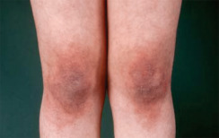
    3. May also involve the back and shoulders (shawl sign), neck and chest (V-shape), knees (Fig. E1), and elbows
    4. Photosensitivity
    5. Gottron papules (violaceous papules overlying dorsal interphalangeal or metacarpophalangeal areas, elbow or knee joints)
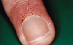
    6. Nail cracking, thickening, and irregularity (Fig. E2) with periungual telangiectasia
    7. Associated with increased risk of interstitial lung disease
  • Antisynthetase syndrome is associated with:
    1. Interstitial lung disease5
    2. Arthritis5
    3. Raynaud phenomenon5
    4. Mechanic’s hands (dry, cracked skin on hands)5

Figure E1 Violaceous Plaques on the Knees in a Patient with Dermatomyositis (Gottron Sign)

From Hochberg MC et al: Rheumatology, ed 5, St Louis, 2011, Mosby.

Figure E2 Enlarged nail fold capillaries in a patient with dermatomyositis.

From Hochberg MC et al: Rheumatology, ed 5, St Louis, 2011, Mosby.

Etiology

Mechanisms of muscle fiber damage in myositis are illustrated in Fig. E3.

DM: Complex, immune-mediated microangiopathy. Adaptive immune response via humorally mediated complement attack

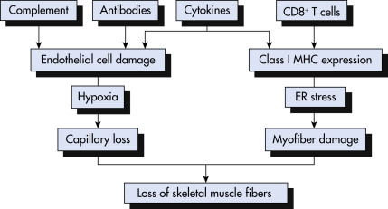
Figure E3 Mechanisms of Muscle Fiber Damage in Myositis

ER, Endoplasmic Reticulum; MHC, Major Histocompatibility Complex.

!!flowchart!!

From Firestein GS et al: Firestein & Kelley’s textbook of rheumatology, ed 11, Philadelphia 2021, Elsevier.

PM: Unknown:

  • Cell-mediated immune major histocompatibility-I (MHC-1) process directed against muscle fibers is likely, given biopsy features.

Diagnosis ⬆ ⬇

TABLE 2 Histologic Features of Idiopathic Inflammatory Myopathies

FeatureDermatomyositisPolymyositisInclusion Body Myositis
Necrosis of muscle fibers+++
Variation in fiber diameter+++
Regeneration of muscle fibers+++
Proliferation of connective tissue+++
Infiltration of mononuclear cells∗+++
Perivascular and perimysial inflammation+–/+–/+
Endomysial inflammation–/+++
Perifascicular atrophy+––
Abnormally dilated capillaries+–/+–
Reduced capillary density+–/+–
Deposition of complement on vessel walls+–/+–
Microinfarcts+––
Invasion of nonnecrotic fibers by cytotoxic T lymphocytes and macrophages–++
Expression of major histocompatibility complex class I on muscle fibers–/+++
Rimmed vacuoles with amyloid deposits and tubulofilaments†––+
Angulated or atrophic and hypertrophic fibers––+
Ragged red or cytochrome oxidase-negative fibers––+

∗Inflammation is absent in a small proportion of polymyositis and dermatomyositis biopsies.

† Also seen in chronic neurogenic conditions and distal myopathies.

From Firestein GS et al: Kelley’s textbook of rheumatology, ed 9, Philadelphia, 2013, Saunders, Elsevier.

TABLE E1 Classification of Inflammatory Myopathies

DisorderAge RangeClinical FeaturesMuscle Pathology
DermatomyositisJuvenile and adult formsProximal weakness plus skinPerimysial and perivascular inflammation, perifascicular atrophy
Antisynthetase syndromeChildren and adultsProximal weaknessPerifascicular necrosis and macrophagocytosis
PolymyositisAdult (rare in childhood)Proximal weaknessEndomysial inflammation with invasion of nonnecrotic muscle fibers
Immune-mediated necrotizing myopathyAdultProximal weaknessMultifocal necrotic muscle fibers
Inclusion body myositisAdult >40 yr oldProminent quadriceps and finger flexor weakness; treatment refractoryEndomysial inflammation with invasion of nonnecrotic muscle fibers plus rimmed vacuoles
  • Overlap syndromes
  • Adult
  • Myositis plus defined connective tissue disease
  • Nonspecific inflammation
  • Other (granulomatous myositis, eosinophilic myositis)
  • All ages
  • Proximal or distal weakness
  • Specific to type (e.g., granulomas present with granulomatous myositis)

From Goldman L, Schafer AI: Goldman-Cecil medicine, ed 26, Philadelphia, 2019, Elsevier.

Differential Diagnosis (TABLE E3

  • IBM
  • Muscular dystrophies
  • Amyloid myoneuropathy
  • Amyotrophic lateral sclerosis
  • Myasthenia gravis
  • Eaton-Lambert syndrome
  • Drug-induced myopathies (e.g., quinidine, NSAIDs, penicillamine, HMG-CoA-reductase inhibitors)
  • Diabetic amyotrophy
  • Guillain-Barré syndrome
  • Hyperthyroidism or hypothyroidism
  • Lichen planus
  • Amyopathic DM (rash without weakness)
  • DM sine rash (weakness with characteristic biopsy, but no rash)
  • Systemic lupus erythematosus (SLE)
  • Contact atopic or seborrheic dermatitis
  • Psoriasis

TABLE E3 Differential Diagnosis of Inflammatory Myopathies

DiseaseKey Diagnostic Features
Dystrophic Myopathies
Dysferlinopathy (Miyoshi myopathy and LGMD2B)Mutations in dysferlin gene
Progressive proximal (LGMD2B) and distal (Miyoshi myopathy) muscle weakness
Onset in late teens to early 20s
Increased creatine kinase (CK) levels
Inflammation in muscle biopsy
Nonresponsiveness to steroids
Facioscapulohumeral muscular dystrophyPartial deletion in D4Z4 repeats near chromosome 4q telomere at 4q35
Initial facial and shoulder girdle weakness progresses to pelvic girdle and extremities
Normal serum CK levels or modest elevation
Becker dystrophyMutations in dystrophin gene
X-linked recessive disorders
Limb-girdle weakness and cardiomyopathy
High serum CK levels
Proximal myotonic myopathyCCTG expansion in intron 1 of ZNF9 gene
Autosomal dominant
Proximal muscle weakness
Limb-girdle muscular dystrophy (LGMD2D, LGMD2E, LGMD2C, and LGMD2F)Mutations in sarcoglycans (α, β, γ, and Δ)
Limb-girdle weakness and cardiomyopathy
High serum CK levels
Metabolic Myopathies
Acid maltase deficiencyMutations in acid α-glucosidase
Proximal muscle weakness
Respiratory muscle involvement
Abnormal irritability on electromyography (EMG)
Increased serum CK levels
McArdle diseaseMutations in myophosphorylase gene
Exercise intolerance
Fixed proximal muscle weakness
Increased serum CK levels
Mitochondrial MyopathiesMutations in complex I-IV, complex V, and coenzyme Q10 genes
Myopathy with limb-girdle weakness
Exercise intolerance and fatigue
Increased serum CK levels
Endocrine Myopathies
Cushing syndromeInsidious onset
Proximal muscle weakness
Normal serum CK, aspartate aminotransferase (AST), and lactate dehydrogenase (LDH) levels
Thyrotoxic myopathySubacute onset of proximal muscle weakness
Normal serum CK levels or modest elevation
Respiratory muscle weakness
Infectious Myopathies
HIV myopathyProgressive myopathy
Proximal symmetric muscle weakness
Endomysial inflammation
Increased serum CK levels
Parasitic Myopathies
Protozoal myopathyClinical features of idiopathic inflammatory myopathies
Focal or diffuse inflammation
Myocarditis
Increased serum CK levels
Drug-Induced Myopathies
Zidovudine myopathyProximal muscle weakness
Increased serum lactate levels
Ragged red fibers and abnormal mitochondria in muscle
Improves with drug discontinuation
Statin myopathyNecrotizing myopathy
Acute or subacute painful proximal myopathy
Increased serum CK levels
Corticosteroid myopathyProximal and distal weakness
Type 2 atrophy and vacuolar changes in muscle
Increased serum CK levels
d-Penicillamine, interferon-α, and procainamide-induced myopathyProximal muscle weakness and pain
Inflammation and necrosis in muscle
Skin changes
Increased serum CK levels
Neuromuscular Diseases
Motoneuron diseaseUpper and lower motoneuron signs
Asymmetric weakness with denervation atrophy
Fasciculations and fatigability
Fibrillations and enlarged motor unit potentials on EMG
Modest elevation in serum CK levels
Spinal muscular atrophySymmetric muscle weakness and atrophy
Neurogenic changes on EMG and biopsy
Normal serum CK levels
Myasthenia gravisAbnormal weakness and fatigability
Decremental EMG response
Antiacetylcholine receptor antibodies
Positive anticholinesterase drug test

From Firestein GS et al: Firestein & Kelley’s textbook of rheumatology, ed 11, Philadelphia, 2021, Elsevier.

Laboratory Tests

  • Creatine kinase (CK) is the most sensitive muscle enzyme test for muscle breakdown.
  • CK is typically elevated (5 to 50× normal) in active PM.
  • CK may be normal or only slightly elevated in DM.
  • Aldolase, aspartate aminotransferase, alanine aminotransferase, alkaline phosphatase, and lactate dehydrogenase (LDH) may be elevated.
  • Anti-Jo-1 antibodies are seen in myositis with associated interstitial lung disease but are not specific for either DM or PM.
  • Myositis-specific and myositis-associated autoantibodies in adult polymyositis and dermatomyositis and juvenile dermatomyositis are summarized in Table E4.
  • DM: Anti-MDA-5, anti-Mi-2, anti-TIF-1, and anti-NXP2 (implicated in cancer-associated dermatomyositis).
  • PM: Antisynthetase antibodies associated with interstitial lung disease, arthritis, fever, and “mechanic’s hands.”
  • Electrolytes, thyroid-stimulating hormone, Ca, and Mg should be evaluated to exclude other causes of weakness.
  • Check ECG for cardiac involvement.

TABLE E4 Myositis-Specific and Myositis-Associated Autoantibodies in Adult Polymyositis and Dermatomyositis and Juvenile Dermatomyositis

AutoantibodyAutoantigenClinical Features
Jo-1, non-Jo-1 (PL7, PL12, EJ, OJ, KS, Tyr, Zo)Amino-acyl-tRNA synthetaseFever, Raynaud phenomenon, mechanic’s hands, myositis, polyarthritis, ILD
SRPSignal recognition particle (cytoplasmic protein translocation)Severe necrotizing myopathy; predominantly PM
Mi-2HelicaseDM (adult >children); “shawl” sign and other DM rashes
PM-SclNucleolar macromolecular complexOverlap features of myositis and SSc (or either disease alone); mechanic’s hands
U1RNPSmall nuclear ribonucleoproteinOverlap syndromes (MCTD)
SUMO-1 (small ubiquitin-like modifier 1)Small ubiquitin-like modifier enzyme (posttranslational modification)Adult DM, ILD
TIF1-γ (p155/140)Transcriptional intermediary factor 1-γ (TIF1-γ)Cancer-associated myositis in adults; >20% frequency in JDM cohorts; severe cutaneous disease in adult DM and JDM
NXP2/MJ (p140)NXP-2 (SUMO target; possible role in SUMO-mediated transcriptional repression)20%-25% frequency in JDM cohorts; calcinosis; severe disease (atrophy or contractures)
MDA-5 (cADM-140)RNA helicaseADM, ILD; palmar papules and cutaneous ulcerations
PMS1PMS1 (DNA mismatch repair enzyme)Myositis (specifics not known)
Ku170- and 80-kDa nuclear/nucleolar protein complex (DNA break repair and recombination)UCTD and overlap syndromes (Raynaud phenomenon, ILD, myositis, arthritis)
HMGCR (200/100 kDa)2,3HMG CoA reductaseNecrotizing myopathy

ADM, Amyopathic dermatomyositis; DM, dermatomyositis; DNA, deoxyribonucleic acid; HMG CoA, hydroxymethylglutaryl-coenzyme A; ILD, interstitial lung disease; JDM, juvenile DM; MCTD, mixed connective tissue disease; PM, polymyositis; RNA, ribonucleic acid; SSc, systemic sclerosis; tRNA, transfer ribonucleic acid; UCTD, undifferentiated connective tissue disease.

1 Rozelle A et al: Malignancy in the setting of the anti-synthetase syndrome, J Clin Rheumatol 14:285-288, 2008.

2 Zhang L et al: Cardiac involvement in adult polymyositis or dermatomyositis: a systematic review, Clin Cardiol 35(11):686-691, 2012.

3 Oddis CV et al: International consensus guidelines for trials of therapies in the idiopathic inflammatory myopathies, Arthritis Rheum 52(9):2607-2615, 2005.

From Hochberg MC: Rheumatology, ed 7, Philadelphia, 2019, Elsevier.

Imaging Studies

  • Chest x-ray is used to rule out pulmonary involvement. If suspicious for pulmonary interstitial disease, a high-resolution computed tomography scan of the chest may be helpful.
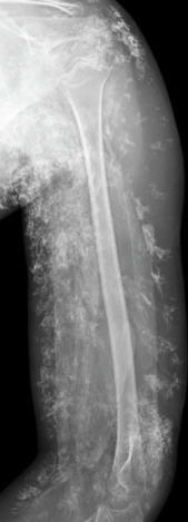
  • Radiography is an efficient means of identifying and characterizing soft-tissue calcinosis (Fig. E4).
  • Although MRI arguably has greater diagnostic value than electromyography or serum enzyme measurements in cases of suspected idiopathic inflammatory myopathy, MRI findings have not been formalized as a diagnostic criterion for idiopathic inflammatory myopathy. The acceptance of MRI as a diagnostic tool in myositis may be inhibited by the high cost and the need for more reliable and validated methods of summarizing the findings of MRI. MRI evaluation before biopsy, however, has become routine at many tertiary care centers. Fascial disease is manifested on MRI by fascial or perifascial hyperintensity on fluid-sensitive sequences. The edema-like signal in the deep subcutis may accompany fasciitis and can indicate associated panniculitis.
  • Video fluoroscopy or barium swallow study to look for upper esophageal dysfunction in patients with dysphagia and DM.
  • Table E5 summarizes affected organs and their evaluation in inflammatory muscle disease.

Figure E4 Advanced Calcinosis

Radiograph of the Arm in a Girl with Juvenile Dermatomyositis, Showing Extensive Amorphous and Wispy Calcification Corresponding to Subcutaneous Calcinosis. Sheetlike Calcification Outlining Muscles Corresponds to Deep Fascial Calcification.

From Pope TL et al: Musculoskeletal imaging, ed 2, Philadelphia, 2015, Saunders.

TABLE E5 Affected Organs and Their Evaluation in Inflammatory Muscle Disease

Organ or SystemModalities of EvaluationPathologic ProcessesFindings
MuscleBiopsyMyofiber degeneration and regenerationMyofiber size variation; vacuolated, necrotic fibers
Large central nuclei, basophilic sarcoplasm
InflammationMononuclear cell infiltration
FibrosisIncreased interstitium and fatty replacement of muscle
EMGMyofiber destructionLow-amplitude, short, polyphasic potential; spontaneous fibrillations; irritability
MRI
T1 imageFibrosisAtrophy of muscle, scarring
STIR imageInflammationBright signal in inflamed muscle
HeartECG, echoMyocarditis, fibrosisArrhythmias, left ventricular hypertrophy
BiopsyMyocarditis, fibrosisMyofiber size variation, mononuclear cell infiltrates, fibrosis
LungsCXR, HRCTInflammation, fibrosisInterstitial markings
PFTsInflammation, fibrosis, restrictive lung diseaseDecreased TLV, FVC, and DLCO
Radionuclide scanInflammation, fibrosisVentilation-perfusion mismatches
BALInflammation, fibrosisAbnormal leukocyte numbers and differentials
BiopsyInflammation, fibrosisMononuclear cell infiltration, destruction of alveolar space and fibrosis
SkinBiopsyInflammationVacuolization of the basal layer; mononuclear cell infiltration
GI systemRadiographic studiesInflammation, fibrosisReflux and uncoordinated peristalsis

BAL, Bronchoalveolar lavage; CXR, chest x-ray examination; echo, echocardiography; DLCO, diffusion capacity for carbon monoxide; ECG, electrocardiography; EMG, electromyography; FVC, forced vital capacity; GI, gastrointestinal; HRCT, high-resolution computed tomography; MRI, magnetic resonance imaging; PFT, pulmonary function tests; STIR, short tau inversion recovery; TLV, total lung volume.

From Hochberg MC: Rheumatology, ed 7, Philadelphia, 2019, Elsevier.

Treatment ⬆ ⬇

Goal: Maintain function, minimize disease/iatrogenic sequelae

Nonpharmacologic Therapy

  • Sun-blocking agents with SPF 15 or greater for skin protection in patients with DM
  • Physical therapy beneficial for gait training and increasing muscle tone and strength
  • Occupational therapy assists with activities of daily living
  • Speech therapy to monitor patients with swallowing dysfunction
Acute General Rx

  • Corticosteroids are the mainstay of therapy. Start prednisone 0.7 to 1 mg/kg per day, up to a maximum dose of 60 mg/day. If severe disease, can give methylprednisolone 1 g intravenous (IV) daily for 3 to 5 days before starting prednisone. Continue until muscle strength improves. Begin tapering by 5 to 10 mg/mo. Consider every-other-day prednisone treatment at same dose (may decrease side effects).6
  • Consider IV immunoglobulin (IVIG) if patient fails to improve on prednisone, or muscle enzymes begin rising when tapering off prednisone. See “Chronic Rx” for specific dosage.
  • Hydroxychloroquine can be used to treat the cutaneous lesions of DM.
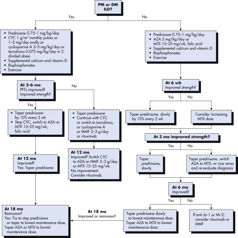
  • A treatment algorithm for adult patients with inflammatory myopathies is illustrated in Fig. 5.
Figure 5 Treatment Algorithm for Adult Patients with Polymyositis (PM) or Dermatomyositis (DM)

Aza, Azathioprine; Cyc, Cyclophosphamide; ILD, Interstitial Lung Disease; Mmf, Mycophenolate Mofetil; Mtx, Methotrexate; Pft, Pulmonary Function Test; SC, Subcutaneous.

!!flowchart!!

From Firestein GS et al: Firestein & Kelley’s textbook of rheumatology, ed 11, Philadelphia, 2021, Elsevier.

Chronic Rx

  • Chronic prednisone therapy may be needed for years, but other immunosuppressive (“steroid-sparing”) agents may be added early to decrease long-term steroid side effects.
  • Azathioprine starting with 50 g daily, titrating to 100 mg/day after 1 week to goal of 2 mg/kg/day.6
  • Methotrexate 5 to 7.5 mg weekly, increased by 2.5 mg/wk to total of 25 mg/wk; consider intramuscular dosing if PO is ineffective. Use cautiously in patients with interstitial lung disease due to possible pulmonary fibrosis.
  • IV immunoglobulin 2 g/kg total dose over 2 to 5 days.
  • IV cyclophosphamide 0.5 to 1 g/m2 monthly for 6 mo is preferred to oral dosing for refractory cases. However, oral dosing of cyclophosphamide is 1 to 2 mg/kg per day PO or 2 to 4 mg/kg per day in conjunction with prednisone.6
  • Cyclosporine A: Initial dose 2.0 to 2.5 mg/kg bid; long-term maintenance is lowest effective dose.
  • Mycophenolate mofetil 500 mg PO bid, titrate to 1500 mg PO bid over 1 to 2 mo.6
Disposition

  • 20% to 30% of patients achieve clinical remission with treatment.6
  • In patients with residual weakness, deficits typically remain stable over long-term follow-up.
  • 10% experience recurrent disease.
  • Serum CK often returns to normal before symptoms improve.
  • During exacerbations, enzymes may rise before clinical symptoms appear.
  • Poor prognostic indicators include delay in diagnosis, older age, recalcitrant disease, malignancy, interstitial pulmonary fibrosis, dysphagia, leukocytosis, fever, and anorexia.
  • Infection, malignancy, and cardiac and pulmonary dysfunction are the most common causes of death.
Referral

Neurology or rheumatology referral should be made to help establish the diagnosis and implement treatment.

Pearls & Considerations ⬆ ⬇

Related Content

Dermatomyositis and Polymyositis (Patient Information)

Inclusion Body Myositis (Related Key Topic)

Necrotizing Autoimmune Myopathy (Related Key Topic)

Related Content ⬆

  1. Selva-O’Callaghan A. : Classification and management of adult inflammatory myopathiesLancet Neurol. ;17:816-828, 2018.
  2. DeWane M.E. : Dermatomyositis: clinical features and pathogenesisJ Am Acad Dermatol. ;82:267-281, 2020.
  3. Madan V. : Defining cancer risk in dermatomyositis. Part 1Clin Exp Dermatol. ;34:451-455, 2009.
  4. Amato A.A. : Inflammatory myopathies, Continuum. ;19:1615-1633, 2013.
  5. Opinc A.H. : Antisynthetase syndrome-much more than just a myopathySemin Arthritis Rheum. ;51:72-83, 2021.
  6. McGrath E.R. : Autoimmune myopathies: updates on evaluation and treatmentsNeurotherapeutics. ;15:976-994, 2018.