AUTHOR: Lydia Sharp, MD
Inclusion body myositis (IBM) is the most common acquired idiopathic myopathy after the age of 50. The idiopathic inflammatory myopathies include polymyositis (PM), dermatomyositis (DM), autoimmune necrotizing myopathy, and IBM. They are associated with inflammatory involvement of muscle fibers and recurrent weakness. Although IBM is classified among the inflammatory myopathies, its underlying pathophysiology has not yet been delineated, and antiinflammatory medications do not improve weakness.
Thorough neurologic examination with emphasis on the motor examination. Nerve conduction studies should be performed to exclude other causes and electromyography performed to document a myopathy.
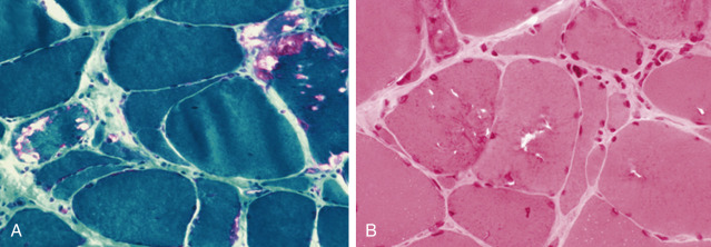
The diagnosis of definite IBM requires the following features on muscle biopsy (Fig. E1): (1) Inflammation, (2) inflammatory cells invading healthy muscle fibers, (3) vacuoles, and (4) amyloid deposits by Congo red staining, TDP-43 sarcoplasmic staining, or tubulofilaments on electron microscopy.2
MRI may reveal atrophy and signal abnormalities in volar forearm muscle groups and quadriceps atrophy with relative preservation of the rectus femoris muscle.
Table E1 Clinical and Laboratory Features of Idiopathic Inflammatory Myopathy Subgroups
| Clinical Features | Dermatomyositis | Polymyositis | Inclusion Body Myositis |
|---|---|---|---|
| Age | Children and adults | Adultsa | Adults >50 yr |
| Disease onset | Subacute | Subacute | Chronic |
| Muscle weakness | Proximal | Proximal | Selective patternb |
| Symmetry | Symmetric | Symmetric | Asymmetric |
| Systemic features | Yesc | Yesc | Yesd |
| Skin changes | Yese | No | No |
| Calcinosis | Yesf | Rarely | No |
| Associated connective tissue disease | Yesg | Yesg | Yesh |
| Associated malignancy | Yes | Yes | Yes |
| Laboratory features | |||
| Serum enzymesi | Normal to high | Normal to high | Normal to high |
| Abnormal EMGj | Yes | Yes | Yes |
| Abnormal muscle biopsy | Perifascicular atrophy, capillary depletion, patchy MHC class I expression and microinfarcts | CD8+ T cell invasion of nonnecrotic fibers and MHC class I expression on fibers | CD8+ T cell invasion, MHC expression, vacuolated fibers, and tubulofilamentous inclusions in fibers |
CD8+T cell, Cytotoxic T lymphocyte cell; EMG, electromyogram; MHC, major histocompatibility complex.
b Early involvement of finger flexor, wrist flexor, or wrist extensor weakness, and involvement of quadriceps femoris.
c Some patients have dysphagia, synovitis, and interstitial lung disease.
d Some patients have dysphagia.
e Gottron sign and heliotrope rash.
g Overlap with scleroderma, systemic lupus erythematosus, rheumatoid arthritis, Sjögren syndrome, and mixed connective tissue disease.
h Associated with Sjögren syndrome but less frequently associated with other connective tissue diseases.
i Serum creatine kinase, aspartate transaminase, lactate dehydrogenase, and aldolase vary from normal to very high levels.
j Myopathic motor unit potentials with spontaneous discharges in dermatomyositis, with and without spontaneous discharges in PM, and mixed pattern of short- and long-duration motor unit potentials in inclusion body myositis.
From Firestein GS et al: Kelleys textbook of rheumatology, ed 9, Philadelphia, 2013, Saunders.
Some patients choose to self-treat with creatine supplementation, coenzyme Q10, or lithium. There is no evidence supporting these treatments.
Life expectancy is not significantly altered in this late-onset, slowly progressive disorder. On average, individuals require use of assistive device for ambulation 7 to 10 years from symptom onset, and a wheelchair 13 to 15 years from onset.3
Patient information and support groups can be found at https://www.ninds.nih.gov/Disorders/All-Disorders/Inclusion-Body-Myositis-Information-Page and https://www.myositis.org/.