AUTHORS: Joseph Edmund, MD and Bharti Rathore, MD


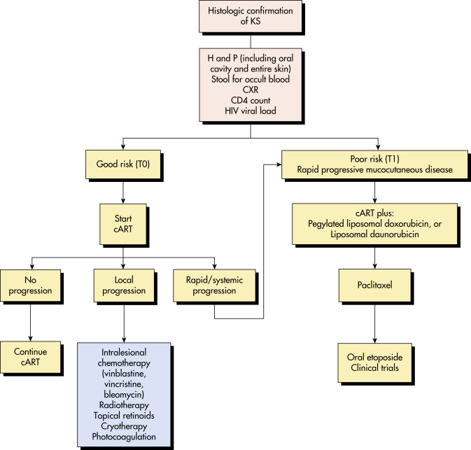
Kaposi sarcoma (KS) is a low-grade vascular tumor associated with Kaposi sarcoma herpes virus (KSHV)/Human gammaherpes virus 8 (HHV-8) infection and is an AIDS-defining illness. Kaposi sarcoma lesions present predominantly at mucocutaneous sites but may involve all organs and anatomic locations.
| ||||||||||||||||||||||||||||||||||||||||||||
- In the U.S., epidemic or acquired immunodeficiency syndrome (AIDS)-associated KS is the most common presentation; however, transplant or iatrogenic immunosuppression-related KS is on the increase. In clinical practice, classic and endemic KSs are also encountered.
- AIDS-related KS affects about 47 per 100,000 person-yr with HIV.
- Highest incidence is in homosexual men.
- KS lesions evolve from early (patch stage) macules into plaques (plaque stage) that may subsequently develop into larger nodules (tumor stage).1 These tumors may ulcerate, cause marked lymphedema, present as exophytic growths (e.g., cutaneous horns), or invade subjacent tissues (e.g., underlying bone). Different stages can coexist in the same individual at the same time.
- Although KS commonly presents at mucocutaneous sites, tumors may involve lymph nodes as well as visceral organs, most notably the respiratory and gastrointestinal tracts.
- Extracutaneous KS has been described in unusual anatomic locations, including the musculoskeletal system, nervous system, heart, wounds, within pemphigus lesions, and in blood clots.
- AIDS-related KS: Multifocal and widespread red-purple (Fig. E1) or dark plaques and/or nodules on cutaneous or mucosal surfaces. This is a more aggressive illness with early dissemination.2
- Generalized lymphadenopathy at the time of diagnosis is present in >50% of patients with AIDS-related KS; the initial lesions have a rust-colored appearance; subsequent progression to red or purple nodules or plaques occurs (Fig. E2).
- Most frequently affected areas are the face, trunk, oral cavity, and upper and lower extremities.
- The GI tract is the most frequent site of internal involvement in classic KS.
- In AIDS-associated KS, 25% of patients have cutaneous involvement alone, whereas 29% have visceral lesions only (lymph nodes 50%, GI tract 50%, lungs 37%).
- In most patients with HIV-associated KS who initiate highly active antiretroviral therapy (HAART), the KS lesions stabilize with decreasing HIV plasma viremia and immune reconstitution, or even resolve completely without any specific treatment.
Figure E1 Kaposi sarcoma: Epidemic human immunodeficiency virus associated.
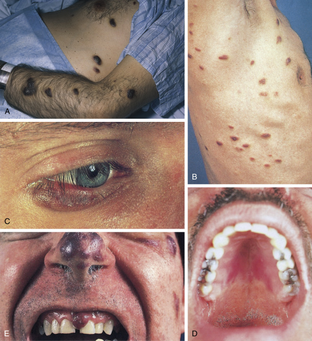
A and B, Plaque lesions. C, Violaceous lesion affecting the lateral lower eyelid. D, Confluent plaque on the hard palate. E, Nodular lesion on the gingiva and nose.
From Swartz MH: Textbook of physical diagnosis, history and examination, ed 7, Philadelphia, 2014, Saunders.
Figure E2 Classic Kaposi sarcoma with involvement of the lower extremities.
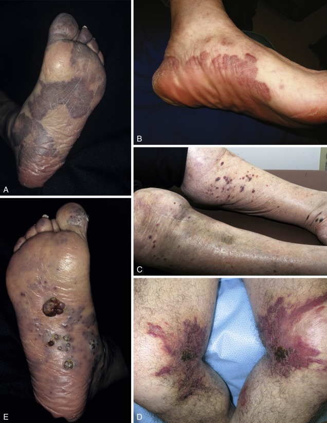
Violaceous patches become plaques (A, B) and may develop a nodular component (C, E) or verrucous appearance (D, E).
- KSHV/HHV-8 is thought to be the causative agent and can be transmitted through sexual (homosexual or heterosexual activities) or nonsexual contact such as maternal-infant transmission (common in African countries).3 KSHV establishes lifelong infection in the host, and its DNA is found in all KS lesions. In most cases fulminant KS is accompanied and preceded by a rise in KSHV viral load. KS is primarily the consequence of systemic viral reactivation from a latent reservoir, most likely a lymph node resident B cell. KS develops in response to severe T-cell depletion or inactivation. Infant immunodeficiency or aging-, chemical-, or HIV-induced immune deficiency is an essential cofactor for the development of KS.
- It can be divided into the following four subsets:1,4
- Classic KS: Occurs mostly in elderly Eastern European and Mediterranean males. It consists initially of violaceous macules and papules with subsequent development of plaques and red-purple nodules. Growth is slow, and most patients die of unrelated causes.
- Epidemic (AIDS-related) KS: Most frequently occurs in HIV-infected persons. Lesions are generally multifocal and widespread (see Fig. E1). Lymphadenopathy may be associated.
- Endemic KS: Usually affects sub-Saharan African children and adults. An aggressive lymphadenopathic form affects African children in particular.
- Organ transplant-associated, KS: Usually associated with immunosuppressive therapy. Transplant can transmit HHV-8.