AUTHOR: Fred F. Ferri, MD


Definition
- A hamartomatous polyp is a benign intestinal growth that may contain all components of the intestinal mucosa. In Peutz-Jeghers syndrome (PJS), hamartomas are found primarily in the small bowel but can also be present in the colon and stomach. In GI polyposis, multiple such polyps coexist within the intestinal tract, and associated manifestations are usually also present.
- Juvenile polyps are benign polyps composed of cystic dilations of glandular structures within the fibroblastic stroma of the lamina propria. They may cause bleeding or intussusception.
- Commonly recognized syndromes are Peutz-Jeghers syndrome, juvenile polyposis syndrome, Cowden disease, Bannayan-Ruvalcaba-Riley syndrome, and Cronkhite-Canada syndrome. Other, lesser-known inherited hamartomatous polyposis syndromes are hereditary mixed polyposis syndrome, intestinal ganglioneuromatosis and neurofibromatosis (variant of von Recklinghausen syndrome), Devon family syndrome, basal cell nevus syndrome, and tuberous sclerosis (may involve GI tract). Table E1 describes general features of some inherited colorectal cancer syndromes.
| ||||||||
Epidemiology & Demographics
- The incidence of PJS is 1 in 200,000.
- Colonic adenomas, the precursors of nearly all colorectal cancers, are found in nearly 40% of patients by age 60 yr.
- 25% of men and 15% of women who undergo colonoscopy are found to have one or more adenomas.
- Detection of any adenoma in patients <60 yr confers an increased risk of colorectal cancer (by a factor of 2.6) in their first-degree relatives.
Physical Findings & Clinical Presentation
Peutz-Jeghers Syndrome
- Transmission: Autosomal dominant with incomplete penetrance. The syndrome is caused in the majority of patients by a germline mutation of the STK11/LKB1tumor suppression gene on chromosome 19P13.
- Disease expression:
- Stomach, small and large intestinal hamartomas with bands of smooth muscle in the lamina propria
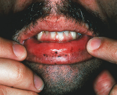
- Pigmented lesions around mouth (lips and buccal mucosa [Fig. E2]), nose, hands, feet, genitals, and perineal areas
- Ovarian tumors
- Sertoli cell testicular tumors
- Airway polyps
- Pancreatic cancer
- Breast cancer
- Urinary tract polyps
- Cumulative lifetime cancer risk:
- Clinical manifestation:
Juvenile Polyposis Syndrome
- Transmission: Autosomal dominant
- Disease expression:
- Cumulative cancer risk is increased (may be as high as 50%)
- Clinical manifestation:
TABLE E1 General Features of Some Inherited Colorectal Cancer Syndromes
| Syndrome | Polyp Histology | Polyp Distribution | Age of Onset | Risk of Colon Cancer | Genetic Lesion | Clinical Manifestations | Associated Lesions |
|---|---|---|---|---|---|---|---|
| Familial adenomatous polyposis (Fig. E1) | Adenoma | Large intestine, duodenum | 16 yr (range, 8-34 yr) | 100% | 5q (APC gene) | Rectal bleeding, abdominal pain, bowel obstruction | Desmoids, CHRPE |
| Peutz-Jeghers syndrome | Hamartoma | Large and small intestine | First decade | Slightly above average | 19p (STK11 gene) | Possible rectal bleeding, abdominal pain, intussusception | Orocutaneous melanin pigment spots, other tumors |
| MUTYH-associated polyposis | Adenoma | Large intestine, duodenum | 45-50 yr (range, 13-60 yr) | 75% (range, 50%-100%) | 1p (MYH gene) | Rectal bleeding, abdominal pain, bowel obstruction | CHRPE, osteomas |
| Juvenile polyposis | Hamartoma (rarely adenoma) | Large and small intestine | First decade | ∼9% | PTEN, SMAD4, BMPR1 | Possible rectal bleeding, abdominal pain, intussusception | Pulmonary AVMs |
| Hereditary nonpolyposis colon cancer | Adenoma | Large intestine | 40 yr (range, 18-65 yr) | 30% | Mismatch repair genes+∗ | Rectal bleeding, abdominal pain, bowel obstruction | Other tumors (e.g., ovary, uterus, pancreas, stomach) |
AVM, Arteriovenous malformation; CHRPE, congenital hypertrophy of the retinal pigment epithelium; MUTYH, mutY homolog (Escherichia coli).
∗Including hMSH2, hMSH3, hMSH6, hMLH1, hPMS1, and hPMS2.
From Goldman L, Schafer AI: Goldman’s Cecil medicine, ed 24, Philadelphia, 2012, Saunders.
Cowden Disease
- Transmission: Autosomal dominant, rare
- Disease expression:
- Juvenile intestinal polyposis
- Orocutaneous hamartomas
- Fibrocystic breast disease and breast cancer
- Goiter and thyroid cancer
- Facial tricholemmomas (papules) in 83%
- Cumulative cancer risk:
Figure E1 Familial adenomatous polyposis.
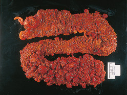
The disorder is marked by the development of hundreds of large bowel adenomas, as seen in this segment of large bowel covered with adenomas of various sizes. It usually arises in the second and third decades.
(From Skarin AT: Atlas of diagnostic oncology, ed 4, St Louis, 2010, Mosby.)
Cronkhite-Canada Syndrome
- Transmission: Acquired
- Age of onset: Midlife
- Disease expression:
- Diffuse GI juvenile polyposis (50% to 95% of cases)
- Chronic diarrhea and protein-losing enteropathy (the entire intestinal mucosa may be inflamed), which leads to abdominal pain, weight loss, and various complications of malnutrition
- Dystrophic nails
- Alopecia
- Hyperpigmentation
- Cumulative cancer risk: Same as the average population


