section name header

Basic Information

AUTHOR: Ritesh Rathore, MD

Definition

Colorectal cancer (CRC) is a malignant neoplasm arising from the luminal surface of the large bowel with locations including the descending colon (40% to 42%), rectosigmoid and rectum (30% to 33%), cecum and ascending colon (25% to 30%), and transverse colon (10% to 13%).

Synonym

CRC

ICD-10CM CODES
C18Malignant neoplasm of colon
C18.2Malignant neoplasm of colon, ascending colon
C18.4Malignant neoplasm of colon, transverse colon
C18.6Malignant neoplasm of colon, descending colon
C18.7Malignant neoplasm of colon, sigmoid colon
C19Malignant neoplasm of rectosigmoid junction
Epidemiology & Demographics

  • CRC is the fourth most common cancer and the second leading cause of cancer deaths in the U.S. (estimated 151,030 new cases and 52,580 deaths in 2022).1 Distant metastatic disease is present in 18% to 22% of patients at time of diagnosis.
  • Worldwide, CRC is the third most common cancer and accounted for an estimated 1.9 million new cases and approximately 900,000 deaths in 2020. The highest incidence is in North America, Australasia, Europe, and South Korea.
  • The peak incidence is in the seventh decade of life. The lifetime risk for development of CRC is 1 in 17, with 90% of cases occurring after age 50 yr.
  • An alarming increase in cases of early onset colorectal cancer, defined as diagnosis in patients younger than 50 yr of age, has occurred in the United States and other high income countries over the past few decades.1a
  • Risk factors (Table 1):
    1. Hereditary polyposis syndromes
    2. Familial polyposis (high risk)
    3. Gardner syndrome (high risk)
    4. Turcot syndrome (high risk)
    5. Peutz-Jeghers syndrome (low to moderate risk)
    6. Inflammatory bowel disease (IBD), both ulcerative colitis and Crohn disease
    7. Family history of “cancer family syndrome”
    8. Heredofamilial breast cancer and colon carcinoma
    9. Pelvic irradiation history
    10. First-degree relatives with colorectal carcinoma
    11. Age >45 yr
    12. Dietary factors (diet high in fat or red meat, alcohol use, low vegetable intake)
    13. Hereditary nonpolyposis colon cancer (HNPCC): Autosomal dominant disorder characterized by early age of onset (mean age 44 yr) and right-sided or proximal colon cancers, synchronous and metachronous colon cancers, mucinous and poorly differentiated colon cancers; accounts for 1% to 5% of all cases of CRC; Criteria for diagnosis of HNPCC are summarized in Table 2
    14. Previous endometrial or ovarian cancer, particularly when diagnosed at an early age

TABLE 1 Recognized Risk Factors for Colorectal Cancer

Family History
  • Colorectal cancer (CRC)
  • Inherited syndromes (e.g., familial adenomatous polyposis [FAP])
  • Racial and ethnic background (e.g., African American, Ashkenazi Jews)
Personal History
  • Age
  • Male sex
  • Previous colonic polyps or CRC
  • History of inflammatory bowel disease
  • Diabetes mellitus
Lifestyle
  • Obesity
  • High consumption of alcohol
  • Diet high in red meat and fat, low in fiber

From Niederhuber JE: Abeloff’s clinical oncology, ed 6, Philadelphia, 2020, Elsevier.

TABLE 2 Criteria for Diagnosis of Hereditary Nonpolyposis Colon Cancer (HNPCC)

Amsterdam Criteria1
  • At least three relatives have colorectal cancer, and all of the following are present:
    1. One is a first-degree relative (parent, sibling, or child) of the other two
    2. At least two successive generations are involved
    3. At least one relative had colorectal cancer when he or she was younger than 50
    4. Familial adenomatous polyposis has been excluded
Revised Bethesda Guidelines2
(Indicates tumors to select for microsatellite instability testing)
  • Colorectal cancer in a patient younger than 50 yr
  • A second synchronous or metachronous colorectal cancer or cancer associated with HNPCC
  • Presence of high-level microsatellite instability histologically in a patient younger than 60 yr
  • One or more first-degree relatives with either colorectal cancer or HNPCC-associated tumor diagnosed at younger than 50 yr
  • Colorectal cancer in two or more first- or second-degree relatives with HNPCC-related tumors at any age
Either all of the Amsterdam criteria or one of the Bethesda criteria is used to identify an individual at risk of HNPCC
  • HNPCC-associated tumors include endometrial, gastric, ovarian, pancreatic, ureter, renal pelvis, biliary tract, small bowel, and brain

1 Vasen HF et al: New clinical criteria for hereditary nonpolyposis colorectal cancer (HNPCC, Lynch syndrome) proposed by the International Collaborative group on HNPCC, Gastroenterology 116:1453-1456, 1999.

2 Umar A et al: Revised Bethesda Guidelines for hereditary nonpolyposis colorectal cancer (Lynch syndrome) and microsatellite instability, J Natl Cancer Inst 96:261-268, 2004.

From Niederhuber JE: Abeloff’s clinical oncology, ed 6, Philadelphia, 2020, Elsevier.

Physical Findings & Clinical Presentation

  • Physical examination may be completely unremarkable.
  • Digital rectal examination can detect approximately 50% of rectal cancers.
  • Palpable abdominal masses may indicate metastasis or complications of colorectal carcinoma (abscess, intussusception, volvulus).
  • Abdominal distention and tenderness are suggestive of colonic obstruction.
  • Hepatomegaly is indicative of hepatic metastasis.
Etiology

CRC can arise through either of two mutational pathways: Microsatellite instability or chromosomal instability. Germline genetic mutations are the basis of inherited colon cancer syndromes; an accumulation of somatic mutations in a cell is the basis of sporadic colon cancer. Fig. E1 illustrates the molecular carcinogenesis of colon cancer. Approximately 10% to 15% of CRC lack one or more mismatch repair enzymes (mismatch repair deficient [dMMR]-CRC).

Figure E1 Accumulation of molecular changes that precede colorectal carcinoma (CRC).
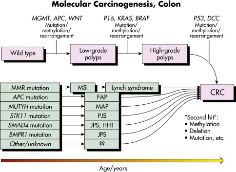
Sporadic tumors are initiated by a serial accumulation of somatic mutations that may eventuate in CRC. The initiating event in these polyposes is the mutation present at birth. Second hits may include a variety of molecular changes, such as tumor suppressor gene promoter methylation, mutations, and copy number changes. Mismatch repair (MMR) includes hMLH1, hMLH2, hMSH2, hPMS2, and EpCAMP genes. FAP, Familial adenomatous polyposis; HHT, hereditary hemorrhagic telangiectasia; JPS, juvenile polyposis syndrome; MAP, MUTYH-associated polyposis; MSI, microsatellite instability; PJS, Peutz-Jeghers syndrome.

From Niederhuber JE: Abeloff’s clinical oncology, ed 6, Philadelphia, 2020, Elsevier.

Diagnosis

Differential Diagnosis

  • Diverticular disease
  • Strictures or adhesions
  • IBD
  • Infectious or inflammatory lesions
  • Arteriovenous malformations
  • Metastatic carcinoma
  • Extrinsic masses (cysts, abscesses)
Workup

The clinical presentation of colorectal malignancies may consist of nonspecific symptoms (weight loss, anorexia, malaise) or of specific symptoms related to mass effect or bleeding. It is useful to divide colon cancer symptoms into those usually associated with the right- or left-sided cancers because the clinical presentation can vary with the location.

  • Right side of colon:
    1. Anemia (from chronic blood loss).
    2. Abdominal pain may be present, or the patient may be completely asymptomatic.
    3. Rectal bleeding is often missed because blood is mixed with feces.
    4. Obstruction and constipation are unusual because of large lumen and more liquid stools.
  • Left side of colon:
    1. Change in bowel habits (constipation, diarrhea, tenesmus, pencil-thin stools).
    2. Rectal bleeding (bright red blood coating the surface of the stool).
    3. Intestinal obstruction is frequent because of small lumen.
Classification and Staging

(Table 3)

TABLE 3 World Health Organization Classification of Colorectal Carcinoma

  • Adenocarcinoma
  • Cribriform comedo-type adenocarcinoma
  • Medullary carcinoma
  • Micropapillary carcinoma
  • Mucinous adenocarcinoma
  • Serrated adenocarcinoma
  • Signet ring cell carcinoma
  • Adenosquamous carcinoma
  • Spindle cell carcinoma
  • Squamous cell carcinoma
  • Undifferentiated carcinoma
  • Neuroendocrine carcinoma (NEC):
    1. Large NEC
    2. Small NEC
  • Mixed adenoneuroendocrine carcinoma

From Niederhuber JE: Abeloff’s clinical oncology, ed 6, Philadelphia, 2020, Elsevier.
American Joint Committee on Cancer 8th edition classification for CRC:

  1. Confined to the mucosa-submucosa (stage I)
  2. Invasion of muscularis propria (stage II)
  3. Local node involvement (stage III)
  4. Distant metastasis (stage IV)

TNM Classification:

StageTNM Classification
IT1-2, N0, M0
IIAT3, N0, M0
IIBT4a, N0, M0
IICT4b, N0, M0
IIIAT1-2, N1, M0; T1, N2a, M0
IIIBT3-4a, N1, M0; T2-3, N2a, M0
T1-2, N2b, M0
IIICT4a, N2a, M0; T3-4a, N2b, M0
T4b, N1-2, M0
IVAT(any), N(any), M1a
IVBT(any), N(any), M1b
IVCT(any), N(any), M1c
Laboratory Screening Tests

  • Positive fecal occult blood test (FOBT): Many primary care physicians use single digital FOBT as their primary screening test for CRC. Single FOBT has low specificity for detecting human hemoglobin, is a poor screening method for CRC (sensitivity, 4.9%), and is inappropriate by itself since negative results do not decrease the odds of advanced neoplasia. The American College of Gastroenterology recommends the fecal immunochemical test (FIT), which measures intact human globin protein (as opposed to heme) in the stool and detects more advanced adenomas than FOBT.
  • Fecal DNA testing is a screening method that detects colonic cells shed into the fecal stream that possess specific genetic or epigenetic changes. The technique has a reported sensitivity of 97% and a specificity of 90% for CRC stages I to III. In trials involving asymptomatic persons at average risk for CRC, multitarget stool DNA testing detects significantly more cancers than FIT but has more false-positive results. High cost and rate of false positives are the main obstacles inhibiting broader adoption of fecal DNA testing.
  • Molecular markers (Table 4) including abnormal DNA from cancerous cells can be detected in stool. FIT combined with stool DNA test (FIT-DNA) has been approved by the FDA for colorectal screening. One study showed that one-time FIT-DNA had a higher sensitivity for detection of CRC than one-time FIT alone (92.3% vs. 73.8%), but specificity was lower (86.6% vs. 94.9%).2
  • Plasma carcinoembryonic antigen (CEA) level is not useful for screening because it can be increased in nonmalignant conditions (smoking, IBD, alcoholic liver disease). A normal CEA result does not exclude the diagnosis of CRC.

TABLE 4 Molecular Biomarkers Used in Clinical Standard-of-Care Decision Making in Colorectal Cancer

BiomarkerPurpose
APC mutation detectionDiagnosis of FAP
MMR protein expression (MSH2, MLH1, MSH6, PMS2)Diagnosis of HNPCC
MSI analysis
MMR mutation detection (MSH2, MLH1, MSH6, PMS2)
BRAF mutation detection
MYH mutation detectionDiagnosis of MYH-associated polyposis
LKB1, SMAD4, BMPR1A, PTEN mutation detectionDiagnosis of hamartomatous polyp syndromes
KRAS mutation analysisMolecular stratification for treatment with EGFR inhibitors
BRAF mutation analysis
Thymidylate synthase protein expressionIdentification of response to 5-FU
MSIIdentification of response to 5-FU
Gene expression signaturePrognostication
PD-1Stratification for response to PD-L1 blockade

EGFR, Epidermal growth factor receptor; FAP, familial adenomatous polyposis; 5-FU, 5-fluorouracil; HNPCC, hereditary nonpolyposis colorectal cancer; MMR, mismatch repair; MSI, microsatellite instability.

From Niederhuber JE: Abeloff’s clinical oncology, ed 6, Philadelphia, 2020, Elsevier.
Imaging Studies

  • Colonoscopy with biopsy (primary assessment tool): The American College of Physicians (ACP) recommends screening colonoscopy beginning at age 50, and it should be repeated every 10 yr in average-risk patients. Screening is recommended in African Americans beginning at age 45 yr. Persons with only one first-degree relative with CRC or advanced adenomas diagnosed at 60 yr or older may be screened as at average risk. In 2021, updated guidelines from the U.S. Preventive Services Task Force expanded routine colonoscopy screening to between ages 45 and 75 yr while maintaining that screening should not be routinely recommended in persons older than 75 yr, and it should not be recommended at all in persons older than 85 yr. If persons between the ages of 75 and 85 yr have never undergone screening, the decision about screening should be individualized according to health status.3 The ACP recommends that clinicians stop screening for CRC in adults over age 75 yr or in adults with a life expectancy of <10 yr. In addition, the American College of Gastroenterology (ACG) and the American Cancer Society (ACS) both recommend expanded screening eligibility starting at age 45 in routine-risk patients.4 Table 5 describes CRC screening and surveillance recommendations.
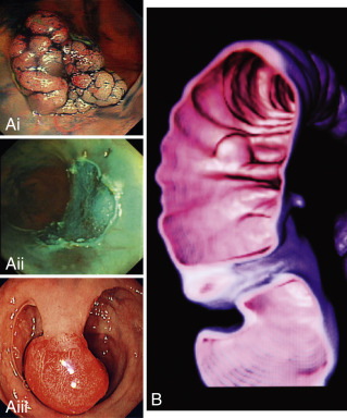
  • Computed tomography colonoscopy (CTC) virtual colonoscopy (VC) uses helical (spiral) CT scanning to generate a 2D or 3D virtual colorectal image (Fig. E2). CTC does not require sedation, but, like optical colonoscopy, it requires some bowel preparation (either bowel cathartics or ingestion of iodinated contrast medium with meals during the 48 h before CT) and air insufflation. It also involves substantial exposure to radiation. In addition, patients with lesions detected by VC will require traditional colonoscopy. Compared with colonoscopy, CTC sensitivity for detection of polyps >10 mm ranges from 70% to 96%, and specificity ranges from 72% to 96%. CTC has replaced double-contrast barium enema as the radiographic screening alternative when patients decline colonoscopy.
  • Capsule endoscopy allows visualization of the colonic mucosa but is not recommended as a screening procedure because its sensitivity for detecting colonic lesions is low compared with colonoscopy.
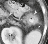
  • CT scanning of the abdomen (Fig. E3), pelvis, and chest assists in preoperative staging.
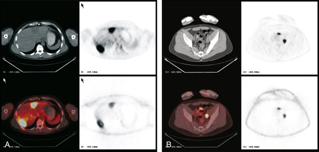
  • PET scanning (Fig. E4) can display functional information and is accurate in the detection of CRC and its distant metastases. Colonography composed of a combined modality of PET and CT is a newer diagnostic modality that can provide whole-body tumor staging in a single session.

TABLE 5 Colorectal Cancer (CRC) Screening and Surveillance Recommendations

IndicationRecommendations
Average riskACP guidelines:
Beginning at age 50 yr: Colonoscopy every 10 yr; computed tomographic colonography every 5 yr; flexible sigmoidoscopy every 5 yr; double-contrast barium enema every 5 yr; stool blood testing annually or stool; DNA testing acceptable but not preferred
USPSTF, ACS, and ACG guidelines:
Starting above recommendations for testing beginning at age 45 yr
One or two first-degree relatives with CRC at any age or adenoma at age <60 yrColonoscopy every 5 yr beginning at age 40 yr, or 10 yr younger than earliest diagnosis, whichever comes first
Hereditary nonpolyposis CRCGenetic counseling and screening. Colonoscopy every 1-2 yr beginning at age 25 yr and then yearly after age 40 yr.
Familial adenomatous polyposis and variantsGenetic counseling and testing. Flexible sigmoidoscopy yearly beginning at puberty
Personal history of CRCColonoscopy within 1 yr of curative resection; repeat at 3 yr and then every 5 yr if normal
Personal history of colorectal adenomaColonoscopy every 3-5 yr after removal of all index polyps
Inflammatory bowel diseaseColonoscopy every 1-2 yr beginning after 8 yr of pancolitis or after 15 yr if only left-sided disease

ACG, American College of Gastroenterology; ACP, American College of Physicians; ACS, American Cancer Society; DNA, deoxyribonucleic acid; USPSTF, U.S. Preventative Services Task Force.

Recommendations proposed by the American Cancer Society and U.S. Multi-Society Task Force on Colorectal Cancer; recommendations for average-risk patients also endorsed by the American College of Radiology.

Screening colonoscopy at age 45 recommended by USPSTF.

Whenever possible, affected relatives should be tested first because of potential false-negative results.

Screening recommendation for individuals with positive or indeterminate tests as well as for those who refuse genetic testing.

Figure E2 Colon Polyps Seen at Colonoscopy (Ai-iii) and Computed Tomography (B) Colonography

Aii, After Endoscopic Resection of the Polyps in Ai.

From Ballinger A: Kumar & Clark’s essentials of medicine, ed 5, Edinburgh, 2012, Saunders.

Figure E3 Colon carcinoma: Wall thickening.

A carcinoma of the descending colon near the splenic flexure causes thickening of the colon wall (arrowhead) and narrowing of the lumen. Stranding densities (arrow) extending into the pericolonic fat suggest tumor extension through the bowel wall.

From Webb WR et al: Fundamentals of body CT, ed 4, Philadelphia, 2015, Saunders.

Figure E4 Colon cancer.

A, Positron emission tomography (PET) and computed tomography (CT) image display of a patient with two fluorine-18 (18F)-fluorodeoxyglucose (FDG)-avid lesions in the liver. These are seen on the CT scan (upper left), the attenuation-corrected PET scan (upper right), the nonattenuation-corrected PET scan (lower right), and fused images (lower left). B, PET and CT images of the pelvis, oriented as in A, show increased FDG uptake in a left external iliac lymph node metastasis.

From Niederhuber JE: Abeloff’s clinical oncology, ed 6, Philadelphia, 2020, Elsevier.
Treatment

General Rx

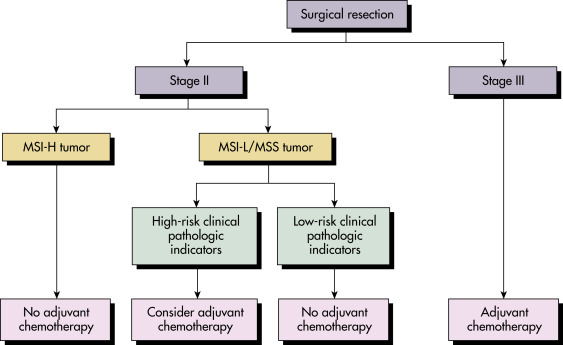
  • Surgical resection is the definitive and curative upfront treatment for stages I to III colon cancers. Selected patients (high-risk stage II, all stage III) are recommended to receive adjuvant chemotherapy. Microsatellite instability can be used alongside clinicopathologic factors in stage II and III CRC to guide therapy (Fig. 5).
  • The standard chemotherapy regimen for adjuvant therapy of resected CRC is the combination of oxaliplatin with a fluoropyrimidine (5-fluorouracil or capecitabine) for a period of 3 to 6 mo. Older patients and patients with significant comorbidities have more toxicity with combination chemotherapy; treatment with single-agent fluoropyrimidine therapy is a reasonable option in these patients.
  • Neoadjuvant combination chemotherapy (6 to 8 cycles) followed by chemoradiotherapy is used to downsize and downstage rectal cancers before definitive resection and to improve chemotherapy delivery preoperatively. This approach is called total neoadjuvant therapy (TNT) and is a recommended option for patients with larger tumors (T3-4) or node-positive (N1-3) disease. This new approach improves overall survival and local disease control in these high-stage cancers.5 Patients with smaller tumors or those who are unfit for this intensive approach can be treated with neoadjuvant chemoradiotherapy alone as before.
  • Adjuvant chemotherapy in stage II colon cancer provides a modest improvement in overall survival by 3% to 4%, with current 5-yr survival rates in the 80% range. As such, current guidelines recommend consideration of adjuvant chemotherapy only in high-risk stage II patients. The magnitude of survival benefit is significantly higher in stage III patients, and combination chemotherapy is associated with 5-yr overall survival rate in the 70% range with wide variation in the subgroups. More recent data have revealed that low-risk stage III colon cancer patients may have equivalent survival with adjuvant multiagent chemotherapy of only 3 mo in duration.6
  • Circulating tumor DNA levels have been utilized to make informed treatment decisions in patients with stage II colon cancer. After resection, patients were randomly assigned to have treatment decisions guided by either ctDNA results or standard clinicopathologic features. A ctDNA-positive result after surgery prompted chemotherapy use while patients who were ctDNA-negative were not treated. The results showed that a ctDNA-guided approach reduced adjuvant chemotherapy use without compromising recurrence-free survival.7
  • The outlook for patients with metastatic and relapsed CRC has improved dramatically in the past few years. Median overall survival in patients with unresectable metastatic CRC now is in the 30- to 36-mo range with modern chemotherapeutic regimens. In patients with limited, resectable metastases in sites such as the liver, the 5-yr median overall survival is in the 50% range.
  • Chemotherapy agents used in the metastatic setting include 5-fluorouracil (5-FU), capecitabine, irinotecan, oxaliplatin, and mitomycin. Chemotherapy regimens using a combination of antimetabolite (5-FU or capecitabine) in combination with either oxaliplatin or irinotecan form the backbone of systemic chemotherapy.
  • Molecularly targeted therapy against the epidermal growth factor receptor (EGFR) and the angiogenesis pathway are used in combination with the chemotherapy backbone in metastatic CRC. Antiangiogenic agents (bevacizumab, aflibercept, and ramucirumab) and the EGFR receptor blockers (cetuximab and panitumumab) are utilized in combination with standard chemotherapy regimens in metastatic CRC patients.
  • The oral multitargeted kinase inhibitor regorafenib and the oral antimetabolite drug TAS-102 provide modest survival benefit in patients who have failed standard chemotherapy approaches.
  • Patients with tumors harboring BRAF mutations can now have improved survival with the use of combination molecularly targeted therapy consisting of encorafenib, binimetinib, and cetuximab.8
  • The liver is generally the initial and most common site of CRC metastases. Resection of liver-limited metastases followed by systemic combination chemotherapy is curative in more than 30% of selected patients. Metastasectomy of limited pulmonary metastases can also be considered in selected cases.
  • Unresectable multiple liver metastases are often approached by locoregional therapeutic approaches such as transarterial chemoembolization, selective internal radiation therapy using yttrium-90 brachytherapy, or hepatic arterial infusional chemotherapy.
  • In patients with pathologically confirmed microsatellite instability in their cancers (see Fig. 5), the checkpoint inhibitors (pembrolizumab, nivolumab) are effective options after failure of standard therapies and have been recently approved in this setting. Recent data have shown that upfront treatment with pembrolizumab is superior to conventional chemotherapy and results in doubling of progression-free survival in newly diagnosed patients with microsatellite instability-driven colorectal cancers.9 Smaller studies have shown benefit with dual immunotherapy combination of nivolumab and ipilimumab, but survival data are not available currently.
  • Reviews of randomized trials in metastatic CRC have demonstrated that right-sided cancers are associated with shorter overall survival when compared with left-sided cancers.
Figure 5 Proposed Algorithm for Using Microsatellite Instability (MSI) Alongside Clinicopathologic Factors in Stages II and III Colorectal Cancer

!!flowchart!!

ASCO guidelines: Inadequate samples nodes, T4 lesions, perforation, poorly differentiated histology. MSI-H, High-level microsatellite instability; MSI-L/MSS, low-level microsatellite instability/microsatellite stability.

From Niederhuber JE: Abeloff’s clinical oncology, ed 6, Philadelphia, 2020, Elsevier.

Chronic Rx

Follow-up is indicated with:

  • Physician visits with a focus on clinical and disease-related history, directed physical examination, coordination of follow-up, and counseling every 3 to 4 mo for the first 3 yr and then every 6 mo for 2 yr.
  • Colonoscopy at end of first yr, then after 3 yr, and subsequently every 5 yr.
  • Malignant potential and surveillance of colonic polyps is summarized in Table 6.
  • Baseline CEA level, if elevated, can be used after surgery as a measure of completeness of tumor resection. It is used to monitor tumor recurrence and is obtained every 3 to 6 mo for up to 5 yr.
Disposition

The 5-yr survival rate varies with the stage of the carcinoma:

TNM Stage5-yr Survival Rate (%)
I>90
IIA-C60-85
IIIA-C25-65
IV5-10

  • Overall, the 5-yr disease-free survival rate has increased from 50% to 63% during the past two decades.
  • High-frequency microsatellite instability (MSI-H) in CRC is independently predictive of a relatively favorable outcome and reduces the likelihood of metastases.
  • In patients with high-risk stage II and with stage III CRC, there is improved 5-yr survival among patients treated with adjuvant chemotherapy.
  • Expression patterns of microRNA are systemically altered in colon adenocarcinomas. High miR-21 expression is associated with poor survival and poor therapeutic outcome.
  • The optimal timing from surgery to initiation of adjuvant chemotherapy is 4 to 8 wk. A longer time to initiation of adjuvant chemotherapy is associated with worse survival rates.
  • Regular aspirin use after the diagnosis of CRC has been reported to be associated with lower risk for CRC-specific and overall mortality, especially among individuals with tumors that overexpress cyclooxygenase-2. Regular aspirin use is associated with lower BRAF-wild type CRC but not with BRAF-mutated cancer risk. All aspirin doses starting with 75 mg daily had similar effects on CRC incidence and mortality.10

TABLE 6 Malignant Potential and Surveillance of Colonic Polyps

Polyp TypeMorphologyMalignant Potential/Surveillance Requirements
Tubular adenomaBranched tubules<1 cm: 1%
1-2 cm: 10%
>2 cm: 34%
Surveillance every 5-10 yr (1-2 small polyps with low-grade dysplasia after polypectomy) or every 3 yr if 3-10 adenomas or one >10 mm
Villous adenomaFrond-like pattern<1 cm: 4%
1-2 cm: 20%
>2 cm: >50%
Surveillance every 3 yr if 1 or more villous/tubulovillous adenomas
Tubulovillous adenomaMixed pattern<1 cm: 4%
1-2 cm: 9%
>2 cm: 45%
Surveillance every 3 yr
Hyperplastic polyp confined to rectosigmoidIncreased glandular cells
Reduced cytoplasm
Nuclear atypia, stratification and hyperchromatism is absent
Small and distally located
Do not appear to be associated with increased risk of colorectal cancer
Surveillance every 10 yr
Serrated polyps
  1. Sessile serrated polyp without dysplasia
Surveillance every 5 yr if <10 mm, every 3 yr if large (or dysplasia)
  1. Traditional serrated adenoma with dysplasia
Surveillance every 3 yr
  1. Mixed: sessile serrated and tubular adenoma
Surveillance every 3 yr
  1. Serrated polyposis syndrome
1 cm or more, 30 or more polyps, and mixed adenomatous features have increased risk of colorectal cancerSurveillance every 1 yr

From Talley NJ et al: Essentials of internal medicine, ed 4, Chatswood, NSW, 2021, Elsevier Australia.

Referral

Multidisciplinary referral to colorectal surgery or surgical oncology, medical oncology, radiation oncology

Pearls & Considerations

Comments

  • Metastases of tumor cells to regional lymph nodes is the single most important prognostic factor in patients with colon cancer.
  • Decreased fat intake to 30% of total energy intake, increased fiber through fruit and vegetable consumption may reduce CRC risk.
  • Chemoprophylaxis with aspirin (81 mg/day) reduces the incidence of colorectal adenomas in persons at risk.9
  • The National Cancer Institute has published consensus guidelines for universal screening for HNPCC in patients with newly diagnosed CRC. Tumors in mutation carriers of HNPCC typically exhibit microsatellite instability, a characteristic phenotype caused by expansion or contraction of short nucleotide repeat sequences. These guidelines (Bethesda Guidelines) are useful for selective patients for microsatellite instability testing. Screening patients with newly diagnosed CRC for HNPCC is cost effective, especially if the benefits to their immediate relatives are considered.
  • The use of either annual or biennial FOBT significantly reduces the incidence of CRC.
  • The detection of mutations in the APC gene from stool samples is a promising new modality for early detection of colorectal neoplasms.
Related Content

Colon Cancer (Patient Information)

Familial Adenomatous Polyposis and Gardner Syndrome (Related Key Topic)

Lynch Syndrome (Related Key Topic)

Peutz-Jeghers Syndrome and Other Polyposis Syndromes (Related Key Topic)

Related Content

    1. Siegel R.L. : Cancer statistics 2022 CA Cancer J Clin. ;72(1):7-33, 2022.
    2. Sinicrope F.A. : Increasing incidence of early-onset colorectal cancerN Engl J Med. ;386(16):1547-1558, 2022.
    3. Imperiale T.F. : Multitarget stool DNA testing for colorectal-cancer screeningN Engl J Med. ;370:1287-1297, 2014.
    4. Inadomi J.M. : Screening for colorectal neoplasiaN Engl J Med. ;76:149-156, 2017.
    5. U.S. Preventive Services Task Force : Screening for Colorectal Cancer: U.S. Preventive Services Task Force recommendation statementJAMA. ;325(19):1965-1977, 2021.
    6. Conroy T. : Neoadjuvant chemotherapy with FOLFIRINOX and preoperative chemoradiotherapy for patients with locally advanced rectal cancer (UNICANCER-PRODIGE 23): a multicentre, randomised, open-label, phase 3 trialLancet Oncol. ;22(5):702-715, 2021.
    7. Grothey A. : Duration of adjuvant chemotherapy for stage III colon cancerN Engl J Med. ;378(13):1177-1188, 2018.
    8. Tie J. : Circulating tumor DNA analysis guiding adjuvant therapy in stage II colon cancerN Engl J Med. ;386(24):2261-2272, 2022.
    9. Kopetz S. : Encorafenib, binimetinib, and cetuximab in BRAF V600E-mutated colorectal cancerN Engl J Med. ;381(17):1632-1643, 2019.
    10. Cohen R. : Immune checkpoint inhibition in metastatic colorectal cancer harboring microsatellite instability or mismatch repair deficiencyCancers (Basel). ;13(5), 2021.
    11. Chuba K.J. : Aspirin for the prevention of cancer: incidences and mortality: systemic evidence reviews for the U.S. Preventive Services Task ForceAnn Intern Med. ;164:814-815, 2016.