section name header

Basic Information ⬇

AUTHOR: Joseph S. Kass, MD, JD, FAAN

Definition

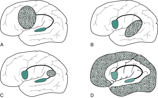
  • Aphasia is an acquired disorder of language resulting from brain damage to the dominant (typically left) hemisphere. Brain lesions causing aphasia are illustrated in Fig. E1. The damage may be vascular, traumatic, neurodegenerative, neoplastic, infectious, or inflammatory. Stroke is by far the most common cause of aphasia.
  • Aphasia is not the same as dysarthria (a disorder of the neuromuscular control of speech) dysphonia (a disorder of the voice), apraxia of speech (disorder of programming of articulation of sequences of phonemes, especially consonants), aphemia (muteness with normal reading, writing, and comprehension), or stuttering.
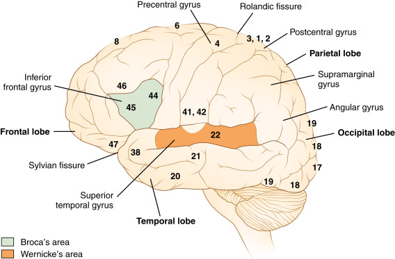
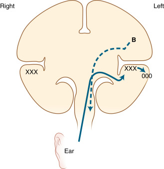
  • Language has a specific neuroanatomy, and aphasia manifests differently depending on the part of the language system that is damaged. Fig. E2 depicts most of the major nodes of the language system. Fig. E3 is a schematic of how the brain processes and produces language.

Figure E1 (A) Lesions Causing Nonfluent Aphasia are Typically Located in the Frontal Lobe and Encompass Broca Area and the Adjacent Cortex Motor Strip

(B) Those Causing Fluent Aphasia are in the Temporoparietal Region. Neurodegenerative Illnesses Also May Damage Wernicke Areas and More Posterior Regions and Cause Fluent Aphasia. (C) Lesions Causing Conduction Aphasia, Which are Relatively Small, Interrupt the Arcuate Fasciculus in the Parietal or Posterior Temporal Lobe. (D) Those Causing Mixed Transcortical (Isolation) Aphasia Involve the Watershed Region, Which Encircles the Perisylvian Language Arc.
From Kaufman DM et al: Kaufman’s clinical neurology for psychiatrists, ed 9, Philadelphia, 2023, Elsevier.

Figure E2 The Lateral Surface of the Left Hemisphere, Showing a Simplified Gyral Anatomy and the Relationships Between Wernicke Area and Broca Area

Not Shown is the Arcuate Fasciculus, Which Connects the Two Cortical Speech Centers by the Deep, Subcortical White Matter.
From Jankovic J et al: Bradley and Daroff’s neurology in clinical practice, ed 8, Philadelphia, 2022, Elsevier.

Figure E3 Coronal Plane Diagram of the Brain, Indicating the Inflow of Auditory Information from the Ears to the Primary Auditory Cortex in Both Superior Temporal Regions (xxx) and Then to the Wernicke Area (Ooo) in the Left Superior Temporal Gyrus

The Motor Outflow of Speech Descends from the Broca Area (B) to the Cranial Nerve Nuclei of the Brain Stem by the Corticobulbar Tract (Dashed Arrow). Actually, the Broca Area is Anterior to the Wernicke Area, and the Two Areas Would Not Appear in the Same Coronal Section.
From Jankovic J et al: Bradley and Daroff’s neurology in clinical practice, ed 8, Philadelphia, 2022, Elsevier.

Synonyms

  • Aphasia conductive
  • Aphasia, expressive (difficulty speaking)
  • Aphasia, expressive and receptive
  • Aphasia, receptive
  • Aphasia-angular gyrus syndrome
  • Conduction aphasia
  • Expressive dysphasia
  • Global aphasia
  • Psycho-sensory aphasia
ICD-10 CM CODES
R47.01Aphasia
I69.320Aphasia following cerebral infarction
I69.020Aphasia following nontraumatic subarachnoid hemorrhage
I69.120Aphasia following nontraumatic intracerebral hemorrhage
I69.920Aphasia following unspecified cerebrovascular disease
Epidemiology & Demographics
Prevalence

The United States has >1 million survivors of stroke-related aphasia. Aphasia is common after acute stroke, with 21% to 38% of stroke patients experiencing aphasia and almost 80% still experiencing aphasia at 12 mo after the stroke.

Diagnosis ⬆ ⬇

TABLE 4 Principal Aphasia Syndromes Due to Stroke

TypeLesion SiteFluencyComprehensionRepetitionNamingOther Signs
Broca (expressive)Inferior frontal lobe↓Good↓↓Contralateral weakness
Wernicke (receptive)Posterior superior temporal lobeGood↓↓↓Homonymous hemianopia
Transcortical motorInferior frontal gyrus↓GoodGoodMay be normalMay be contralateral weakness
Transcortical sensoryMiddle temporal gyrus, thalamusGood↓GoodUsually normalMay be normal
ConductionSupramarginal gyrusGoodGood↓↓None
GlobalFrontal lobe (large)↓↓↓↓Hemiplegia

↓, Reduced.

From Wing EJ, Schiffman FJ: Cecil essentials of medicine, ed 10, Philadelphia, 2022, Elsevier.

TABLE 1 Nonfluent Aphasias

ComprehensionRepetition
Broca’sIntactLost
Transcortical motorIntactIntact
Mixed transcorticalLost (isolation)Intact
GlobalLostLost

From Kaufman DM et al: Kaufman’s clinical neurology for psychiatrists, ed 9, Philadelphia, 2023, Elsevier.

TABLE 2 Fluent Aphasias

ComprehensionRepetition
Wernicke’sLostLost
Transcortical sensoryLostIntact
ConductionIntactLost
AnomicIntactIntact

From Kaufman DM et al: Kaufman’s clinical neurology for psychiatrists, ed 9, Philadelphia, 2023, Elsevier.

TABLE 3 Salient Features of the Nonfluent and Fluent Aphasias

FeatureNonfluentFluent
Other termsExpressive
Motor
Brocass
Receptive
Sensory
Wernickeee
Spontaneous speech
ContentPaucity of words, mostly nouns and verbsComplete sentences with normal syntax
ArticulationDysarthric, slow, stutteringGood
ErrorsTelegraphic speechParaphasic errors, circumlocutions, tangentialities, clang associations
Associated deficitsRight hemiparesis (arm, face, leg)Hemianopsia, hemisensory loss
Localization of lesionFrontal lobeTemporal or parietal lobe Occasionally diffuse

From Kaufman DM et al: Kaufman’s clinical neurology for psychiatrists, ed 9, Philadelphia, 2023, Elsevier.

BOX 1 Clinical Evaluation for Aphasia

Spontaneous speech: Fluent versus nonfluent

  • Verbal tests
    • Comprehension
      • Ability to follow simple requests, e.g., “Please, pick up your hand”
      • Ability to follow complex requests, e.g., “Please, show me your left ring finger, and stick out your tongue”
    • Naming
      • Common objects: Tie, keys, pen
      • Uncommon objects: Watchband, belt buckle
    • Repetition
      • Simple phrases: e.g., “The boy went to the store”
      • Complex phrases: e.g., “No if's, and's, or but's”
      • Reading and writing tests

From Kaufman DM et al: Kaufman’s clinical neurology for psychiatrists, ed 9, Philadelphia, 2023, Elsevier.

Differential Diagnosis
Neurodegenerative Causes of Aphasia

  • Language function is among the cognitive domains affected by Alzheimer disease (AD) even in early clinical stages. Patients develop difficulty understanding others, will sometimes use the incorrect word, and will have trouble getting words out and finding the correct word.
  • Primary progressive aphasias (PPAs) are a group of neurodegenerative disorders in which an aspect of the language network is the primary target of degeneration. The PPAs include nonfluent/agrammatic variant of PPA (nfvPPA) and the semantic variant of PPA (svPPA), both classified under the umbrella of frontotemporal lobar degeneration (FTLD), as well as logopenic progressive aphasia (lvPPA), an atypical variant of AD pathology. Table 5 outlines the clinical features and localization.
  • In nfvPPA, patients present with nonfluent, agrammatic speech, with some also experiencing apraxia of speech. Degeneration of the left posterior frontoinsular association cortex is the cause of this language-based dementia.
  • In svPPA, patients develop difficulty naming, loss of object knowledge, and surface dyslexia (the inability to read irregular words properly) typically to the result of left anterior temporal lobe degeneration. However, this variant can begin in the right anterior temporal lobe, causing difficulty recognizing faces and meaning of facial expressions, before spreading to the left anterior temporal lobe and causing semantic language dysfunction.
  • In lvPPA, patients have impaired naming and sentence repetition, develop long pauses in their speech, and experience phonologic errors in their speech. This variant arises from left posterior perisylvian or parietal association cortex degeneration.

TABLE 5 Clinical Characteristics of Primary Progressive Aphasia

Clinical Features∗Cortical AtrophyPathologic Changes†Alternative Nomenclature
Nonfluent/agrammatic PPAGrammatical simplification and errors in language production
Effortful, halting speech with speech sound errors
Two or more of the following: Impaired syntactic comprehension, spared content word comprehension, or spared object knowledge
Left inferior frontal and insulaFTLD-tau (52%), AD (25%), FTLD-TDP (19%), and other (4%)Progressive nonfluent aphasia (PNFA)
Agrammatic PPA (PPA-G)
Semantic variant PPAPoor confrontation naming
Impaired single-word comprehension
Three or more of the following: Poor object or person knowledge or both; surface dyslexia; spared repetition; or spared motor speech
Anterior and ventral temporal lobeFTLD-TDP (69%), AD (25%), and FTLD-tau (6%)Semantic dementia (SD)
Semantic PPA (PPA-S)
Logopenic variant PPAImpaired single-word retrieval
Impaired repetition of phrases and sentences
Three or more of the following: Speech sound errors, spared motor speech, spared single-word comprehension and object knowledge, or absence of agrammatism
Left posterior superior temporal and inferior parietalAD (50%), FTLD-TDP (38%), and FTLD-tau (12%)Logopenic progressive aphasia (LPA)
Logopenic PPA (PPA-L)
Progressive mixed aphasia (PMA)

AD, Alzheimer disease; FTLD-tau, frontotemporal lobar degeneration with tau-positive pathology; FTLD-TDP, frontotemporal lobar degeneration with ubiquitin-positive and TDP-43-positive pathology; PPA, primary progressive aphasia.

∗Based on expert consensus.

† From a literature review of confirmed pathologic changes in patients with PPA recruited without a-priori bias.

Reprinted with permission from Elsevier. From Grossman M: The non-fluent/agrammatic variant of primary progressive aphasia, Lancet Neurol 11(6):545-555, 2012. https://doi.org/10.1016/s1474-4422(12)70099-6.

Workup

  • Targeted to the underlying etiology of the aphasia but will always include neuroimaging, preferably MRI of the brain.
  • In nonstroke aphasia and in patients without significant kidney disease, MRI should be done with and without contrast.
  • In stroke-related aphasia, acute neuroimaging will include a brain computed tomography (CT) and CT angiogram of the head and neck. An MRI can be performed after acute interventions have been performed.
  • Advanced neuroimaging techniques such as fluorodeoxyglucose (FDG)-PET may be appropriate for neurodegenerative aphasias.
Laboratory Tests

Targeted to the underlying etiology of the aphasia. In stroke-related aphasia, the laboratory workup will include an assessment of vascular risk factors. In a neurodegenerative aphasia, the assessment will be similar to that for AD.

Treatment ⬆ ⬇

Nonpharmacologic Therapy

Speech language therapy. Commonly used techniques include script training, response elaboration training, constraint-induced aphasia therapy, speech entrainment, and melodic intonation therapy.

Acute General Rx

Target the underlying cause of the aphasia

Chronic Rx

Target the underlying cause of aphasia and include speech language therapy.

Referral

  • Neurology
  • Speech language pathology
  • Neuropsychology
  • Physical medicine and rehabilitation
  • Psychiatry

Pearls & Considerations ⬆ ⬇

Comments

  • Recovery from poststroke aphasia is very difficult to predict. The most important factors correlating with recovery are the lesion location and size, aphasia type and severity, and to some extent the nature of early hemodynamic response to acute interventions such as thrombolysis, and treatment received.
  • Most studies attest that recovery is inversely related to the size of lesion, with a preserved left superior temporal gyrus as one of the most important factors for satisfactory recovery and intact basal ganglia also contributing significantly to better recovery.
  • Broca and conduction aphasia enjoy better recovery than global and anomic aphasia.
Prevention

Prevention of stroke-related aphasia involves modification of vascular risk factors.

Patient/Family Education

Teach families of aphasia patients about supportive communication strategies (Table 6)

BOX 2 Stimulation-Facilitation Approaches for Aphasia Therapy

  • Gestural expression and pointing
  • Word-to-picture matching
  • Yes/no response reliability
  • Oral-motor imitation
  • Phoneme, then word repetition
  • Verbal cueing for words and sentence completion
  • Contextual cueing
  • Phonemic and semantic word-retrieval strategies
  • Priming for responses
  • Auditory processing at phrase level and then sentence level
  • Word-, phrase-, then sentence-level reading
  • Melodic stimulation
  • Graphic tasks: Tracing, copying, word completions
  • Calculations
  • Pragmatic linguistic and nonlinguistic conversational skills
  • Psychosocial supports

From Jankovic J et al: Bradley and Daroff’s neurology in clinical practice, ed 8, Philadelphia, 2022, Elsevier.

TABLE 6 Supportive Communication Strategies for Aphasia

Verbal StrategiesEnvironmental StrategiesVisual Strategies
  • Speak slowly but with a natural tone and rhythm
  • Use short, simple sentences
  • Gently give two or three options when providing choices
  • Ask yes or no questions
  • Use writing, gestures, or drawing in addition to speaking
  • Give the person more time to speak
  • Repeat your question or instructions if the person asks or seems unsure
  • Repeat what the person says to make sure you understood correctly
  • Ignore simple mistakes that do not change the meaning of the message (do not correct the person)
  • Remove distractions (e.g., turn off the TV, close the door)
  • Adjust lighting so the person can see you easily
  • Sit or stand facing the person when speaking or listening to the person; make eye contact
  • Keep communication tools nearby (e.g., pen/paper, communication book, picture)
  • Limit group conversations to only a few people
  • Use direct eye contact
  • Use gestures to enhance your message (e.g., thumbs up/down)
  • Write important words you are saying to help the person understand
  • Write notes when communication breakdown occurs and return to them later
  • Point to objects/pictures as you talk to give the person a visual of what you are saying
  • If written information is being provided, leave extra white space on the paper, use short sentences, and use simple language

From Armour M et al: Supportive communication for individuals with aphasia, Arch Phys Med Rehabil 102(7);1437-1439, 2021. https://doi.org/10.1016/j.apmr.2021.03.008.

Related Content

Stroke, Ischemic (Related key Topic)

Alzheimer Disease (Related key Topic)

Frontotemporal Dementia (Related key Topic)

Suggested Readings ⬆

.

    1. Gorno-Tempini M.L. : Classification of primary progressive aphasia and its variantsNeurology. ;76(11):1006-1014, 2011.
    2. Grossman M. : The non-fluent/agrammatic variant of primary progressive aphasiaLancet Neurol. ;11:545-555, 2012.
    3. Grossman M. : Primary progressive aphasia: clinical-pathological correlationsNat Rev Neurol. ;6:88-97, 2010.
    4. Supportive communication for patients with aphasiaArch Phys Med Rehab. ;102:1437-1439, 2021.
    5. Kirshner HS, Wilson SM: Aphasia and aphasic syndromes. In Jankovic J et al (eds): editors: Bradley and Daroff’s neurology in clinical practice, 8th ed, Philadelphia, 2022, Elsevier, pp 133-148.e2.
    6. Peterson R, Graff-Radford J: Alzheimer disease and other dementias. In Jankovic J et al (eds): editors: Bradley and Daroff’s neurology in clinical practice, 8th ed, Philadelphia, 2022, Elsevier, pp 1452-1497.e12.
    7. Sharma VK, Wong LKS: Middle cerebral artery disease. In Grotta JC et al (eds): editors: Stroke: pathophysiology, diagnosis, and management, ed 7, Philadelphia, 2022, Elsevier, pp 317-346.e9.
    8. Watila M.M., Balarabe S.A. : Factors predicting post-stroke aphasia recoverJ Neurol Sci. ;352:12-18, 2015.