section name header

Basic Information

AUTHOR: Bharti Rathore, MD

Definition

Breast cancer refers to epithelial carcinoma arising from the ducts (ductal) or the lobules (lobular) of the breast that is initially in situ but can progress to become invasive in nature.

Classification1

Carcinomas make up the majority of breast malignancies and originate in the epithelium of the collecting ducts (ductal) or the terminal lobular ducts (lobular). Sarcomas are rare, constituting less than 1% of primary breast cancers, and arise from stromal or connective tissue. Tumor grade is based on tubule formation, nuclear pleomorphism, and mitotic counts using the Nottingham score to determine low, intermediate, or high grade. Higher grade is associated with a poorer long-term prognosis.

Initially, breast carcinoma may be divided into invasive and in situ lesions (Table 1). Invasive ductal carcinoma accounts for the majority, approximately 80%, of invasive carcinomas. Invasive lobular carcinomas constitute approximately 10% to 15% of cases. Other subtypes include mucinous, tubular, medullary, micropapillary, and papillary. Both in situ and invasive carcinomas are often found in the same quadrant of the breast. Additionally, multifocal carcinomas are not uncommon, and bilateral breast carcinomas occur in 1% to 2% of newly diagnosed cases.

TABLE 1 Simplified Classification of Breast Carcinoma Based on Histology

Type of CarcinomaPercentage of All Cases Diagnosed
Ductal carcinoma
In situ5
Infiltrating70
Infiltrating with uniform histologic appearance10
Medullary, colloid, comedo, tubular, papillary
Lobular carcinoma
In situ3
Infiltrating9
Inflammatory carcinoma2
Paget disease1

From Gershenson DM et al: Comprehensive gynecology, ed 8, Philadelphia, 2022, Elsevier.

Genomic Profiling1

Historically, the treatment of breast cancer was based on tumor histologic characteristics, axillary node status, tumor size, receptor patterns, and grade of differentiation. In addition to simplified histologic classification, a classification based on gene expression or profiling, including the presence of hormone receptors, has evolved. Identification of tumor receptor status is critical because endocrine therapy is used both for adjuvant therapy and in the management of advanced disease. The genomic analysis of tumors has led to the molecular subtyping of breast cancers. In the early 2000s, breast tumors were classified into four different molecular subtypes: luminal, basal, HER2, and normal. Subsequently, the luminal group was further differentiated into luminal-A and luminal-B subgroups. Basal-like tumors include triple-negative tumors, tumors that are estrogen, progesterone, and HER2 negative by immunohistochemistry. A more aggressive subtype of triple-negative tumors, claudin-low tumors, has also been described. These divisions are detailed in Table 2.

TABLE 2 Classification of Breast Carcinoma Based on Gene Profiling and Hormone Receptor

Expression TypeGradeCharacteristic BehaviorHormone Receptor Status
Luminal AUsually low gradeGood prognosisE and P+
Luminal BAll gradesMixed prognosisE and P+, Her2 (Neu)+
Her2 (Neu)Higher gradesPoor prognosisE and P-, Her2 (Neu)+
BasalUsually grade 3Poor prognosisTriple negative
Normal breastUsually low gradeGood prognosisTriple negative

E, Estrogen receptor; P, progesterone receptor.

Normal breast does not express gene profiling of basal elements and myoepithelial gene expression.

From Gershenson DM et al: Comprehensive gynecology, ed 8, Philadelphia, 2022, Elsevier.

Synonym

Carcinoma of the breast

ICD-10CM CODES
C50.911Malignant neoplasm of unspecified site of right female breast
C50.912Malignant neoplasm of unspecified site of left female breast
C50.919Malignant neoplasm of unspecified site of unspecified female breast
C50.921Malignant neoplasm of unspecified site of right male breast
C50.922Malignant neoplasm of unspecified site of left male breast
C50.929Malignant neoplasm of unspecified site of unspecified male breast
Epidemiology & Demographics

  • In the U.S., there were an estimated 290,560 new patients and an estimated 43,780 deaths in 2022.2
  • Hormone-receptor-positive cancers are the most common type and their incidence has increased, particularly among young women.
  • Only 1% of breast cancers occur in males.
  • Table 3 describes risk factors for breast cancer.
  • Factors associated with a decreased risk for breast cancer are summarized in Table 4.
  • Known genetic mutations in breast cancer are described in Table 5. Genetically defined group of women with BRCA1 or BRCA2 genes (Table 6) carry lifetime risk as high as 85%.

TABLE 5 Known Genetic Mutations in Breast Cancer and Their Management

Genetic MutationBreast Cancer RiskManagement
ATMIncreased by 15%-40%Annual mammography starting at age 40 with consideration for breast tomosynthesis/magnetic resonance imaging (MRI)
BARD1Limited evidence for increased risk but stronger for triple-negative breast cancerAnnual mammography starting at age 40 with consideration for breast tomosynthesis/MRI
BRCA1
BRCA2
Both carry increased absolute risk greater than 60%Breast awareness starting at age 18
Clinical breast examination every 6-12 mo starting at age 25 yr
Breast screening:
Age 25-29 yr: Annual breast MRI screening with contrast or mammogram with consideration of tomosynthesis, only if MRI is unavailable or individualized based on family history if a breast cancer diagnosis before age 30 is present
Age 30-75 yr: Annual mammogram with consideration of tomosynthesis and breast MRI screening with contrast
Age >75 yr: Management should be individualized
Consider risk-reducing mastectomy
BRIP1Potential increase in riskManagement based on family history
CDH1Increased absolute risk 41%-60%Annual mammogram with consideration of tomosynthesis or breast MRI with contrast starting at age 30
CHECK2Increased absolute risk 15%-40%Annual mammography starting at age 40 with consideration for breast tomosynthesis/MRI
MSH2
MLH1
MSH6
PMS2
EPCAM
Limited evidence of increased risk, absolute risk is <15%Management based on family history
NBNIncreased risk of breast cancer with variant 657del5Management based on family history
NF1Increased absolute risk 15%-40%Annual mammography starting at age 30 with consideration for breast tomosynthesis/MRI
PALB2Increased absolute risk 41%-60%Annual mammography starting at age 30 with consideration for breast tomosynthesis/MRI
Consider risk-reducing mastectomy
PTENIncreased absolute risk 40%-60%Breast awareness starting at age 18
Clinical breast examination every 6-12 mo starting at age 25 yr (or 5-10 yr before earliest known breast cancer in the family)
Breast screening:
Age 25-29 yr: Annual breast MRI screening with contrast or mammogram with consideration of tomosynthesis, only if MRI is unavailable or individualized based on family history if a breast cancer diagnosis before age 30 is present
Age 30-75 yr: Annual mammogram with consideration of tomosynthesis and breast MRI screening with contrast
Age >75 yr: Management should be individualized
Consider risk-reducing mastectomy
RAD51CIncreased absolute risk 15%-40%Management based on family history
RAD51DIncreased absolute risk 15%-40%Management based on family history
STK11Increased absolute risk 40%-60%Annual mammography alternating with breast MRI every 6 mo starting at age 30 and clinical breast examination every 6 mo
TP53Increased absolute risk >60%Breast awareness starting at age 18
Clinical breast examination every 6-12 mo starting at age 20 yr
Breast screening:
Age 20-29 yr: Annual breast MRI screening with contrast or mammogram with consideration of tomosynthesis, only if MRI is unavailable or individualized based on family history if a breast cancer diagnosis before age 30 is present
Age 30-75 yr: Annual mammogram with consideration of tomosynthesis and breast MRI screening with contrast
Age >75 yr: Management should be individualized
Consider risk-reducing mastectomy

From Gershenson DM et al: Comprehensive gynecology, ed 8, Philadelphia, 2022, Elsevier.

TABLE 6 Major Inherited Gene Mutation Syndromes Associated With Breast Cancer

SyndromeGeneIncidenceLifetime Breast Cancer RiskAssociated Cancer Risks
BRCA1BRCA11/500-1/100085%Ovary and pancreas
BRCA2BRCA2Unclear85%Ovary and pancreas
CowdenPTEN1/100,000-1/200,00050%Thyroid and endometrium
Li-FraumeniTP531/20,00090%Sarcoma, brain, and leukemia

Other syndromes, including Peutz-Jeghers syndrome, ataxia telangiectasia, CHEK2 gene mutation, and Fanconi syndrome, have much smaller lifetime risks with poorer penetrance.

From Gershenson DM et al: Comprehensive gynecology, ed 8, Philadelphia, 2022, Elsevier.

TABLE 4 Factors Associated With a Decreased Risk for Breast Cancer

DemographicQualificationRelative Risk
Born and living outside Western countries
Late menarcheAfter age 14
OophorectomyYes vs. no0.3
Lactation>16 mo vs. none0.73
Parity5 vs. 00.73
Postmenopausal body mass (kg/m2)<22.9 vs. >30.70.63
Physical activityYes vs. no0.70
Vitamin DLow levels associated with risk
Intake of vitamin DAssociated with decreased risk
Olive oil and omega-3 fatty acids
Low-fat dietResults suggestive but not yet conclusive
Aspirin>1×/wk for 16 mo vs. no use0.79

From Gershenson DM et al: Comprehensive gynecology, ed 8, Philadelphia, 2022, Elsevier.

TABLE 3 Risk Factors for Breast Cancer

Risk FactorQualificationRelative Risk
Age49 yr
50-59 yr
Age 60-69 yr
70 yr
2.0
2.3
3.5
6.7
GeographicCommon in Western countries
Age at menarche>14 yr (low risk) vs. <12 yr1.5
Age at first full-term pregnancy<20 yr (low risk) vs. >30 yr1.9-3.5
Late menopause<45 yr (low risk) vs. >55 yr (high risk)2.0
Hormone replacement therapyNo use vs. current1.2
Contraceptive pill useNone vs. past or current use1.07-1.2
Alcohol useNone vs. 2-5 drinks/day1.4
Postmenopausal weight gainWomen with a higher body mass index (BMI)1.1 per 5 BMI units
Bone densityLowest vs. highest quartile2.7-3.5
Nightshift workExposed to nightshift work1.48
SmokingHistory of smoking1.10
Benign breast diseaseNone vs. positive biopsy result1.7
Breast density (as measured by mammography)0% vs. 75%1.8-6.0
Hyperplasia with atypiaNone vs. positive biopsy result3.7
Multiple relatives, not first degree, with breast cancer
One first-degree relative with breast cancer (mother or sister)None vs. yes2.6
Two or more first-degree relativesIncreased risk if the cancers are premenopausal
Deleterious BRCA1/BRCA2 genesNegative vs. positive2.0-7.0
Mantle radiation for treatment of malignancyVery high risk, which increases with age

Summary relative risk.

From Gershenson DM et al: Comprehensive gynecology, ed 8, Philadelphia, 2022, Elsevier.

Physical Findings & Clinical Presentation

  • Increasing number of small breast cancers are found by mammograms, in which case patients are usually completely free of symptoms or physical findings.
  • Palpable lump or mass which can be self- or physician-detected.
  • Skin and/or nipple retraction and skin edema, erythema, ulcer, satellite nodule.
  • Nodal enlargement in axilla and supraclavicular areas.
  • Nipple discharge may be serous or bloody.
  • Generalized symptoms and signs, including fatigue, weight loss, jaundice, and anorexia, may be present in metastatic cases.
Etiology

  • Endogenous and exogenous estrogen exposure is key to the development of receptor-positive breast cancer
  • Breast cancer is no longer perceived as a single disease. Molecular classification based on gene expression profiling has shown breast cancer to be of the following types3:
    1. Luminal A type: Endocrine responsive with favorable prognosis
    2. Luminal B type: Endocrine responsive with less favorable prognosis
    3. Normal type: Resemble normal epithelium and prognosis similar to luminal B type
    4. HER2 amplified type: HER2 gene amplification occurs in 15% to 20% of cases
    5. Basal type: Hormone receptor and HER2 negative cancers with poor prognosis
  • Approximately 10% of all women with breast cancer have a germline mutation of BRCA1, BRCA2, P53, or other mutations
  • Possibly interaction of ovarian estrogen, nonovarian estrogen, estrogens of exogenous origin with breast tissue of varied carcinogenic susceptibility to develop cancer
  • Other known or suspected variables: Childbearing, breastfeeding practice, diet, physical activity, body mass, alcohol intake
Diagnosis

Differential Diagnosis

Benign disease is summarized in Table 7. Noninvasive neoplasms of the breast were previously broadly divided into two major types, LCIS and DCIS (Box 1). LCIS is no longer regarded as a neoplasm of the breast in the eighth edition of the AJCC staging system but is regarded as a risk factor for the development of breast cancer.

BOX 1 Transverse Rectus Abdominis Muscle Flap Reconstruction

  • Indications
    • Breasts of all sizes
    • Breast ptosis
  • Relative Contraindications
    • Smoking
    • Abdominal liposuction
    • Previous abdominal surgery
    • Pulmonary disease
    • Obesity
  • Contraindications
    • Previous abdominoplasty
    • Patient unable to tolerate 4- to 6-week recovery period
    • Patient unable to tolerate longer procedure

From Townsend CM et al: Sabiston textbook of surgery, ed 21, St Louis, 2022, Elsevier.

TABLE 7 American Board of Pathology Histologic Classification of Benign Disease

Histopathologic FindingsApproximate Relative Risk
NonproliferativeNo added risk
Cysts
Duct ectasia
Calcification
Fibroadenoma
Milk ductal epithelial hyperplasia
Sclerosing adenosisNo added risk
PapillomatosisSlight added risk
Radial scars
Complex sclerosing lesions?
Moderate florid hyperplasia1.5:1-2:1
Atypical hyperplasia (ductal and lobular)4:1
Extensive ductal involvement of atypical hyperplasia7:1
Lobular carcinoma in situ10:1
Ductal carcinoma in situ10:1

From Niederhuber JE: Abeloff’s clinical oncology, ed 6, Philadelphia, 2020, Elsevier.

The following nonmalignant breast lesions can simulate breast cancer on both physical and mammogram examinations:

  • Fibrocystic changes
  • Fibroadenoma
  • Hamartoma
Workup

  • Initial workup:
    1. Mass and axillary nodal assessment by medical professional.
    2. Diagnostic mammogram followed by breast ultrasound for suspicious lesions.
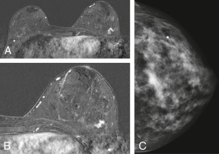
    3. MRI (Fig. 1) detects suspicious lesions better than mammography in women with dense breasts or inherited predisposition to breast cancer.4 When MRI is used for screening, it should be used in addition to screening mammography. Although MRI is more sensitive than mammography, it may still miss some malignancies that a mammogram would detect. For routine MRI screening, the American Cancer Society has recommended a risk-adjusted model:
      1. Annual screening beginning at age 30 yr for women at high lifetime risk for breast cancer development (approximately 20% to 25% or greater) (Box 2).
      2. Discussion with physicians regarding the benefits and limitations of adding MRI screening for women at moderately increased lifetime risk (15% to 20%).
      3. MRI is not recommended for women with a lifetime risk of developing breast cancer of less than 15%.
      4. Table 8 summarizes guidelines for evaluation and treatment of nonpalpable ductal carcinoma in situ.
  • Diagnosis:
    1. Positive aspiration cytology on a clinically and mammographically suspicious mass is highly accurate, but requires open biopsy confirmation.
    2. Stereotactic, ultrasound-guided core-needle biopsy procedures are accurate and have low complication rates. Indications for stereotactic core biopsy are summarized in Box 3. Box 4 describes contraindications to stereotactic core biopsy.
    3. Excisional surgical biopsy establishes diagnosis.

BOX 3 Indications for Stereotactic Core Biopsy

  • Certain probably benign lesions, BI-RADS 3, depending on clinical suspicion, patient or physician preference, or when short-term follow-up is not practical
  • Lesions suspicious, BI-RADS 4
  • Lesions highly suspicious, BI-RADS 5
  • New suspicious microcalcifications, developing asymmetries, or architectural distortions
  • Nonpalpable asymmetry, focal asymmetry, or solid mass on mammogram not seen on ultrasound
  • Mammographic lesions corresponding to suspicious areas of enhancement on MRI

From Cameron JL, Cameron AM: Current surgical therapy, ed 12, Philadelphia, 2017, Elsevier.

BOX 4 Contraindications to Stereotactic Core Biopsy

  • Patient unable to lie prone or cooperate
  • Patient’s weight
  • Lesion location near nipple, too superficial to skin, or too posterior to chest wall
  • Lesion mammographically occult
  • Patient has severe kyphosis or movement disorders
  • Lack of breast tissue thickness for adequate compression

From Cameron JL, Cameron AM: Current surgical therapy, ed 12, Philadelphia, 2017, Elsevier.

Figure 1 A 42-yr-old woman with BRCA2 mutation.

Magnetic resonance imaging (MRI) screening showed a 1.2-cm invasive ductal cancer in the lower outer left breast seen only on MRI (A and B) and subsequently on second-look ultrasound. C, The mammogram showed only dense breast tissue.

From Cameron JL, Cameron AM: Current surgical therapy, ed 12, Philadelphia, 2017, Elsevier.

TABLE 8 Guidelines for Evaluation and Treatment of Nonpalpable Ductal Carcinoma In Situ

  1. Careful multiview mammography with or without ultrasound and including magnification views
    • Document extent of disease
    • Identify other areas of microcalcification
  2. Suspicious microcalcifications and densities cleared with needle localization biopsy
  3. Specimen radiography with magnification techniques
  4. Radiograph-directed histopathologic evaluation with orientation of specimen by surgeon using multicolored inked margins
  5. Complete pathologic description to include:
    • Type of DCIS and size of tumor
    • Relation to microcalcifications
    • Distance of lesion from inked margins
    • Presence of multifocality
    • Presence or risk of microinvasion
  6. Repeat mammography with magnification to confirm successful clearing of suspicious areas
  7. Repeat breast excision if:
    • Residual microcalcifications are found
    • Margins are unacceptable

DCIS, Ductal carcinoma in situ.

From Niederhuber JE: Abeloff’s clinical oncology, ed 6, Philadelphia, 2020, Elsevier.

BOX 2 American Cancer Society MRI Breast Cancer Screening Recommendations

Women at High Lifetime Risk (Risk Criteria for Breast Magnetic Resonance Imaging Screening. 20%-25% or Greater) of Breast Cancer

  • Known BRCA1 or BRCA2 gene mutation
  • First-degree relative with BRCA1 or BRCA2 gene mutation, but have not had genetic testing themselves
  • Lifetime risk of breast cancer of 20%-25% or greater
  • Radiation therapy to the chest between the ages of 10 and 30
  • Li-Fraumeni syndrome or Cowden syndrome or a first-degree relative with one of these syndromes

Women at Moderately Increased (15%-20%) Lifetime Risk

  • Lifetime risk of breast cancer of 15%-20% according to risk assessment tools based mainly on family history
  • Personal history of breast cancer, ductal carcinoma in situ, lobular carcinoma in situ, atypical ductal hyperplasia, or atypical lobular hyperplasia
  • Extremely dense breasts or unevenly dense breasts when viewed by mammograms

From Townsend CM et al: Sabiston textbook of surgery, ed 21, St Louis, 2022, Elsevier.

Staging

Table 9 describes the pathologic staging of breast cancer.

TABLE 9 Pathologic Staging of Breast Cancer

TXPrimary Tumor Cannot Be Assessed
T0No evidence of primary tumor
TisCarcinoma in situ
Tis (DCIS)Ductal carcinoma in situ
Tis (LCIS)Lobular carcinoma in situ
Tis (Paget)Paget disease of the nipple NOT associated with invasive carcinoma and/or carcinoma in situ (DCIS and/or LCIS) in the underlying breast parenchyma. Carcinomas in the breast parenchyma associated with Paget disease are categorized based on the size and characteristics of the parenchymal disease, although the presence of Paget disease should still be noted
T1Tumor 20 mm in greatest dimension
T1miTumor 1 mm in greatest dimension
T1aTumor >1 mm but 5 mm in greatest dimension
T1bTumor >5 mm but 10 mm in greatest dimension
T1cTumor >10 mm but 20 mm in greatest dimension
T2Tumor >20 mm but 50 mm in greatest dimension
T3Tumor >50 mm in greatest dimension
T4Tumor of any size with direct extension to the chest wall and/or to the skin (ulceration or skin nodules)
T4aExtension to chest wall, not including only pectoralis muscle adherence/invasion
T4bUlceration and/or ipsilateral satellite nodules and/or edema (including peau d’orange) of the skin, which do not meet the criteria for inflammatory carcinoma
T4cBoth T4a and T4b
T4dInflammatory carcinoma
NXRegional lymph nodes cannot be assessed (e.g., previously removed)
N0No regional lymph node metastasis
N1Metastasis to movable ipsilateral level I, II axillary lymph node(s)
N2Metastases in ipsilateral level I, II axillary lymph nodes that are clinically fixed or matted or in clinically detected ipsilateral internal mammary nodes in the absence of clinically evident axillary lymph node metastasis
N2aMetastases in ipsilateral level I, II axillary lymph nodes fixed to one another (matted) or to other structures
N2bMetastases only in clinically detected ipsilateral internal mammary nodes and in the absence of clinically evident level I, II axillary lymph node metastases
N3Metastases in ipsilateral infraclavicular (level III axillary) lymph node(s), with or without level I, II axillary node involvement, or in clinically detected ipsilateral internal mammary lymph node(s) and in the presence of clinically evident level I, II axillary lymph node metastasis; or metastasis in ipsilateral supraclavicular lymph node(s), with or without axillary or internal mammary lymph node involvement
N3aMetastasis in ipsilateral infraclavicular lymph node(s)
N3bMetastasis in ipsilateral internal mammary lymph node(s) and axillary lymph node(s)
N3cMetastasis in ipsilateral supraclavicular lymph node(s)
M0No clinical or radiographic evidence of distant metastasis
cM0(i+)No clinical or radiographic evidence of distant metastases, but deposits of molecularly or microscopically detected tumor cells in circulating blood, bone marrow, or other nonregional nodal tissue that are no larger than 0.2 mm in a patient without symptoms or signs of metastases
M1Distant detectable metastases as determined by classic clinical and radiographic means and/or histologically proven >0.2 mm
GXGrade cannot be assessed
G1Low combined histologic grade (favorable)
G2Intermediate combined histologic grade (moderately favorable)
G3High combined histologic grade (unfavorable)
Stage Groupings in Breast Cancer
StageTNM
0TisN0M0
IAT1N0M0
IBT0-T1N1miM0
IIAT0-T1N1M0
T2N0M0
IIBT2N1M0
T3N0M0
IIIAT0-T2N2M0
T3N1-N2M0
IIIBT4N0-N2M0
IIICAny TN3M0
IVAny TAny NM1

From Goldman L, Schafer AI: Goldman’s Cecil medicine, ed 24, Philadelphia, 2012, Saunders; AJCC 8th ed., 2018.

Imaging Studies

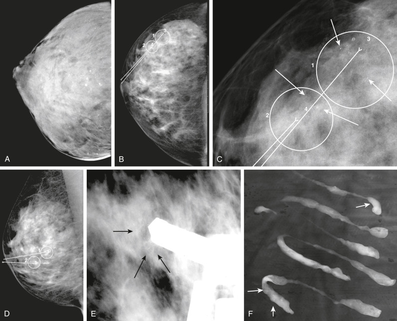
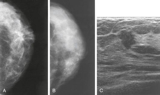
Mammograms (Fig. E2, Fig. E3): 30% to 50% of breast cancers are detected by screening mammograms as a spiculated mass, a mass with or without microcalcifications, or a cluster of microcalcifications. MRI is particularly useful in patients with breast implants and when there is a strong family history of breast cancer. MRI is better for assessing response to neoadjuvant chemotherapy and for identifying the primary tumor in patients presenting with axillary adenopathy.

Figure E2 Mammogram and ultrasound findings of breast disease.

A, A stellate mass in the breast. The combination of a density with spiculated borders and distortion of surrounding breast architecture suggests a malignancy. B, Clustered microcalcification. Fine, pleomorphic, and linear calcifications that cluster together suggest the diagnosis of ductal carcinoma in situ (DCIS). C, An ultrasound image of breast cancer. The mass is solid, containing internal echoes, and displaying an irregular border. Most malignant lesions are taller than they are wide.

From Townsend CM et al [eds]: Sabiston textbook of surgery, ed 17, Philadelphia, 2004, Saunders.

Figure E3 Digital mammography and workup of breast calcifications.

A, Craniocaudal (CC) view in a routine screening of a 43-yr-old woman. Digital mammography penetrates dense breast tissue better than film-screen mammography, clearly showing diffuse benign pattern of large and small calcifications throughout the breast. B, CC view in a routine screening of a 46-yr-old woman. Digital mammography shows two areas of very faint calcification in the outer breast that require further workup. C, Close-up view of B, showing the calcifications (arrows) to be variable in size and shape, which is worrisome for malignancy, particularly ductal carcinoma in situ. Such tiny calcifications in a background of dense breast stroma are easier to see on digital mammography than on film-screen mammography. Computer-assisted diagnosis programs also help the radiologist to locate even tiny groups of faint calcifications such as these. D, Mediolateral view in the same patient as in B, showing that the calcifications are in the lower breast. E, Image from stereo core biopsy procedure in the same patient as in B, showing the core needle immediately proximal to one of the groups of calcifications. Stereo core biopsy allows histologic sampling of tiny groups of calcifications, which can be very helpful in planning surgical approach. This interventional procedure can decrease the total number of surgeries that a patient must undergo to achieve definitive treatment. F, Radiograph of specimen from stereo core biopsy procedure on the same patient as in B, showing that there are several tiny calcifications (arrows) within some of the core samples. Pathologic analysis of the core biopsy revealed ductal carcinoma in situ, high grade, with comedo features.

From Skarin AT: Atlas of diagnostic oncology, ed 4, Philadelphia, 2010, Elsevier.

Treatment

Nonpharmacologic Therapy

  • These approaches include various types of surgical resection and reconstruction (Box 5) as well as adjuvant radiotherapy.
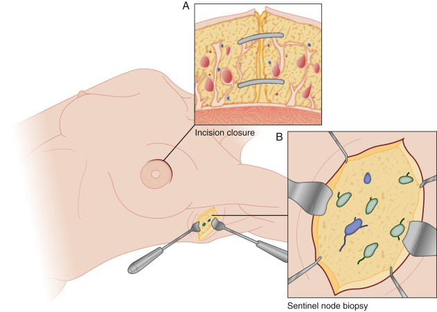
  • For early breast cancer, the primary therapy is usually surgical with a choice between modified radical mastectomy and breast-conserving treatments, and axillary staging with sentinel node biopsy or axillary dissection. Breast-conserving surgery removes the malignancy with a surrounding rim of grossly normal breast parenchyma. This procedure is depicted in Fig. E4, which shows the completed lumpectomy and skin incision for the axillary component of the procedure. The breast specimen that is removed is oriented and its edges are inked before sectioning. Specimen radiography should be performed for all nonpalpable lesions or if there are microcalcifications associated with the palpable tumor. If a margin appears to be close or is positive histologically on intraoperative assessment, reexcision to achieve a clear margin is required. Orientation of the surgical specimen allows focal reexcision of involved margins rather than global reexcision and improves the cosmetic result by reducing the amount of normal breast parenchyma that is excised.
  • Table 10 compares ductal versus lobular carcinoma in situ. Adjuvant treatment guidelines for patients with early-stage invasive breast cancer are described in Table 11.
  • Among patients with limited sentinel lymph nodes (SLN) who are treated with breast conservation and systemic therapy, survival with sentinel lymph node dissection (SLND) is not inferior to axillary lymph node dissection (ALND).5
  • DCIS: Local breast-conserving therapy (lumpectomy plus radiation therapy) or mastectomy followed by endocrine therapy in estrogen receptor-positive cases.
  • Invasive breast cancer: Mastectomy or lumpectomy, along with sentinel lymph node evaluation followed by radiation therapy for large tumors.
  • Invasive breast cancer may require systemic therapy options including endocrine therapy and/or chemotherapy. Current guidelines use stage and biologic characteristics in the development of treatment recommendations to guide decisions regarding systemic therapy for breast cancer (Table 12). Endocrine therapy is recommended alone or after chemotherapy in patients with hormone-receptor positive tumors. Breast cancer intrinsic subtypes are summarized in Table 13. Treatment according to breast cancer subtypes is described in Table 14. Adjuvant hormone therapy with antiestrogen drugs reduces disease recurrence and mortality in women with breast cancer. Aromatase inhibitors (anastrozole, letrozole, exemestane, fulvestrant) decrease the agonist effect of estrogen by inhibiting estrogen synthesis and have become preferred first-line hormonal treatment agents over the selective estrogen receptor modulator tamoxifen.
  • The FDA recently approved the addition of the CDK inhibitor abemaciclib as adjuvant treatment for patients with hormone receptor-positive, HER2-negative, node-positive early breast cancer who are at high risk of recurrence.6
  • There is an improvement in disease-free survival with the adjuvant use of the PARP inhibitor Olaparib in addition to standard endocrine therapy in breast cancer patients with germline BRCA1/BRAC2 mutations.7
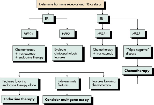
  • Standard adjuvant chemotherapy used in the U.S. include AC (cyclophosphamide plus doxorubicin), AC-T (doxorubicin and cyclophosphamide followed by a taxane), and TC (docetaxel and cyclophosphamide) regimens. In patients with HER2 expressing breast cancers, standard adjuvant regimens also include the addition of trastuzumab and pertuzumab for 1-yr duration. Fig. E5 illustrates considerations for adjuvant chemotherapy in breast cancer.
  • Neoadjuvant chemotherapy (same regimens as used in the adjuvant setting) results in pathologic complete responses in significant number of cases, causes downstaging, provides an assessment of chemosensitivity, and provides no deleterious effect on survival.
  • In patients with early-stage, triple-negative breast cancer, the FDA has approved the PD-1 inhibitor immunotherapy agent pembrolizumab in combination with chemotherapy as neoadjuvant treatment and then continued as a single agent as adjuvant treatment after surgical resection.
  • The benefit of adjuvant chemotherapy or hormone therapy can be assessed by commercially available multigene assays (OncotypeDx, Mammaprint), which help in determining prognostic and predictive benefit associated with the use of both hormonal therapy and chemotherapy in breast cancer. Results from the prospective TAILORx trial support the routine use of multigene assays to identify intermediate-risk score patients in which chemotherapy can be safely omitted.8 Among premenopausal women with one to three positive lymph nodes and a recurrence score of 25 or lower based on the 21-gene breast cancer assay, those who received chemoendocrine therapy had longer invasive disease-free survival and distant relapse-free survival than those who received endocrine-only therapy, whereas postmenopausal women did not have any benefit from adjuvant chemotherapy.
  • Metastatic breast cancer is treated based on the extent of bone-only or visceral disease sites as well as the rate of symptomatic progression. All management of metastatic breast cancer is palliative. Table 15 summarizes median survival of patients with metastatic breast cancer by location of metastases.
  • Typically, bone-only metastatic disease is approached with upfront, sequential hormonal therapy with tamoxifen or aromatase inhibitors. The combination of CDK4/CDK6 inhibitors (palbociclib, ribociclib, abemaciclib) with aromatase inhibitors is typically used as up-front therapy and improves overall survival. Second-line therapy with aromatase inhibitor plus mTOR inhibitors (everolimus) is used.
  • Among patients with PIK3CA-mutated, hormone-receptor-positive, HER2-negative advanced breast cancer who had received endocrine therapy previously, treatment with the combination of alpelisib plus fulvestrant prolonged progression-free survival, leading to the approval of this combination approach.9 Commonly used cytotoxic chemotherapy and anti-HER2 therapy drugs in metastatic breast cancer are summarized in Table 16.
  • Patients with progressive bone disease or those with visceral disease are treated with typically single-agent chemotherapy and occasionally with combination chemotherapy regimens. The chemotherapy agents are the same as those used in early stages of disease. Sequential chemotherapy with different classes of chemotherapy agents are usually used to provide palliation with improvement in survival and symptoms.
  • Patients whose tumors have germline BRCA1/2 mutations can be treated with the PARP inhibitors olaparib or talazoparib.
  • Patients with triple-negative breast cancer whose tumors express the program death ligand-1 receptor derive a survival benefit with the use of the combination of immune checkpoint inhibitor pembrolizumab plus chemotherapy.10
  • Sacituzumab govitecan is an antibody-drug conjugate composed of an antibody targeting the human trophoblast cell-surface antigen 2 (Trop-2), which is expressed in most breast cancers, coupled to SN-38 (topoisomerase I inhibitor). Progression-free and overall survival were significantly longer with sacituzumab govitecan than with single-agent chemotherapy among relapsed triple-negative breast cancer patients leading to its approval recently.11
  • In patients with metastatic HER2/neu-positive breast cancer, the addition of the monoclonal antibody pertuzumab to the standard regimen of trastuzumab plus taxane chemotherapy has been demonstrated to significantly improve the median overall survival to >5 yr.12 Also, the antibody-drug conjugates TDM-1 and trastuzumab dexrutecan improve survival in this group of patients.11 The chimeric antibody margetuximab, which incorporates an engineered Fc region to increase immune activation, results in significant improvement in progression-free survival when used with chemotherapy in patients previously treated with two or more prior anti-HER2/neu therapies. In addition, oral HER2-directed inhibitors such as neratinib and tucatinib are active in previously treated patients with metastatic disease.
  • The FDA has approved elacestrant, an oral estrogen receptor antagonist for treatment of ER-positive, HER2-negative, ESR1-mutated advanced or metastatic breast cancer in postmenopausal women or men who had disease progression following endocrine therapy.

TABLE 16 Commonly Used Cytotoxic Chemotherapy and Anti-HER2 Therapy Drugs in Metastatic Breast Cancer

Cytotoxic Chemotherapy
Albumin-bound paclitaxel
Capecitabine
Cisplatin
Carboplatin
Docetaxel
Doxorubicin
Epirubicin
Eribulin
Gemcitabine
Ixabepilone
Vinorelbine
Anti-HER2 Therapy
Ado-trastuzumab emtansine
Lapatinib
Pertuzumab
Trastuzumab

HER2, Human epidermal growth factor receptor 2.

From Niederhuber JE: Abeloff’s clinical oncology, ed 6, Philadelphia, 2020, Elsevier.

TABLE 15 Median Survival in Months of Patients With Metastatic Breast Cancer by Location of Metastases

LocationSurvival (Months)
Liver (>30% replacement)3
Lung (lymphangitic)3
Lung (nodular)22
Skin27
Bones36+

From Townsend CM et al: Sabiston textbook of surgery, ed 21, St Louis, 2022, Elsevier.

TABLE 12 Decision-Making for Systemic Therapy

StageSystemic TherapyComments
I (<1 cm)
Hormone receptor-positiveEndocrine therapy ± chemotherapyConsider genomic testing
Hormone receptor-negativeConsider chemotherapy
HER-2-positiveStrongly consider trastuzumab and chemotherapy
I (>1 cm)
Hormone receptor-positiveEndocrine therapy ± chemotherapyConsider genomic testing
Hormone receptor-negativeChemotherapy
HER-2-positiveTrastuzumab and chemotherapy
II (Lymph Node-Negative)
Hormone receptor-positiveEndocrine therapy ± chemotherapyConsider genomic testing
Hormone receptor-negativeChemotherapy
HER-2-positiveTrastuzumab and chemotherapy
II (Lymph Node-Positive), III
Hormone receptor-positiveChemotherapy + endocrine therapyEndocrine therapy should be recommended for all patients
Hormone receptor-negativeChemotherapyDecision-making for chemotherapy may be influenced by results from ongoing clinical trials
HER-2-positiveTrastuzumab and chemotherapyConsider neoadjuvant chemotherapy with dual HER-2-targeted therapy

HER-2, Human epidermal growth factor receptor 2.

From Townsend CM et al: Sabiston textbook of surgery, ed 21, St Louis, 2022, Elsevier.

TABLE 13 Breast Cancer Intrinsic Subtypes

SubtypeCharacteristicsMarkers
Luminal ALow grade. High ER 40% of all breast cancer
Good prognosis
ER+, PR+, HER2-Low Ki-67 (<14%)
Luminal BHigher grade. Lower ER 20% of all breast cancer
Poorer prognosis than luminal A
ER+, PR+/-, HER2+/-High Ki-67 (>14%)
HER2-enrichedHigh grade. Often node positive P53 mutations
10%-15% of all breast cancer
ER-, PR-, HER2+
Basal-likeHigh proliferation. BRCA dysfunction 15%-20% of all breast cancer
Poor prognosis
ER-, PR-, HER2-CK5/6
Or EGFR+

CK, Cytokeratin; EGFR, epidermal growth factor receptor; ER, estrogen receptor; HER2, human epidermal growth factor receptor 2; PR, progesterone receptor.

From Cameron JL, Cameron AM: Current surgical therapy, ed 12, Philadelphia, 2017, Elsevier.

TABLE 14 Treatment According to Breast Cancer Subtype

SubtypeTreatment Response and Prognosis
Luminal ARespond to endocrine therapy
  • Premenopausal: SERMs (tamoxifen)
  • Postmenopausal: AIs (exemestane, anastrozole, letrozole)
Luminal BResponse to endocrine therapy lower. Response to chemotherapy greater than luminal A
HER2-enrichedRespond to anti-HER2 agents (trastuzumab, pertuzumab, lapatinib)
Basal-likeNo response to endocrine therapy or anti-HER2 agents. Chemotherapy only treatment outside of a clinical trial

AIs, Aromatase inhibitors; HER2, human epidermal growth factor receptor 2; SERMs, selective estrogen receptor modulators.

From Cameron JL, Cameron AM: Current surgical therapy, ed 12, Philadelphia, 2017, Elsevier.

TABLE 10 Carcinoma In Situ: Lobular Versus Ductal

FeatureLobular Carcinoma In SituDuctal Carcinoma In Situ
AgeYoungerOlder
Palpable massNoUncommon
Mammographic appearanceNot detected on mammographyMicrocalcifications, mass
ImmunophenotypeE-cadherin negativeE-cadherin positive
Usual manifestationIncidental finding on breast biopsyMicrocalcifications on mammography or breast mass
Bilateral involvementCommonUncertain
Risk and site of subsequent breast cancer25% risk for invasive breast cancer in either breast over remaining lifespanAt site of initial lesion; 0.5% risk/yr of invasive breast cancer in opposite breast
PreventionConsider tamoxifen or raloxifeneConsider tamoxifen or raloxifene if estrogen-receptor positive
TreatmentYearly mammography and breast examinationLumpectomy ± radiation; mastectomy for large or multifocal lesions

From Goldman L, Schafer AI: Goldman’s Cecil medicine, ed 24, Philadelphia, 2012, Saunders.

TABLE 11 Adjuvant Treatment Guidelines for Patients With Early-Stage Invasive Breast Cancer

Patient GroupTreatment
Hormone Receptor-Positive and HER2 Positive Breast Cancer
<0.5 cmConsider adjuvant endocrine therapy
0.6-1 cmAdjuvant endocrine therapy
Consider adjuvant chemotherapy and trastuzumab
>1 cmAdjuvant endocrine therapy
Adjuvant chemotherapy and trastuzumab
Node positiveAdjuvant endocrine therapy
Adjuvant chemotherapy with pertuzumab and trastuzumab
Hormone Receptor-Positive and HER2 Negative Breast Cancer
<0.5 cmNo adjuvant therapy
>0.5 cmAdjuvant hormonal therapy
Consider adjuvant chemotherapy based on 21-gene recurrence score
Node-positiveAdjuvant hormonal therapy + adjuvant chemotherapy
Hormone Receptor-Negative and HER2 Positive Breast Cancer
<0.5 cmConsider adjuvant chemotherapy and trastuzumab
0.6-1 cmConsider adjuvant chemotherapy and trastuzumab
>1 cmAdjuvant chemotherapy and trastuzumab
Node positiveAdjuvant endocrine therapy
Adjuvant chemotherapy with pertuzumab and trastuzumab
Hormone Receptor-Negative and HER2 Negative Breast Cancer
0.5 cmNo adjuvant therapy
0.6-1.0 cmConsider adjuvant chemotherapy
>1 cm or node positiveAdjuvant chemotherapy

HER2, Human epidermal growth factor receptor 2.

Modified from National Comprehensive Cancer Network Guidelines. Available at www.nccn.org.

BOX 5 Options for Breast Reconstruction

Autogenous

  • Abdominal-based flaps
  • Transverse rectus abdominis muscle (TRAM)
  • Single pedicle
  • Double pedicle
  • Free flap
  • Deep inferior epigastric perforator flap

Latissimus dorsi musculocutaneous flap

Gluteal flap

  • Superiorly based
  • Inferiorly based

Rubens flap

Thoracoepigastric flap

Lateral thigh flap

Breast-splitting procedure

Alloplastic

  • Silicone gel implant
  • Silicone implant with saline fill
  • Smooth wall
  • Textured wall
  • Round
  • Anatomic shaped
Combination procedures

  • Latissimus dorsi flap with implant
  • TRAM flap with implant

From Townsend CM et al: Sabiston textbook of surgery, ed 21, St Louis, 2022, Elsevier.

Figure E4 Breast-conserving surgery.

A, Incisions to remove malignant tumors are placed directly over the tumor or around the areola. After the partial mastectomy has been completed, the parenchymal defect is closed (inset) to prevent a cosmetic deformity. B, A transverse incision below the axillary hairline is used for sentinel node biopsy or axillary dissection. The boundaries of the axillary dissection are the axillary vein superiorly, the latissimus dorsi muscle laterally, and the chest wall medially. The inferior dissection enters the tail of Spence (the axillary tail of the breast). In sentinel node biopsy, a similar transverse incision is made, which may be located by percutaneous mapping with the gamma probe to detect a hot spot from the radiolabeled colloid. It is extended through the clavipectoral fascia, and the true axilla is entered. The sentinel node is located by staining with blue dye (inset), radioactivity, or both and is dissected free as a single specimen.

From Townsend CM et al: Sabiston textbook of surgery, ed 21, St Louis, 2022, Elsevier.

Figure E5 Management of recurrent and metastatic breast cancer.

!!flowchart!!

Considerations for adjuvant chemotherapy. ER, Estrogen receptor; HER2, human epidermal growth factor receptor 2.

From Cameron JL, Cameron AM: Current surgical therapy, ed 10, Philadelphia, 2011, Saunders.

Chronic Rx

Follow-up after treatment of early breast cancer stages includes:

  • Patient instruction in monthly breast self-examination.
  • Regular clinical evaluations as delineated by medical oncologist or surgeon.
  • Annual mammograms and breast MRI as indicated.
  • Laboratory tests as indicated.
  • Tumor markers and CT scans for surveillance are not recommended.
  • Adjuvant therapy: In premenopausal women with hormone receptor-positive early breast cancer, use of aromatase inhibitor exemestane plus ovarian suppression, compared with tamoxifen plus ovarian suppression, significantly reduced recurrence. In postmenopausal women with hormone receptor-positive breast cancer, aromatase inhibitor therapy improves survival outcomes compared with tamoxifen. Women who take tamoxifen for 10 yr lower their recurrence risk by 25% and their dying of breast cancer risk by 27% compared with those who took it for just 5 yr. In postmenopausal women with hormone-receptor positive breast cancer, recent trial data showed that in women treated with 5 yr of adjuvant endocrine therapy, extending hormone therapy by 5 yr provided no benefit over a 2-yr extension but was associated with more bone fractures.
  • Retrospective analyses suggest that occult lymph-node metastases are an important prognostic factor for disease recurrence or survival; however, recent data indicate that the magnitude of the difference in outcome at 5 yr is small (1.2 percentage points) and do not favor additional evaluation of initially negative sentinel nodes.
  • Adding zoledronic acid to adjuvant endocrine therapy improves disease-free survival in premenopausal patients with estrogen-responsive early breast cancer.
Referral

Referral to a multidisciplinary team consisting of a breast surgeon, reconstructive surgeon, medical oncologist, and radiation oncologist is necessary as soon as breast cancer is suspected.

Requires microsurgical procedure and historical note only.

Pearls & Considerations

Breast Cancer In Pregnancy and Lactation

  • Frequency in women 40 yr or younger reported to be 15%
  • May carry worse prognosis because disease discovery delayed by engorged and nodular breast changes and/or because disease progression more rapid in pregnancy
  • Survival is similar to nonpregnant early-stage patients in same age group
  • Expedient workup recommended, including mammography and sonography
  • Choice of mastectomy or lumpectomy with axillary dissection for treatment
  • Adjuvant chemotherapy and radiotherapy delayed until third trimester or after delivery
  • Other contraindications to radiation are summarized in Box 6

BOX 6 Contraindications to Radiation

Absolute

  • Pregnancy
Relative

  • Systemic scleroderma
  • Active systemic lupus erythematosus
  • Prior radiation to breast or chest wall
  • Severe pulmonary disease
  • Severe cardiac disease (if tumor is left sided)
  • Inability to lie supine
  • Inability to abduct arm on affected side
  • p53 mutation

From Townsend CM et al: Sabiston textbook of surgery, ed 21, St Louis, 2022, Elsevier.

Ductal Carcinoma In Situ (Dcis, Intraductal Carcinoma) (See TABLE 10

  • Discovered by mammogram as cluster of microcalcifications and/or density
  • Presents less often as a palpable mass or nipple discharge
  • With widespread mammogram screening, DCIS accounts for 15% to 20% of all breast cancers
  • Treated with lumpectomy with cure rates 98% to 99%
  • Higher-risk cases require breast radiation and adjuvant hormone therapy
  • Mastectomy is required with multifocal and/or high-grade DCIS
Inflammatory Carcinoma

  • Rare, rapidly progressive, and often lethal form of breast cancer
  • Presents as erythematous and edematous breast resembling mastitis
  • Biopsy required, including that of the skin
  • Treatment with upfront combination chemotherapy followed by surgery and radiotherapy
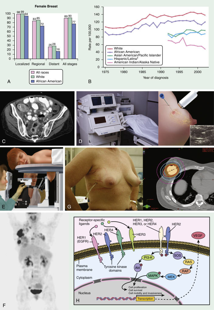
  • Prognosis is improved with 5-yr disease-free survival approaching 50% (Fig. E6)

Figure E6 A, 5-Yr Relative Survival by Race and Stage at Diagnosis of Breast Cancer (SEER Data, 1996-2002)

B, Female Breast Cancer Incidence by Race and Ethnicity (SEER Data). C, When Lobular Breast Cancer Metastasizes, It Can Often Infiltrate Serosal Surfaces, Mimicking Ovarian Cancer. This Patient Presented with Abdominal Bloating, Tightness, and Narrowing in Her Stool Caliber 9 Yr after the Diagnosis of a Stage I Breast Cancer. Note the Diffuse Thickening of the Rectal and Colonic Wall, Peritoneal Carcinomatosis, and Ascites. Fig. E6 (Continued)A Colonoscopy was Performed, and Biopsy Confirmed Diffuse Involvement with Metastatic Adenocarcinoma Consistent with a Breast Primary. On Restaging, She was Also Noted to have Multiple Osseous Metastases. D, In an Ultrasound-Guided Needle Biopsy, the Ultrasound Probe is Used to Localize the Lesion that was Identified Either on Physical Examination or on Mammogram. A Biopsy Needle is Passed through the Lesion Several Times to Obtain Tissue. Compared to a Stereotactic Biopsy, an Ultrasound-Guided Biopsy is Faster and Better Tolerated by Most Patients. However, Not All Lesions May Be Amenable to an Ultrasound-Guided Biopsy. E, The Premise Behind Stereotactic Needle Biopsy is that a Lesion Can Be Localized in 3D by Evaluating its Changes in Position in a Series of Angled Radiographic Views. First, a Radiograph Localizes the Suspicious Area; Then Two Additional Views, Angled 15 Degrees to Either Side of the Lesion, are Obtained. A Computer Calculates How Much the Lesion’s Position Appears to have Changed on Each of the Angled Views and Uses These Data to Estimate the Lesion’s Location Within 3D Space. With the Advent of Digital Mammography, These Images are Commonly Acquired Digitally. F, Positron Emission Tomography (PET) Involves Injection of a Substance Labeled with a Positron-Emitting Isotope (Commonly, Fluorine-18 Bound to D-Glucose, Called FDG for 2-([18F]fluoro-2-Deoxy-D-Glucose)). Metabolically Active Cells, Especially Malignant Ones, Preferentially Take Up Glucose and Therefore FDG, as Compared with Nonneoplastic Tissue. Sensitivity of PET Can Vary Considerably by Tumor Type and Size. False-Positive Results Can Occur in Areas of Inflammation or Infection. Many Machines Now Acquire CT Images in Tandem with PET Images, Which Can Then Be Fused Together to Provide Anatomic Correlation by CT with Metabolic Activity Measurements by PET. This Patient Presented with Palpable Axillary Adenopathy and a Large Breast Mass with Associated Erythema, Skin Edema, and Nipple Retraction. Note the Extremely Intense Areas of Uptake Within the Right Breast and Axilla Corresponding to the Patient’s Known Locally Advanced Breast Cancer. Also Note the Intense Uptake in the Right Supraclavicular, Paratracheal, Prevascular, Precarinal, and Hilar Lymph Nodes Suspicious for Metastatic Disease. Uptake in the Kidney, Bladder, and Ureters is Physiologic and Due to FDG Excretion. Uptake in the Right Adnexa and Jaw is Most Likely Physiologic and Benign. G, Panels 1 and 2: Accelerated Partial Breast Irradiation (APBI) Encompasses Techniques Including Intracavitary and Interstitial Brachytherapy as Well as 3D-Conformal, Intensity-Modulated, and Intraoperative External-Beam Radiation Therapy. One of the More Commonly Used Brachytherapy Methods in the U.S., the Mammosite Brachytherapy System (Hologic, Massachusetts), Involves Insertion of a Catheter with a Balloon Tip into the Lumpectomy Cavity at the Time of Surgery or Shortly Thereafter (Panel 1). The Balloon is Filled with Saline, and a High-Dose-Rate Radioactive Source is Introduced Twice Per Day for 5 Days by Computed Axial Tomography Scan-Based Treatment Planning, Permitting a Highly Conformal Dose to Be Delivered to the First Centimeter of Remaining Breast Tissue with Optimal Sparing of the Remaining Tissue and Other Regional Organs (Panel 2). The Balloon Catheter is Removed Upon Completion. APBI is an Option Only for Selected Patients, Mainly Older Women with Smaller, Node-Negative “low-Risk” Tumors and with Negative Margins. H, The HER Family of Receptors (Human Epidermal Growth Factor Receptor, Also Called ErbB, is a Group of Transmembrane Tyrosine Kinase Receptors that Regulate Cell Growth, Survival, and Differentiation Via a Variety of Pathways, Including RAS (Rat Sarcoma), RAF (Receptor Activation Factor), MAPK (Mitogen-Activated Protein Kinase), and MEK (Mitogen Extracellular Signal Kinase). The Tyrosine Kinase Domains are Activated by Dimerization. Current Therapeutics Involve Tyrosine Kinase Inhibitors (E.g., Lapatinib) and Antibodies Directed Against the HER2 Protein and VEGF (Vascular Endothelial Growth Factor) (E.g., Trastuzumab and Bevacizumab). Akt, Protein Kinase B; Pi3-K, Phosphatidylinositol 3-Kinase; SOS, Son of Sevenless.

From Skarin AT: Atlas of diagnostic oncology, ed 4, Philadelphia, 2010, Elsevier. Image courtesy of Drs. Pamela Dipiro and Wendy Chen, Dana Farber Cancer Institute, Boston, MA. Image courtesy of Robyn L. Birdwell, MD, Brigham and Women’s Hospital, Boston, MA, and Diagnostic Imaging Breast, Amirsys, Inc., Salt Lake City, UT, 2006. Courtesy of Phillip M. Devlin, MD, Dana Farber/Brigham and Women’s Cancer Center, Harvard Medical School, Boston, MA.

Comments

  • The U.S. Preventive Services Task Force (USPSTF) now recommends against automatic “routine” screening of younger women (age range 40 to 49). The task force recommends biennial screening mammography for all middle-aged women (age range 50 to 74). It also states that current evidence is insufficient to assess the benefits and harms of screening mammography in older women (aged 75 and older). The task force also discourages women from performing breast self-examination. Several other U.S. organizations, such as the American Congress of Obstetricians and Gynecologists, however, still recommend annual screening beginning at age 40 yr. Breast cancer screening recommendations by organization are summarized in Table 17.
  • The American Cancer Society screening guidelines are summarized in the following13:
  • Women should continue screening mammography as long as their overall health is good and they have a life expectancy of 10 yr or longer. (Qualified recommendation)
  • Women with an average risk of breast cancer should undergo regular screening mammography as below:
    1. Women should have the opportunity to begin annual screening between the ages of 40 and 44 yr. (Qualified recommendation)
    2. Women aged 45 to 54 yr should be screened annually. (Qualified recommendation)
    3. Women 55 yr and older should transition to biennial screening or continue screening annually. (Qualified recommendation)
  • Women who are at high risk for breast cancer based on certain factors should get an MRI and a mammogram every year, typically starting at age 30 yr. This includes women who:
    1. Have a lifetime risk of breast cancer of about 20% to 25% or greater, according to risk assessment tools that are based mainly on family history.
    2. Have a known BRCA1 or BRCA2 gene mutation (based on genetic testing).
    3. Have a first-degree relative (parent, brother, sister, or child) with a BRCA1 or BRCA2 gene mutation and have not had genetic testing themselves.
    4. Had radiation therapy to the chest between the ages of 10 and 30 yr.
    5. Have Li-Fraumeni syndrome, Cowden syndrome, or Bannayan-Riley-Ruvalcaba syndrome, or have first-degree relatives with one of these syndromes.
  • Physicians should be familiar with the risks and benefits of these various competing recommendations in order to better counsel patients.
  • Breast radiologic evaluation, evaluation of nipple discharge, and evaluation of palpable mass are described in Section III.
  • Exposure of the heart to ionizing radiation during breast cancer radiotherapy increases risk of ischemic heart disease. The increased rate of ischemic heart disease begins within a few years of exposure and continues for at least 20 yr. The increase is proportional to the mean radiation dose to the heart.

TABLE 17 Breast Cancer Screening Recommendations by Organization

OrganizationWhen to Initiate ScreeningFrequency of ScreeningWhen to Stop Screening
American Academy of Family Physicians (AAFP)Follow U.S. Preventive Services Task Force (USPSTF) recommendationsFollow USPSTF recommendationsFollow USPSTF recommendations
American Cancer Society (ACS)Opportunity to begin screening at ages 40-44
Regular screening starting at age 45
Annually from age 45-54
Biennially starting at age 55 with opportunity to continue annually
Continue screening mammography as long as overall health is good, and life expectancy is 10 yr or longer
American College of Obstetricians and Gynecologists (ACOG)Annual screening starting at age 40AnnuallyNot specified
American College of Physicians (ACP)Individualized for women ages 40-49
Regular screening starting at age 50
BienniallyAge 75 yr or older
Women of any age with life expectancy <10 yr
American College of Radiology (ACR)Annual screening starting at age 40AnnuallyShould be considered as long as the patient is in good health and is willing to undergo additional testing if an abnormality is detected
National Comprehensive Cancer Network (NCCN)Annual screening starting at age 40AnnuallyUpper age limit is not yet established
Consider comorbid conditions limiting life expectancy (e.g., 10 yr) and whether therapeutic interventions are planned
U.S. Preventive Services Task Force (USPSTF)Individualized for women ages 40-49
Regular screening beginning at age 50
BienniallyInsufficient evidence to recommend for or against screening at age 75 or older

From Niederhuber JE: Abeloff’s clinical oncology, ed 6, Philadelphia, 2020, Elsevier.

Risk Reduction Strategies

  • Prophylactic bilateral mastectomy reduces the risk for invasive breast cancer by >90%.
  • Counseling points for women with average risk interested in contralateral prophylactic mastectomy (CPM) are summarized in Box 7.
  • Selective estrogen receptor modulators (SERM) reduce the incidence of hormone receptor-positive invasive breast cancer by 50%.
  • Ovarian failure is a common toxic effect of chemotherapy. Administration of the gonadotropin-releasing hormone (GnRH) agonist goserelin appears to protect against ovarian failure, reducing the risk of early menopause and improving prospects for fertility.

BOX 7 Counseling Points for Women With Average Risk Interested in Contralateral Prophylactic Mastectomy (CPM)

  • There is a low annual contralateral breast cancer risk in women with average risk factors.
  • Risk of contralateral breast cancer is decreasing with use of adjuvant therapy.
  • Removing the contralateral breast does not decrease the risk for developing distant metastases.
  • Breast cancer does not commonly metastasize from one breast to the other.
  • CPM does not improve breast cancer-specific survival.
  • CPM does not decrease local recurrence.
  • Contralateral breast cancers tend to be at a lower stage than the initial primary cancer.
  • CPM increases the surgical complication risk.
  • Choice of CPM may influence reconstruction options.
  • There are alternatives to CPM, including chemoprevention and surveillance.

From Cameron JL, Cameron AM: Current surgical therapy, ed 12, Philadelphia, 2017, Elsevier.

Related Content

Breast Cancer (Patient Information)

Breast Cancer: For Men (Patient Information)

Breast Abscess (Related Key Topic)

Fibrocystic Breast Disease (Related Key Topic)

Related Content

    1. Rock D.T. : Detection, management, and surveillance of breast disease Gershenson D.M., editors : Comprehensive gynecology,. ed 8Elsevier-Philadelphia, 2022.
    2. Siegel R.L. : Cancer statistics, 2022, CA CancerJ Clin. ;72(1):7-33, 2022.
    3. Gradishar WJ et al: Breast cancer, version 3.2020, NCCN clinical practice guidelines in oncology, J Natl Compr Canc Netw 18(4):452-478, 2020.
    4. Morrow M. : MRI for breast cancer screening, diagnosis, and treatmentLancet. ;378:1804-1811, 2011.
    5. Giuliano A.E. : Axillary dissection vs no axillary dissection in women with invasive breast cancer and sentinel node metastasisJAMA. ;305(6):569-575, 2011.
    6. Martin M. : Treatment with adjuvant abemaciclib plus endocrine therapy in patients with high-risk early breast cancer who received neoadjuvant chemotherapy: a prespecified analysis of the monarchE randomized clinical trialJAMA Oncol. ;8(8):1190-1194, 2022.
    7. Turk A.A., Wisinski K.B. : PARP inhibitors in breast cancer: bringing synthetic lethality to the bedsideCancer. ;124(12):2498-2506, 2018.
    8. Sparano J.A. : Adjuvant chemotherapy guided by a 21-gene expression assay in breast cancerN Engl J Med. ;379(2):111-121, 2018.
    9. Ellis H., Ma C.X. : PI3K inhibitors in breast cancer therapyCurr Oncol Rep. ;21(12), 2019.
    10. Cortes J. : Pembrolizumab plus chemotherapy in advanced triple-negative breast cancerN Engl J Med. ;387(3):217-226, 2022.
    11. Adams E. : Sacituzumab govitecan and trastuzumab deruxtecan: two new antibody-drug conjugates in the breast cancer treatment landscapeESMO Open. ;6(4), 2021.
    12. Miles D. : Final results from the PERUSE study of first-line pertuzumab plus trastuzumab plus a taxane for HER2-positive locally recurrent or metastatic breast cancer, with a multivariable approach to guide prognosticationAnn Oncol. ;32(10):1245-1255, 2021.
    13. Oeffinger K.C. : Breast cancer screening for women at average risk, 2015 guideline update from the American Cancer SocietyJAMA. ;314(15):1599-1614, 2015.