section name header

Basic Information

AUTHOR: Jessica E. Shill, MD

Definition

Diabetic ketoacidosis (DKA) is a life-threatening complication of diabetes mellitus. It results from an absolute or relative insulin deficiency that results in insulin resistance when paired with counterregulatory hormone and free fatty acid excess. DKA is characterized by the presence of an anion gap metabolic acidosis, ketonemia, and hyperglycemia.

Synonym

DKA

ICD-10CM CODES
E13.10Diabetic ketoacidosis
E10.10Type 2 diabetes mellitus with other specified complication
E10.11Diabetic ketoacidosis with coma associated with type 1 diabetes mellitus
E08.10Diabetes mellitus due to underlying condition with ketoacidosis without coma
E08.11Diabetes mellitus due to underlying condition with ketoacidosis with coma
E13.11Diabetic ketoacidosis with coma
Epidemiology & Demographics

DKA is the most common hyperglycemic emergency among patients with type 1 (T1D) and type 2 diabetes (T2D). In the past decade, the frequency of DKA has increased in the U.S., with more than 160,000 hospital admissions in 2017.1 Social and racial-ethnic disparities are remarkable, with Black race/ethnicity and lower income individuals at heightened risk of DKA.2 DKA most commonly occurs in individuals with T1D, with about one third of cases occurring in those with T2D. Those with ketosis-prone T2D are especially vulnerable. Overall, prevalence of DKA has increased, yet mortality has decreased to <5%, which is significantly lower than mortality from hyperglycemic hyperosmolar syndrome. Mortality from DKA in children and adolescents is most commonly due to cerebral edema, whereas in adults it is usually related to the precipitating illness (e.g., sepsis, cardiac or central nervous system ischemia, pneumonia). Older adult patients may present with multiple comorbidities that can complicate DKA even further.3 The most common precipitating factor for DKA in older adults is related to insulin therapy nonadherence and underlying comorbidities. These patients often present with sepsis and, frequently, with atrial fibrillation. The combination of diabetes with atrial fibrillation increases morbidity and mortality associated with atrial fibrillation. In addition, older adult patients may be prescribed antipsychotic medications for underlying dementia, and this situation has been associated with a higher incidence of DKA admissions.

Physical Findings & Clinical Presentation

  • Polyuria, polydipsia, polyphagia, weight loss, weakness
  • Signs of dehydration (tachycardia, hypotension, dry mucous membranes, sunken eyes, poor skin turgor)
  • Nausea, vomiting, abdominal tenderness, ileus
  • Mental obtundation (can range from full alertness to coma)
  • Tachypnea with air hunger (Kussmaul respirations)
  • Fruity breath (caused by acetone)
  • Evidence of precipitating factors (e.g., ischemia or infection)
Etiology

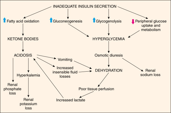
Hyperglycemia occurs from relative insulinopenia for the degree of transient insulin resistance plus an increase in counterregulatory hormones, which leads to increased hepatic gluconeogenesis and glycogenolysis. The resulting lipolysis and fatty acid oxidation produce ketonemia and metabolic acidosis. Both hyperglycemia and ketonemia result in an osmotic diuresis, which can lead to hypovolemia and subsequent decline in renal function. The pathophysiology of diabetic ketoacidosis is illustrated in Fig. E1.

DKA can be precipitated by various conditions:

  • Infection (commonly of the respiratory tract, such as COVID-19, or the urinary tract or skin)
  • Insulin deficiency (undiagnosed diabetes, medication nonadherence/inadequacy, insulin pump malfunction/disconnect, diabulimia)
  • Inflammatory conditions (e.g., acute pancreatitis)
  • Ischemia/infarction (e.g., myocardial infarction, stroke, bowel ischemia)
  • Severe extracellular fluid volume depletion
  • Drugs (e.g., steroids; thiazides; atypical antipsychotics; SGLT2 inhibitors; alcohol; sympathomimetics, including cocaine; and cancer treatment involving immune checkpoint inhibitors)

Figure E1 Pathophysiology of diabetic ketoacidosis.

From Marcdante KJ et al: Nelson Essentials of Pediatrics, ed 9, Philadelphia, 2023, Elsevier.

Diagnosis

Differential Diagnosis4

  • Hyperosmolar nonketotic state
  • Alcoholic/starvation ketoacidosis
  • Lactic acidosis
  • Acute kidney injury/chronic kidney disease
  • Metabolic acidosis caused by exogenous poisons (e.g., methanol, ethylene glycol, paraldehyde)
  • Salicylate poisoning
  • Hypovolemic or septic shock
Workup

After initial history is obtained, perform physical examination, including evaluation of airway, breathing, circulation, mental status, volume status, and signs suggestive of precipitating event(s).

Laboratory Tests

  • Serum glucose level: Generally >250 mg/dl. However, “euglycemic DKA” (EDKA) presenting with glucose <200 to 250 can occur in 10% of patients with DKA (e.g., exogenous insulin injection en route to hospital, food restriction, administration of an SGLT inhibitor [SGLTi],5 pregnancy, prolonged fasting, h/o bariatric surgery, gastroparesis, insulin pump failure, cocaine intoxication, chronic liver disease, and glycogen storage disease).
  • Arterial blood gas (demonstrating metabolic acidosis): Arterial pH <7.30 (<7.00 in severe cases).
  • Serum beta hydroxybutyrate and urine ketones: Positive (beta hydroxybutyrate >3 mmol/L; 2+ urine ketones).
  • Serum electrolytes:
    1. Serum bicarbonate concentration: <15 mmol/L (mild DKA may reduce levels to <18 mmol/L).
    2. Serum potassium concentration: Levels may initially measure as normal or high from extracellular shift with insulin deficiency and hyperosmolality. However, overall total body potassium depletion occurs from urinary losses and vomiting.
    3. Serum sodium concentration: May be low, normal, or high. Hyperglycemia increases plasma osmolality, which attracts intracellular water to the extracellular compartment and decreases the serum sodium level.6 Correct the serum sodium concentration: Add 1.6 mmol/L to the measured serum sodium level for every 100 mg/dl increase of serum glucose >100 mg/dl and <400 mg/dl, and then increase the sodium level by 4 mmol/L for each glucose increment of 100 mg/dl above 400 mg/dl.
    4. Serum calcium, magnesium, and phosphorus: May be significantly low and may decrease further with DKA treatment.
    5. Anion gap: Na – (Cl + HCO3). Anion gap is increased (>10 mmol/L) from elevated ketones.
    6. Blood urea nitrogen (BUN) and creatinine: Generally, reveals acute kidney injury.
  • Hemoglobin A1c if not performed within the preceding 3 mo.
  • CBC with differential: May indicate underlying infection (leukocytosis >25,000/mm3),4 inflammatory condition, or hemoconcentration. A leukocytosis of 10,000 to 15,000/mm3 is expected from the stress of illness alone.7
  • Urinalysis, urine/blood cultures: As indicated based on exam findings.
  • Pregnancy test: Perform in all female patients of reproductive age. DKA in pregnancy bodes significant fetal morbidity and mortality.8
  • Lipase/liver enzymes: Obtain if abdominal pain is present. Elevated lipase can occur without underlying pancreatitis.
Imaging Studies

ECG, chest x-ray examination, and other imaging studies as indicated to evaluate the precipitating cause(s).

Treatment

Nonpharmacologic Therapy

  • Monitor mental status, vital signs, and urine output hourly until improved.
  • Monitor serum glucose hourly and serum electrolytes, BUN, and creatinine every 2 to 4 h until DKA resolves.
Acute General Rx (Fig. 24

Figure 2 Management of diabetic ketoacidosis.

!!flowchart!!

BUN, Blood urea nitrogen; DKA, diabetic ketoacidosis; ECG, electrocardiogram; IV, intravenous; Rx, prescription; SC, subcutaneous.

From Nyenwe EA et al: The evolution of diabetic ketoacidosis: an update of its etiology, pathogenesis, and management, Metabolism 65[4]:507-521, 2016.

Fluid Replacement

Fluid therapy is initiated to expand volume and restore renal perfusion for a typical total body water deficit of 8 to 10 L. In the absence of cardiac compromise or severe kidney impairment, infuse 0.9% normal saline (NS) at an initial rate of 1 to 1.5 L/h (alternatively, use 15 to 20 ml/kg per hour) for the first 1 to 2 h. The subsequent fluid choice depends on patient hemodynamics, electrolytes, and urinary output. If corrected serum sodium is normal or high, infuse 0.45% NS at 250 to 500 ml/h. If corrected serum sodium is low, continue 0.9% NS at a similar rate. Once serum glucose decreases to 200 mg/dl, add 5% dextrose to the intravenous (IV) fluid. The recommended sodium decline is 0.5 mmol/L per hour and should not surpass 10 to 12 mmol/L per day. Hyperglycemia (>250 mg/dl) resolves sooner than ketoacidosis (6 vs. 12 h, respectively).

A meta-analysis of three randomized trials8a comparing normal saline to balanced electrolyte solutions (e.g., lactated Ringer's solution) in adults hospitalized with DKA showed that patients who received balanced solutions had shorter time to DKA resolution by 3 hours. Additional trials with larger patient base should help determine if current guidelines for fluid resuscitation in DKA should include balanced crystalloid solutions.

Insulin Administration

Insulin should not be started until after initiation of IV fluid resuscitation and correction of hypokalemia. Once these are addressed, administer initial bolus of IV regular insulin 0.1 units/kg followed by 0.1 units/kg per hour infusion or a continuous infusion of 0.14 units/kg per hour without initial bolus. If the serum glucose declines by less than 50 to 75 mg/dl in the first hour, increase insulin infusion rate hourly until a steady glucose decline is seen. After the serum glucose reaches 200 mg/dl and until DKA resolves, maintain the serum glucose concentration between 150 and 200 mg/dl by decreasing the insulin infusion to 0.02 to 0.05 units/kg per hour, or deliver subcutaneous rapid-acting insulin at 0.1 units/kg every 2 h. An alternative to IV insulin includes use of subcutaneous rapid-acting insulin for those presenting with mild-to-moderate DKA. Potential candidates for subcutaneous insulin include those patients who are alert, do not require admission to a critical care area, have a pH >7.0, are able to tolerate oral intake, and have a bicarbonate level of at least 10 mEq/L. Subcutaneous rapid-acting insulin can be given as an initial bolus of 0.3 units/kg, followed by maintenance doses of 0.2 units/kg every 2 h. Once blood glucose is <250 mg/dl, administer 0.05 to 0.1 units/kg every 2 h until DKA resolves. If anion gap is not closed within 12 h, the patient should be switched to an IV insulin infusion.

Potassium Replacement

Insulin therapy shifts potassium intracellularly, frequently causing hypokalemia. If the serum potassium concentration at presentation is between 3.3 and 5.2 mmol/L, infuse 20 to 30 mmol of potassium chloride (KCl) with each liter of IV fluid to maintain serum potassium at 4 to 5 mmol/L. If serum potassium is <3.3 mmol/L, withhold insulin until the serum potassium level is >3.3 mmol/L, and replace potassium by administering KCl infusion at 20 to 30 mmol/h. If the serum potassium level at presentation is >5.2 mmol/L, monitor the level every 2 h without replacement.

Bicarbonate Replacement

The administration of bicarbonate in DKA is generally not recommended. Bicarbonate does not improve time to resolution of acidosis or discharge, although adverse effects are associated with severe metabolic acidosis (decreased cardiac contractility, cerebral vasodilation). In adult patients with pH <6.9, administer 100 mmol (2 ampules) of sodium bicarbonate in 800 ml of sterile water (isotonic solution) with 20 mmol KCl at 200 ml/h for 2 h until venous pH is >7.0. If pH is still <7 after infusion, repeat infusion every 2 h until pH is >7.

Phosphate Replacement

Phosphate replacement is not routinely recommended, although phosphorus concentrations decrease with insulin administration. In patients with cardiac dysfunction, respiratory depression, or anemia and serum phosphorus <1 mg/dl, add 20 to 30 mmol/L of potassium phosphate to IV fluids to prevent diaphragmatic muscle weakness.

Transition to Subcutaneous Insulin

Resolution of DKA occurs when blood glucose is <200 mg/dl and two of the following occur: Venous pH >7.3, serum bicarbonate >15 mmol/L, and anion gap 12 mmol/L. At this point, patients can transition to subcutaneous insulin but may remain on IV insulin if they have had nothing by mouth. Subcutaneous intermediate- or long-acting insulin should be overlapped with IV insulin by 2 to 4 h to maintain adequate insulin levels and prevent rebound hyperglycemia. When this transition occurs before a meal, the patient may receive a dose of prandial insulin with short- or rapid-acting analogues together with the basal insulin, and the IV insulin may be discontinued in an hour. In patients with a known history of controlled diabetes, their home insulin regimens may be resumed. In patients with known poorly controlled diabetes, the subcutaneous insulin dose can be determined based on stable insulin drip requirements. Insulin-naïve patients may be started on basal-bolus insulin therapy by calculation of total daily dose of 0.5 to 0.8 units/kg (split as half-basal and half-bolus; administer one third of total bolus for each meal) or by stable insulin drip requirements. Further subcutaneous insulin dose titration is based on blood glucose results. Resolution of glucotoxicity and the inciting condition(s) will decrease insulin requirements.

Disposition

In general, patients with DKA should be admitted to an intensive care unit with an insulin infusion. Those with mild DKA may be treated with rapid-acting insulin analogues under observation and then discharged. Timely follow-up with primary care or endocrinology is important, preferably with an appointment made before discharge, as over 40% of patients may be readmitted within 2 wk of hospital discharge.

Pearls & Considerations

Comments

  • DKA is the initial presentation of diabetes in 15% to 20% of adults and 30% to 40% of children. This underscores the importance of educating patients, families, and school administrators regarding early symptoms of diabetes with the aim of early diagnosis.
  • Ketosis-prone diabetes, also referred to as Flatbush diabetes, is found more often in certain ethnic populations, including African American and Asian patients, among others.4,5 Initial therapy with insulin is required for acute management; however, diet alone or oral hypoglycemic agents can be used to achieve glycemic control without the need of insulin.
  • The development of EDKA is characterized by normal or modestly elevated blood sugars with anion gap metabolic acidosis, ketonemia, and ketonuria.9 Management is similar to DKA management. Mainstays of treatment involve correction of electrolytes and dehydration with IV fluids. However, higher percentages of dextrose (10% or 20%) are used to allow for concomitant high-dose insulin required to correct the acidosis.5 If the patient is taking an SGLTi, stop immediately. Patients may resume SGLTi therapy only after DKA has resolved and if the patient is feeling well and discusses their condition with their primary outpatient managing physician.
  • DKA in patients with end-stage renal disease (ESRD) is relatively uncommon secondary to decreased insulin clearance. In fact, low glomerular filtration rate increases the risk for hypoglycemia. However, when patients with ESRD develop DKA, special management considerations are needed.7,10 These patients are usually anuric and do not produce an osmotic diuresis, negating the need for aggressive volume repletion. Vigorous volume resuscitation in this case may lead an to volume overload, pulmonary edema, and respiratory distress. Anuric patients with ESRD do not have urinary potassium loss, thus making potassium replacement unnecessary. Similarly, phosphorus supplementation is not required. In fact, in severe acidosis, transcellular potassium shifts for hydrogen may produce hyperkalemia, prompting some providers to request immediate dialysis. Insulin therapy alone for hyperglycemia may also correct hyperkalemia without the need for dialysis. During DKA in patients with ESRD, dialysis is generally only recommended if there are hyperkalemia-induced electrocardiographic manifestations. A precipitous drop in serum glucose from an insulin infusion and hemodialysis may result in rapid tonicity shifts, predisposing to cerebral edema. Thus, hemodialysis is typically delayed until the serum glucose is corrected. Due to increased risk for hypoglycemia, patients with ESRD require careful and preemptive insulin reduction from the hospital DKA protocol.
  • Address affordability of insulin before discharge. Consider use of neutral protamine Hagedorn (NPH) insulin and regular or aspart insulin for as little as $25 per vial/pen (ReliOn™).
  • Proper instructions for hypoglycemia prevention and management, insulin storage, dosing, meal timing, preparation before injection (e.g., resuspension of NPH), and use of delivery system (syringe/vial, pen, insulin pump) are essential.
Prevention

Many cases of DKA can be prevented by effective patient education and communication. Education of patients regarding sick day management includes early communication with the health care provider, continuing insulin during illness, checking ketones, and continuing an easily digestible liquid diet that contains carbohydrates.

Related Content

Diabetes Mellitus (Related Key Topic)

Hyperglycemic Hyperosmolar Syndrome (Related Key Topic)

Related Content

    1. French E.K. : Diabetic ketoacidosis and hyperosmolar hyperglycemic syndrome: review of acute decompensated diabetes in adult patientsBMJ. ;365, 2019.
    2. McCoy R.G. : Sociodemographic, clinical, and treatment-related factors associated with hyperglycemic crises among adults with type 1 and type 2 diabetes in the United States, 2014-2020JAMA Netw Open1. ;4(9), 2021.
    3. Sehgal V., Ulmer B. : Clinical conundrums in the management of diabetic ketoacidosis in the elderlyJ Transl Int Med. ;7(1):10-14, 2019.
    4. Nyenwe E.A., Kitabchi A.E. : The evolution of diabetic ketoacidosis: an update of its etiology, pathogenesis, and managementMetabolism. ;65(4):507-521, 2016.
    5. Dhatariya K.K. : Defining and characterising diabetic ketoacidosis in adultsDiabetes Res Clin Pract. ;155, 2019.
    6. Verbalis J.G. : Diagnosis, evaluation, and treatment of hyponatremia: expert panel recommendationsAm J Med. ;126(10):S1-S42, 2013.
    7. Seddik A.A. : Challenges in management of diabetic ketoacidosis in hemodialysis patients, case presentation and review of the literatureDiabetes Metab Syndr: Clin Res Rev. ;13:2481-2487, 2019.
    8. Muneer M, Akbar I: Acute metabolic emergencies in diabetes: DKA, HHS and EDKA, Adv Exp Med Biol 85-114, 2020.
    9. Catahay J.A. : Balanced electrolyte solutions versus isotonic saline in adult patients with diabetic ketoacidosis: a systematic review and meta-analysisHeart Lung. ;54:74-79, 2022.
    10. Danne T. : International consensus on risk management of diabetic ketoacidosis in patients with type 1 diabetes treated with sodium-glucose cotransporter (SGLT) inhibitorsDiabetes Care. ;42(6):1147-1154, 2019.
    11. Schaapveld-Davis C.M. : End-stage renal disease increases rates of adverse glucose events when treating diabetic ketoacidosis or hyperosmolar hyperglycemic stateClin Diabetes. ;35(4):202-208, 2017.