section name header

Basic Information

AUTHORS: Frank B. D’ Alessandro, MD, and Fred F. Ferri, MD

Definition

  • Diabetes mellitus (DM) refers to a syndrome of hyperglycemia resulting from many different causes (see “Etiology”). It is broadly classified into type 1 (T1DM) and type 2 DM (T2DM). The terms insulin-dependent and non-insulin-dependent diabetes are obsolete because when a person with type 2 diabetes needs insulin, he or she remains labeled as type 2 and is not reclassified as type 1. Immune-mediated type 1 DM (type 1A) represents 5% to 10% of newly diagnosed diabetics. Tables 1 and 2 provide a general comparison of the two types of DM. One difference is that type 1 has usually complete or near-total knockout of insulin reserves mediated solely by immunogenic responses from carriers of certain genotypes, whereas type 2 is of polygenetic origin and may have patients who may start with hyperinsulinemia but have insulin resistance and through environmental factors such as diet and sedentary lifestyle leads to an imbalance between glucagon and insulin levels, resulting in combination of causes toward hyperglycemia.
  • Some type 1 diabetics also may exhibit high levels of glucagon, and not all type 1 diabetics have complete islet cell destruction.
  • The classification of diabetes also includes:
    1. LADA: Latent autoimmune diabetes of adult onset (sometimes called type 1.5 DM). These individuals are typically not insulin dependent initially and are often misclassified as having type 2 DM.
    2. MODY: Maturity onset diabetes of youth. These have various genetic expressions and can be classified into various subtypes:
      1. MODY 1, 2, 3, 4, and 5 (with 3 being most prevalent: 70% incidence with HNF-1-alpha [12q24] genetic expression).
      2. MODY 7 and 8 (rare).
    3. Ketosis-prone diabetes: Relapsing/remitting beta cell function with slow deterioration over time. It presents with ketoacidosis requiring insulin, then regains beta cell function, and patient is able to discontinue insulin. This for is most common under age 40, in those of African or Afro-Caribbean origin, and in obese or overweight patients.
    4. Secondary diabetes:
      1. Pancreatic disease or resection (e.g., cystic fibrosis).
      2. Chronic excessive corticosteroid exposure or Cushing syndrome.
      3. Glucagonoma.
      4. Acromegaly.
      5. Other rare genetic disorders (e.g., mitochondrial diabetes MELAS syndrome).
    5. Rare autoimmune (e.g., type A and B insulin resistance syndrome).
    6. A classification of diabetes mellitus is shown in Box 1.
  • Diabetes mellitus can be diagnosed by the following tests:
    1. A hemoglobin A1c (HbA1c) value 6.5% is considered diagnostic for diabetes. This test is preferred because of ease of administration and reliability.
    2. A fasting plasma glucose (FPG) 126 mg/dl, which should be confirmed with repeat testing on a different day. Fasting is defined as no caloric intake for at least 8 h.
    3. An oral glucose tolerance test (OGTT) with a plasma glucose 200 mg/dl 2 h after a 75 g (100 g for pregnant women) glucose load.
    4. Symptoms of hyperglycemia and a casual (random) plasma glucose 200 mg/dl are also indicative of DM. Classic symptoms of hyperglycemia include polyuria, polydipsia, and unexplained weight loss. At the time of diagnosis as a diabetic, B-cell function is at 25% to 30%.
  • Individuals with glucose levels higher than normal but not high enough to meet the criteria for diagnosis of DM are considered to have “prediabetes,” the diagnosis of which is made as follows:
    1. A fasting plasma glucose 100 to 125 mg/dl; this is referred to as impaired fasting glucose.
    2. After OGTT, a 2-h plasma glucose of 140 to 199; this is referred to as impaired glucose tolerance. Patients with impaired glucose tolerance or prediabetes have B-cell function at 50% of normal.
    3. A hemoglobin A1c value of 5.7% to 6.4%.
  • Table 3 describes diagnostic categories for DM and at-risk states.

TABLE 3 Diagnostic Categories: Diabetes Mellitus and At-Risk States

Fasting Plasma Glucose Level2-hr (75-g) OGTT Result
<140 mg/dl140-199 mg/dl200 mg/dl
<100 mg/dlNormalIGTDM
100-125 mg/dlIFGIGT and IFGDM
126 mg/dlDMDMDM
HbA1CLevel<5.7%5.7-6.4%6.5%
NormalHigh-riskDM

DM, Diabetes mellitus; HbA1c, glycosylated hemoglobin; IFG, impaired fasting glucose; IGT, impaired glucose tolerance; OGTT, oral glucose tolerance test.

These diagnostic categories are based on the combined fasting plasma glucose level and a 2-h, 75-g oral glucose tolerance test (OGTT) result. Note that a confirmed random plasma glucose level of 200 mg/dl or higher in the appropriate clinical setting is diagnostic of diabetes and precludes the need for further testing.

May be referred to as prediabetes.

From Goldman L, Schafer AI: Goldman’s Cecil medicine, ed 24, Philadelphia, 2012, Saunders.

Box 1 Classification of Diabetes Mellitus

  1. Type 1 diabetes (beta cell destruction, usually leading to absolute insulin deficiency)
    1. Immune mediated
    2. Idiopathic
  2. Type 2 diabetes (may range from predominantly insulin resistance with relative insulin deficiency to a predominantly secretory defect with insulin resistance)
  3. Other specific types
    1. Genetic defects of beta cell function
      1. Chromosome 12, HNF-1α (MODY3)
      2. Chromosome 7, glucokinase (MODY2)
      3. Chromosome 20, HNF-4α (MODY1)
      4. Chromosome 13, insulin promoter factor-1 (IPF-1; MODY4)
      5. Chromosome 17, HNF-1β (MODY5)
      6. Chromosome 2, NeuroD1 (MODY6)
      7. Mitochondrial DNA
      8. Others
    2. Genetic defects in insulin action
      1. Type A insulin resistance
      2. Leprechaunism
      3. Rabson-Mendenhall syndrome
      4. Lipoatrophic diabetes
      5. Others
    3. Diseases of the exocrine pancreas
      1. Pancreatitis
      2. Trauma/pancreatectomy
      3. Neoplasia
      4. Cystic fibrosis
      5. Hemochromatosis
      6. Fibrocalculous pancreatopathy
      7. Others
    4. Endocrinopathies
      1. Acromegaly
      2. Cushing syndrome
      3. Glucagonoma
      4. Pheochromocytoma
      5. Hyperthyroidism
      6. Somatostatinoma
      7. Aldosteronoma
      8. Others
    5. Drug or chemical induced
      1. Vacor
      2. Pentamidine
      3. Nicotinic acid
      4. Glucocorticoids
      5. Thyroid hormone
      6. Diazoxide
      7. β-Adrenergic agonists
      8. Thiazides
      9. Dilantin
      10. γ-Interferon
      11. Others
    6. Infections
      1. Congenital rubella
      2. Cytomegalovirus
      3. Others
    7. Uncommon forms of immune-mediated diabetes
      1. “Stiff-man” syndrome
      2. Antiinsulin receptor antibodies
      3. Others
    8. Other genetic syndromes sometimes associated with diabetes
      1. Down syndrome
      2. Klinefelter syndrome
      3. Turner syndrome
      4. Wolfram syndrome
      5. Friedreich ataxia
      6. Huntington chorea
      7. Laurence-Moon-Biedl syndrome
      8. Myotonic dystrophy
      9. Porphyria
      10. Prader-Willi syndrome
      11. Others
  4. Gestational diabetes mellitus

From McPherson RA, Pincus MR: Henry’s clinical diagnosis and management by laboratory method, ed 23, St. Louis, 2017, Elsevier.

TABLE 1 Characteristic Comparison of Type 1 Versus Type 2 Diabetes Mellitus

CharacteristicType 1Type 2
Nature
Very different
Autoimmune disorder marked by destruction of insulin-producing beta cells and loss of insulin productionA disorder of insulin deficiency involving an interplay between both pancreatic and extrapancreatic contributions to disease
Symptoms
Partial overlap
Rapid onset; very high to extremely high blood glucose levels; polyphagia; polydipsia, polyuria; ketoacidosisMild to moderate onset; modest to high elevations in blood glucose; mild polydipsia/polyuria; fatigue; visual changes/headache
Onset
Very different
Sudden (symptoms for days to weeks)Slower onset (symptoms for mos to yrs)
Risk factors
Typically different but overlap
Family history of autoimmune disease but particularly type 1 diabetes mellitus (10-fold increased risk vs. general population)Overweight/obese; poor diet; sedentary lifestyle; ethnicity (higher in African Americans, Hispanics); family history of type 2 diabetes mellitus; history of gestational diabetes
Onset age
Typically different but overlap
Typically early life through adolescence but can occur at any ageTypically adults but trending toward earlier age of onset
Treatment strategy
Typically different
Absolute requirement for insulin (multiple daily injections or insulin pump); self-management lifestyle modification (monitor food types, exercise, etc.)Dietary modifications and exercise alongside oral agents (for most); increasingly greater percentage of patients require insulin over time
Can it be prevented?
Very different
Not at present (subject of major research efforts); future cases can be predicted by autoantibodies and geneticsYes, for more than half of potential cases, with dietary modifications and exercise
Can it be reversed?
Very different
Not at present (subject of major research efforts)No, but for a limited few; patients can see disease managed and risk for complications reduced through diet modifications, exercise; growing evidence for disease improvements through combination therapies
Complications
Mostly similar, but some variation
Acute emergencies of hypoglycemia and ketoacidosis leading to hypoglycemic unawareness; chronic effects of hyperglycemia can lead to retinopathy, nephropathy, neuropathy, cardiovascular disease, etc.Acute emergencies of hypoglycemia and ketoacidosis leading to hypoglycemic unawareness; chronic effects of hyperglycemia can lead to retinopathy, nephropathy, neuropathy, cardiovascular disease, etc.

From Melmed S et al: Williams textbook of endocrinology, ed 14, Philadelphia, 2020, Elsevier.

TABLE 2 Characteristics of Type 1 and Type 2 Diabetes Mellitus

Type 1 DiabetesType 2 Diabetes
Frequency5%-10%90%-95%
Age of onsetAny, but most common in children and young adultsMore common with advancing age, but can occur in children and adolescents
Risk factorsGenetic, autoimmune, environmentalGenetic, obesity, sedentary lifestyle, race/ethnicity, hypertension, dyslipidemia, polycystic ovarian syndrome
PathogenesisDestruction of pancreatic beta cells, usually autoimmuneNo autoimmunity
Insulin resistance and progressive insulin deficiency
C-peptide levelsVery low or undetectableDetectable
PrediabetesAutoantibodies (GAD65, IA-2, IAA, ZnT8) may be presentAutoantibodies absent
Medication therapyInsulin absolutely necessary; multiple daily injections or insulin pumpOral agents and/or noninsulin injectable hypoglycemic drugs
Insulin commonly needed
Therapy to prevent or delay onset of diabetesTeplizumabLifestyle (weight loss and increased physical activity)
Oral medications (metformin, acarbose) may be helpful.

Modified from McPherson RA, Pincus MR: Henry’s clinical diagnosis and management by laboratory method, ed 23, St. Louis, 2017, Elsevier.

Synonyms

IDDM (insulin-dependent diabetes mellitus)

NIDDM (non-insulin-dependent diabetes mellitus)

Type 1 diabetes mellitus (insulin-dependent diabetes mellitus)

Type 2 diabetes mellitus (non-insulin-dependent diabetes mellitus)

LADA (latent autoimmune diabetes of adult)

MODY (mature onset diabetes of youth)

ICD-10CM CODES
E11.5Type 2 diabetes mellitus with peripheral circulatory complications
E11.7Type 2 diabetes mellitus with multiple complications
E11.8Type 2 diabetes mellitus with unspecified complications
E11.9Type 2 diabetes mellitus without complications
E10.69Type 1 diabetes mellitus with other specified complication
E10.8Type 1 diabetes mellitus with unspecified complications
E10.9Type 1 diabetes mellitus without complications
E11.69Type 2 diabetes mellitus with other specified complication
E11.8Type 2 diabetes mellitus with unspecified complications
E11.9Type 2 diabetes mellitus without complications
Epidemiology & Demographics

  • DM affects 9% to 10% of the U.S. population. Prevalence rates vary considerably by race/ethnicity. T1DM accounts for approximately 5% of diagnosed diabetes cases and is defined by the presence of one or more autoimmune markers.
  • In the U.S., the overall pediatric incidence of T1DM is approximately 25 per 100,000 per year. Incidence a varies according to age (peaking in the pubertal years, ages 10 to 14), season (winter more than summer), geographic location (Finland and other Nordic countries more than equatorial regions), and race and ethnic group (U.S. non-Hispanic White persons more than Native Americans).1
  • Risk factors for type 1 DM are summarized in Table 4.
  • There are three stages of type 1 DM. Patients in stage 1 have two or more diabetes-related autoantibodies, and those in stage 2 have developed asymptomatic dysglycemia but have normal other metabolic indices and do not require insulin treatment. Stage 3 is symptomatic clinical disease. Teplizumab is an anti-CD3 monoclonal antibody recently FDA approved to delay the onset of stage 3 type 1 DM in patients 8 years old who have stage 2 type 1 DM.1a
  • While incidence of diabetes in adolescents is mostly type 1, the rate of type 2 being diagnosed in adolescents has increased by 1.5 times in certain given areas. This seems to correlate with the epidemic of pediatric obesity.
  • Table 5 summarizes epidemiologic determinants of and risk factors for type 2 DM.
  • Incidence rate increases with age, varying from 2% in persons age 20 to 44 yr to 18% in persons 65 to 74 yr. T2DM can have a long presymptomatic phase, leading to a 4- to 7-yr delay in diagnosis. In the U.S. 1.2 million new cases of diabetes are diagnosed each yr, and 86 million have prediabetes. Currently, 30 million Americans have diabetes; with this current trend, it is predicted that by 2050, 1 out of 3 Americans will be diabetic.
  • Diabetes accounts for 8% of all legal blindness in the U.S. and is the leading cause of end-stage renal disease (ESRD). Approximately 40% of patients in a given dialysis center are diabetic.
  • Patients with diabetes are two to four times more likely than nondiabetic patients to experience development of cardiovascular disease (CVD).

TABLE 5 Epidemiologic Determinants of and Risk Factors for Type 2 Diabetes Mellitus

Genetic Factors
Genetic markers
Family history
Demographic Characteristics
Age
Ethnicity
Behavioral and Lifestyle-Related Risk Factors
Obesity (including distribution of obesity and duration)
Physical inactivity
Diet
Stress
Westernization, urbanization, modernization
Medications
Shift work
Metabolic Determinants and Intermediate-Risk Categories of Type 2 Diabetes
Impaired glucose tolerance
Insulin resistance
Gestational diabetes
Offspring of women with diabetes during pregnancy
Intrauterine malnutrition or overnutrition
Microbiome composition

From Melmed S et al: Williams textbook of endocrinology, ed 14, Philadelphia, 2020, Elsevier.

TABLE 4 Risk of Type 1 Diabetes Mellitus

GroupChildhood Annual Incidence
U.S. general population0.3% (15-25/100,000)
Offspring1%
Sibling3.2% (through adolescence); 6% lifetime
Dizygotic twin6%
Mother2%
Father4.6%
Both parents10%
Monozygotic twin50%, but incidence varies with age of index twin

From Melmed S et al: Williams textbook of endocrinology, ed 14, Philadelphia, 2020, Elsevier.

Physical Findings & Clinical Presentation

  • Physical examination varies with the presence of complications and may be normal in early stages
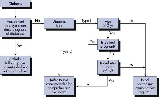
  • Diabetic retinopathy:
    1. Nonproliferative (background diabetic retinopathy):
      1. Initially: Microaneurysms, capillary dilation, waxy or hard exudates, dot and flame hemorrhages, arteriovenous shunts
      2. Advanced stage: Microinfarcts with cotton wool exudates, macular edema
    2. Proliferative retinopathy: Characterized by formation of new vessels, vitreous hemorrhages, fibrous scarring, and retinal detachment
  • Cataracts and glaucoma occur with increased frequency in patients with diabetes
  • Diabetic macular edema: Swelling of the macula leading to loss of sharp vision in this part of the eye. People with this disorder usually already have retinopathy but may present separately as well
  • Diabetic neuropathy:
    1. Distal sensorimotor polyneuropathy:
      1. Symptoms include paresthesia, hyperesthesia, or burning pain involving bilateral distal extremities in a “stocking glove” distribution. This can progress to motor weakness and ataxia
      2. Physical examination may reveal decreased pinprick sensation, decreased sensation to light touch, decreased vibration sense, and loss of proprioception. Motor disturbances such as decreased deep tendon reflexes and atrophy of interosseous muscles can also be seen
    2. Autonomic neuropathy:
      1. GI disturbances: Esophageal motility abnormalities, gastroparesis, diarrhea (usually nocturnal):
        1. Increased gastric emptying seen in type 2
        2. Decreased gastric emptying seen in type 1
      2. Genitourinary (GU) disturbances: Neurogenic bladder (hesitancy, weak stream, and dribbling), impotence
      3. Cardiovascular (CV) disturbances: Orthostatic hypotension, tachycardia, decreased heart rate variability (HRV). Decreased HRV is associated with increased cardiac mortality, independent of ejection fraction
    3. Polyradiculopathy: Painful weakness and atrophy in the distribution of 1 contiguous nerve root
    4. Mononeuropathy involving cranial nerves III, IV, or VI or peripheral nerves can also occur
  • Diabetic nephropathy: Pedal edema, pallor, weakness, uremic appearance
  • Nephrotic syndrome: Proteinuria, hypertriglyceridemia, edema
  • Nephritis: Progressive degeneration of nephrons focal glomerulosclerosis

Type IV renal tubular acidosis, hyperkalemic nephropathy, interstitial nephritis, causing hyporeninemic hypoaldosteronism. It is important to keep in mind that NSAIDs, ACE inhibitors, trimethoprim, and heparin can all reduce aldosterone and can cause or exacerbate the condition. 50% of patients on dialysis have diabetes as primary diagnosis; whereas glycemia is important to prevent nephropathy, the most significant contributing factor is hypertension.

  • Foot ulcers: Occur in 15% of individuals with diabetes (annual incidence rate 2%) and are the leading causes of hospitalization; they are usually secondary to a combination of factors, including peripheral vascular insufficiency, repeated trauma (unrecognized because of sensory loss), and superimposed infection.
    1. Patient symptoms are usually less than would be expected from clinical findings, due to loss of sensation related to peripheral neuropathy.
    2. Comprehensive foot exams include visual inspection, assessment of pedal pulses, and assessment of protective sensation using a 10-g monofilament to test sensation.
    3. Prevention of foot ulcers in an individual with diabetes includes strict glucose control, patient education, prescription footwear, intensive podiatric care, and evaluation for surgical interventions.
    4. Foot examination should be done annually on all diabetics.
  • Neuropathic arthropathy (Charcot joints): Bone or joint deformities from repeated trauma (secondary to peripheral neuropathy; Fig. E1).
  • Necrobiosis lipoidica diabeticorum: Plaque-like reddened areas with a central area that fades to white-yellow, found on the anterior surfaces of the legs (Fig. E2); in these areas, the skin becomes very thin and can ulcerate easily.
  • Other diabetic skin manifestations with diabetes include:
    1. Scleroderma diabeticorum: Thickening of skin and epidermis giving skin a “leather-like texture”; typically affects type 2, mainly on upper back and neck.
    2. Dermatitis herpetiformis: Diffuse petechial rash associated with gluten insensitivity.
    3. Vitiligo: Associated with type 1 and type 2 and affects skin coloration due to autoimmune reaction to pigmentation (at times may be associated with adrenal insufficiency, especially in type 1 patients). Patients should use SPF 30 sunscreen to prevent sunburn.
    4. Acanthosis nigricans: Darkening of skin folds in neck and axilla, at times raised and velvety, thought to be associated with insulin resistance; other conditions such as Cushing and acromegaly can have this as well.
    5. Diabetic dermopathy: Appears as shiny round or oval lesions on thin skin of the lower extremity, also known as “shin spots”; these are usually not painful and do not require treatment.
    6. Eruptive xanthomatosis: Associated with uncontrolled blood sugars and extremely high triglycerides. There is a high risk for pancreatitis in patients with this finding; treatment is aimed at lowering blood sugars with insulin and fibrates.
    7. Digital sclerosis: Skin on toes fingers and hands become waxy, thick, and tight, with joint stiffness; treatment is aimed at lowering blood sugars and encouraging use of lotions and moisturizers.
    8. Bullosis diabeticorum: Rare disorder manifesting with blisters on hands, forearms, toes, and feet, usually painless. Lesions generally heal on their own.
    9. Box 2 summarizes cutaneous associations with diabetes mellitus.

Box 2 Some Cutaneous Associations With Diabetes Mellitus

Established or Probable Association

  • Necrobiosis lipoidica (NL) diabeticorum
  • Diabetic dermopathy
  • Diabetic bullae
  • Acanthosis nigricans
  • Acrochordons (skin tags)
  • Scleroderma diabeticorum
  • Limited joint mobility and waxy skin syndrome
  • Partial lipodystrophy
  • Malignant otitis externa
  • Neuropathic leg ulcers
  • Perforating disorders
  • Eruptive xanthomas
  • Hemochromatosis
  • Carotenemia
  • Pruritus
  • Xerosis and anhidrosis
  • Yellow nails, koilonychia
  • Increased susceptibility to infections:
  • Candida albicans
  • Staphylococcus aureus
  • Group A β-hemolytic streptococcus
  • Pseudomonas aeruginosa
  • Dermatophytes (tinea pedis, onychomycosis)
  • Corynebacteriumminutissimum (erythrasma)
Possible Association

  • Disseminated granuloma annulare
  • Vitiligo

From Paller AS, Mancini AJ: Hurwitz clinical pediatric dermatology: a textbook of skin disorders of childhood and adolescence, ed 5, Philadelphia, 2016, Elsevier.

Figure E1 Diabetic neuropathy of the hindfoot.

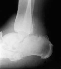
Destruction of the joint with collapse and fragmentation.

From Hochberg MC et al [eds]: Rheumatology, ed 3, St Louis, 2003, Mosby.

Figure E2 Necrobiosis lipoidica diabeticorum.

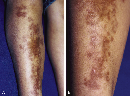
Waxy, yellow-brown plaques (A) were present in this young girl with diabetes mellitus, with subtle atrophy present on closer inspection (B).

From Paller AS, Mancini AJ: Hurwitz clinical pediatric dermatology, a textbook of skin disorders of childhood and adolescence, ed 5, Philadelphia, 2016, Elsevier.

Etiology
Idiopathic Diabetes

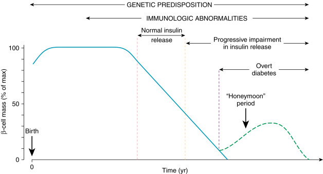
Type 1 DM: Results from autoimmune β-cell destruction, usually leading to absolute insulin deficiency (Fig. 3).

  • Hereditary factors:
    1. Islet cell antibodies (found in 90% of patients within the first yr of diagnosis)
    2. Higher incidence of human leukocyte antigen (HLA) types DR3, DR4
    3. 50% concordance rate in identical twins
  • Environmental factors: Viral infection (possibly coxsackievirus, mumps virus)
Figure 3 Schematic Representation of the Autoimmune Evolution of Diabetes in Genetically Predisposed Individuals

From Marcdante KJ et al: Nelson Essentials of Pediatrics, ed 9, Philadelphia, 2023, Elsevier.

Type 2 DM: Results from insulin resistance and a progressive defect in insulin secretion.

  • Hereditary factors: 90% concordance rate in identical twins
  • Environmental factors: Obesity, sedentary lifestyle, high carbohydrate content in food
Diabetes Secondary to Other Factors

  • Hormonal excess: Cushing syndrome, acromegaly, glucagonoma, pheochromocytoma
  • Drugs: Glucocorticoids, diuretics, oral contraceptives
  • Insulin receptor unavailability (with or without circulating antibodies)
  • Pancreatic disease: Pancreatitis, pancreatectomy, hemochromatosis, cystic fibrosis
  • Genetic syndromes: Maturity onset diabetes of the young (MODY, monogenetic diabetes accounting for 2% to 5% of diabetes), familial hyperlipidemias, myotonic dystrophy, lipoatrophy
  • Gestational diabetes (GDM): Diabetes diagnosed during pregnancy that is due to pregnancy-related insulin resistance

Diagnosis

Differential Diagnosis

  • Diabetes insipidus
  • Stress hyperglycemia
  • Diabetes secondary to hormonal excess, drugs, pancreatic disease
Laboratory Tests

  • Diagnosis of DM is made on the basis of the following tests:
    1. Fasting glucose 126 mg/dl on two occasions.
    2. Non-FPG 200 mg/dl and symptoms of DM.
    3. OGTT (75 g glucose load for nonpregnant individuals) with 2-h value >200 mg/dl.
    4. Glycosylated hemoglobin (HbA1c) 6.5%. HbA1c level reflects average glycemia over previous 3 mo or longer. In known diabetics, this test should be performed at least twice yearly in stable patients and more frequently when therapy changes or patients are not meeting glycemic goals. HbA1c alone does not provide a measure of glycemic variability or hypoglycemia and is affected by the presence of hemoglobin variants, hemolysis, or blood loss.
  • Measurement of autoantibodies glutamic acid decarboxylase (GAD65) and tyrosine phosphatase IA-2 are useful in suspected immune mediated type 1 DM (strong association).
  • Screening for prediabetes and diabetes in asymptomatic patients (see Table 6):
    1. Should be considered in adults of any age who are overweight (body mass index [BMI] >25 kg/m2) or obese (BMI >30) and who have one or more additional risk factors for diabetes.
    2. In those who are without these risk factors, testing should begin at age 45 yr.
    3. If screen is normal, repeat testing should be carried out at least at 3-yr intervals.
    4. ADA recommends screening all Asian Americans with BMI 23 or higher every 2 yr.
  • Detection and diagnosis of gestational diabetes mellitus (GDM):
    1. Screen for GDM using risk factor analysis and use of an OGTT. Pregnant women who are not known to have diabetes should be screened for gestational diabetes at 24 to 28 wks’ gestation with a “1-step” strategy with 75 g oral glucose tolerance test or a “2-step” approach with a 50-g (nonfasting) screen followed by a 100-g oral glucose tolerance test for those who screen positive. A diagnosis of GDM is made if any of the following levels of plasma glucose are exceeded: 92 mg/dl (5.1 mmol/L) when fasting, 80 mg/dl (10 mmol/L) at 1 h, or 153 mg/dl (8.5 mmol/L) at 2 h.
    2. Women with GDM should be screened for diabetes 6 to 12 wk postpartum and should be followed with subsequent screening for the development of diabetes or prediabetes at least every 3 yr. A woman who had GDM during pregnancy has 50% risk of developing diabetes later in life; this is dependent on ethnicity (e.g., Pima Indians, Hispanic, African American).
  • Screening for diabetic nephropathy:
    1. Screening should be done at diagnosis and then yearly for type 2 diabetes and 5 yr after diagnosis, then yearly in type 1 diabetes. 18% of type 1 DM may have early kidney changes, and future guidelines may change screening to 1 yr after diagnosis if poorly controlled, then annually for both type 1 and type 2.
    2. Screening can be performed using an albumin:creatinine ratio (microalbumin) in a random spot urine collection or by measurement of a 24-h urine collection for albumin and creatinine clearance. The urine albumin to creatinine ratio (ACR) is independently associated with mortality at all levels of estimated glomerular filtration rate (eGFR) in older adults with diabetes.
    3. The diagnosis of microalbuminuria (ACR 30 to 299 mg/24 h) should be based on 2 to 3 elevated levels within a 3- to 6-mo period because there is a marked variability in day-to-day albumin excretion. Patients with overt macroalbuminuria (>300 mg albumin/24 h or albumin:creatinine ratio >300) should be followed by urine protein:creatinine ratio.
  • A fasting serum lipid panel, serum creatinine, and electrolytes should be obtained yearly on all adult patients with diabetes.
  • Self-monitoring of blood glucose (SMBG) is crucial for assessing the effectiveness of the management plan. The frequency and timing of SMBG varies with the needs and goals of each patient. In most patients with type 1 DM and pregnant women taking insulin, SMBG is recommended at least 3 times/day. In patients with type 2 DM not on insulin, recommendations are unclear for SMBG, but testing once or twice/day is acceptable in most patients. Glycemic control is best evaluated when SMBG is combined with HbA1c testing.
  • Continuous glucose monitoring (CGM; Fig. E4) is now starting to play a more prominent role in reducing the number of fingersticks. Dexcom CGM has been approved by FDA for use by patients to adjust insulin based on CGM readings. Randomized trials in persons with type 1 DM and high glycated hemoglobin levels have shown that the use of intermittently scanned CGMs with optional alarms for high and low blood glucose levels results in significantly lower glycated hemoglobin levels than levels monitored by fingerstick testing.1b
  • The use of insulin pump and CGM readings and computer software can adjust insulin automatically (known as the “artificial pancreas”).
  • CGM can also be used in patients with type 2 on multiple daily injections.
  • Limitations of CGM are that it cannot predict accuracy of blood glucose when <70 mg/dl.
  • Screening for thyroid dysfunction (TSH level), vitamin B12 deficiency, and celiac disease should be considered in type 1 diabetes due to the increased frequency of other autoimmune diseases in these individuals.
  • Consider screening for autoimmune polyendocrine syndromes (APS-2):
    1. APS-2: Most commonly known as Schmidt syndrome, heterogeneous not linked to one gene (HDLA-DQ2, HDLA-DQ8, AND HLA-DR4). Patients can have IDDM, hyperthyroidism, other autoimmune conditions, B12 deficiency, and myasthenia.

TABLE 6 Criteria for Diabetes Screening in Asymptomatic Individuals

  1. Testing should be considered in all adults who are overweight (BMI >25 kg/m2) and have additional risk factors:
    1. Physical inactivity
    2. A first-degree relative with diabetes
    3. High-risk ethnic population (e.g., African American, Hispanic American, Native American, Asian American, Pacific Islander)
    4. Delivered a baby weighing more than 9 lb or diagnosed with gestational diabetes mellitus
    5. Systemic hypertension (blood pressure >140/90 mm Hg or on antihypertensive therapy)
    6. High-density lipoprotein cholesterol level <35 mg/dl or triglyceride level >250 mg/dl
    7. Polycystic ovary syndrome
    8. Hemoglobin A1c5.7%, impaired glucose tolerance or impaired fasting glucose on prior testing
    9. Other clinical conditions associated with insulin resistance (e.g., severe obesity, acanthosis nigricans)
    10. History of cardiovascular disease
  2. If none of the above criteria are present, screening for diabetes should begin at age 45 yr
  3. If the results are normal, screening should be repeated at least every 3 yr. Depending on initial results and risk status, more frequent testing may need to be considered

In some ethnic groups, such as Asians, at-risk body mass index (BMI) may be lower.

Modified from American Diabetes Association: Diagnosis and classification of diabetes mellitus, Diabetes Care 33(Suppl 1):S14, 2010; and from Goldman L, Schafer AI: Goldman’s Cecil medicine, ed 24, Philadelphia, 2012, Saunders.

Treatment

TABLE 8 American College of Cardiology Foundation/American Heart Association Recommendations for Primary Prevention of Cardiovascular Disease in People With Diabetes

Lifestyle Management
Weight
Structured programs that emphasize lifestyle changes such as reduced fat (<30%-35% of daily energy) and total energy intake and increased regular physical activity, along with regular participant contact, can produce long-term weight loss on the order of 5%-7% of starting weight, with improvement in BP.
For persons with elevated plasma triglycerides and reduced HDL-C, improved glycemic control, moderate weight loss (5%-7% of starting weight), dietary saturated fat restriction, increased physical activity, and modest replacement of dietary carbohydrate (5%-7%) by either monounsaturated or polyunsaturated fats may be beneficial.
Medical Nutrition Therapy
To achieve reductions in LDL-C:
Saturated fats should be less than 7% of energy intake.
Dietary cholesterol intake should be less than 200 mg/day. Intake of trans unsaturated fatty acids should be less than 1% of energy intake.
Total energy intake should be adjusted to achieve body weight goals.
Total dietary fat intake should be moderated (<30%-35% of total calories) and should consist mainly of monounsaturated or polyunsaturated fat.
Ample intake of dietary fiber (14 g/1000 calories consumed) may be of benefit.
If individuals choose to drink alcohol, daily intake should be limited to one drink for adult women and two drinks for adult men. One drink is defined as 12 ounces (oz) of beer, 4 oz of wine, or 1.5 oz of distilled spirits. Alcohol ingestion increases caloric intake and should be minimized when weight loss is the goal.
Individuals with elevated plasma triglyceride levels should limit intake of alcohol because it may exacerbate hypertriglyceridemia.
In both normotensive and hypertensive persons, a reduction in sodium intake may lower BP. The goal should be to reduce sodium intake to 1200-2300 mg/day (50-100 mmol/day), equivalent to 3000-6000 mg/day sodium chloride.
Physical Activity
To improve glycemic control, assist with weight loss or maintenance, and reduce risk for CVD, at least 150 min of moderate-intensity aerobic physical activity or at least 90 min of vigorous aerobic exercise per wk is recommended. The physical activity should be distributed over at least 3 days per wk, with no more than 2 consecutive days without physical activity.
For long-term maintenance of major weight loss, a larger amount of exercise (7 hr of moderate or vigorous aerobic physical activity per wk) may be helpful.
Blood Pressure
BP should be measured at every routine diabetes visit. Patients found to have SBP 130 mm Hg or DBP 80 mm Hg should have BP confirmed on a separate day.
Patients with diabetes should be treated to achieve SBP at least <140 mm Hg and DBP <90 mm Hg, and for patients who can tolerate without adverse symptoms, targets can be as low as SBP <130 mm Hg and DBP <80-85 mm Hg. Patients with SBP of 130-139 mm Hg or DBP of 80-89 mm Hg should initiate lifestyle modification alone (weight control, increased physical activity, alcohol moderation, sodium reduction, and emphasis on increased consumption of fresh fruits, vegetables, and low-fat dairy products) for a maximum of 3 mo. If, after these efforts, targets are not achieved, treatment with pharmacologic agents should be initiated.
Patients with hypertension (SBP 140 mm Hg or DBP 90 mm Hg) should receive drug therapy in addition to lifestyle and behavioral therapy.
All patients with diabetes and hypertension should be treated with a regimen that includes either an ACE inhibitor, or if intolerant to an ACE inhibitor, an ARB. If one class is not tolerated, the other should be substituted. Other drug classes demonstrated to reduce CVD events in patients with diabetes-dihydropyridine calcium channel blockers, thiazide diuretics (chlorthalidone and indapamide), and β-blockers-should be added, in listed order of preference, as needed to achieve BP targets.
If ACE inhibitors, ARBs, or diuretics are used, kidney function and serum potassium levels should be monitored within the first 3 mo. If BP is stable, follow-up could occur every 6 mo thereafter.
Multidrug therapy generally is required to achieve BP targets.
In elderly hypertensive patients, BP should be lowered gradually to avoid complications.
Orthostatic measurement of BP should be performed in people with diabetes and hypertension when clinically indicated.
Patients not achieving target BP despite multidrug therapy should be referred to a physician specializing in the care of patients with hypertension.
Lipids
In adult patients with diabetes, lipid levels should be measured at least annually and more often if needed to achieve goals. In adults with diabetes who are younger than 40 with low-risk lipid values (LDL-C <100 mg/dl, HDL-C >50 mg/dl, triglycerides <150 mg/dl), lipid assessments may be repeated every 2 yr.
Lifestyle modification deserves primary emphasis in all individuals with diabetes. Patients should focus on the reduction of saturated fat and cholesterol intake, weight loss (if indicated), and increases in dietary fiber and physical activity. These lifestyle changes have been shown to improve the lipid profile in patients with diabetes. In persons with diabetes who are older than 40 yr, without overt CVD, statin therapy should be considered for primary prevention with recommendation to use at least moderate-dose and ideally intense-dose statins, independent of baseline LDL-C levels. On maximally tolerated statin, the goal is an LDL-C level <100 mg/dl (2.6 mmol/L), and ideally <70 mg/dl (1.8 mmol/L) for those at highest CVD risk. If LDL-lowering drugs are used, a reduction of at least 50% in LDL-C levels should be obtained.
If baseline LDL-C is <100 mg/dl, statin therapy should be initiated based on risk factor assessment and clinical judgment. Major risk factors in this category include age, sex, race/ethnicity, cigarette smoking, hypertension (BP >140/90 mm Hg or use of antihypertensive medication), high total cholesterol and low HDL-C (<40 mg/dl), and family history of premature CHD (CHD in male first-degree relatives 55 yr of age; CHD in female first-degree relatives 65 yr of age).
In people with diabetes who are younger than 40, without overt CVD, but who are estimated to be at increased risk for CVD either by clinical judgment or by risk calculator, at least moderate-intensity statin therapy is recommended, with an LDL-C goal of <100 mg/dl.
Combination therapy with LDL-lowering drugs (e.g., statins, ezetimibe, PCSK9 inhibitors) and fibrates or niacin may be necessary to achieve lipid targets, but to date, only the addition of ezetimibe to statin therapy has proven incremental CV outcomes benefit.
Beyond the consensus of therapeutic lifestyle intervention, the ADA and AHA guidelines have evolved significantly over recent yrs, no longer recommending pharmacologic treatment of low HDL-C or high triglyceride levels, except for those with extremely high fasting triglyceride levels, to consider fish oil or a fibrate to mitigate pancreatitis risk.
Tobacco
All patients with diabetes should be asked about tobacco use status at every visit.
Every tobacco user should be advised to quit.
The tobacco user’s willingness to quit should be assessed.
The patient can be assisted by counseling and by developing a plan to quit.
Follow-up, referral to special programs, or pharmacotherapy (including nicotine replacement and bupropion) should be incorporated as needed.
Antiplatelet Agents
The ADA and AHA recommend aspirin therapy (75-162 mg/day) for primary prevention in patients with diabetes at increased CV risk (e.g., estimated 10-yr risk >10%), including most age 50 yr who have additional risk factors (e.g., family history of CVD, hypertension, smoking, dyslipidemia, albuminuria). In contrast, the ESC/EASD guidelines discourage aspirin for primary prevention in patients with diabetes, except for those estimated to be at the very highest CV risk, in whom such use may be considered.
People with aspirin allergy, bleeding tendency, existing anticoagulant therapy, recent gastrointestinal bleeding, and clinically active hepatic disease are poor candidates for aspirin, especially for primary prevention. Other antiplatelet agents may be a reasonable alternative for patients with high risk.
Glycemic Control
The HbA1c goal for most patients with diabetes in general is less than 7% in the absence of CVD, with higher targets such as 8% (or higher) endorsed for patients with moderate to severe CVD or other serious comorbidities.
Type 1 Diabetes Mellitus
At present, all of the recommendations listed above for patients with type 2 DM appear to be appropriate for those with type 1 DM as well.

ACE, Angiotensin-converting enzyme; ADA, American Diabetes Association; AHA, American Heart Association; ARB, angiotensin receptor blocker; BP, blood pressure; CHD, coronary heart disease; CV, cardiovascular; CVD, cardiovascular disease; DBP, diastolic blood pressure; DM, diabetes mellitus; EASD, European Association for the Study of Diabetes; ESC, European Society of Cardiology; HbA1c, glycosylated hemoglobin; HDL-C, high-density lipoprotein cholesterol; LDL-C, low-density lipoprotein cholesterol; SBP, systolic blood pressure.

Data are from Fox CS et al: Update on prevention of cardiovascular disease in adults with type 2 diabetes mellitus in light of recent evidence: a scientific statement from the American Heart Association and the American Diabetes Association, Circulation 132:691-718, 2015; Ryden L et al: ESC guidelines on diabetes, pre-diabetes, and cardiovascular diseases developed in collaboration with the EASD. The Task Force on Diabetes, Pre-diabetes, and Cardiovascular Diseases of the European Society of Cardiology (ESC) and developed in collaboration with the European Association for the Study of Diabetes (EASD), Eur Heart J 34:3035-3087, 2013; ADA Standards of Medical Care in Diabetes-2016: abridged for primary care providers, Diabetes Care 34:3-21, 2016; Inzucchi SE et al: Management of hyperglycemia in type 2 diabetes, 2015: a patient-centered approach-update to a position statement of the American Diabetes Association and the European Association for the Study of Diabetes, Diabetes Care 38:140-149, 2013; and Stone NJ et al: 2013 ACC/AHA guideline on the treatment of blood cholesterol to reduce atherosclerotic cardiovascular risk in adults: a report of the American College of Cardiology/American Heart Association Task Force on Practice Guidelines, Circulation 129(Suppl 2):S1-S45, 2014; in Zipes DP: Braunwald’s heart disease: a textbook of cardiovascular medicine, ed 11, Philadelphia, 2019, Elsevier.

TABLE 9 American College of Cardiology Foundation/American Heart Association Recommendations for Secondary Prevention of Cardiovascular Disease Specific to Patients With Diabetes

ClassIndicationLevel of Evidence
ICare for diabetes should be coordinated with the patient’s primary care physician and/or endocrinologist.C
Lifestyle modifications including daily physical activity, weight management, blood pressure control, and LDL cholesterol management are recommended for all patients with diabetes.B
ACE inhibitors (or ARBs for those with ACE inhibitor intolerance) should be started and continued indefinitely in patients with diabetes, unless contraindicated.A
Use of aldosterone blockade in post-MI patients without significant kidney dysfunction or hyperkalemia is recommended in patients who are already receiving therapeutic doses of an ACE inhibitor and β-blocker, who have a left ventricular ejection fraction 40% and diabetes.A
IIaMetformin is an effective first-line pharmacotherapy and can be useful if not contraindicated.A
Individualizing the intensity of blood glucose-lowering interventions based on the individual patient’s risk for hypoglycemia during treatment is reasonable.C
IIbInitiation of pharmacotherapy interventions to achieve target HbA1c may be reasonable.A
A target HbA1c of 7% may be considered, whereas the ADA/EASD endorse a target 8% for those with moderate to severe CVD.C
Less stringent HbA1c goals may be considered for other patients with a history of severe hypoglycemia, limited life expectancy, advanced microvascular complications, or extensive comorbidity, or those in whom the goal is difficult to attain despite intensive therapeutic interventions.C

ACE, Angiotensin-converting enzyme; ADA, American Diabetes Association; ARB, angiotensin receptor blocker; CVD, cardiovascular disease; EASD, European Association for the Study of Diabetes; HbA1c, glycosylated hemoglobin; LDL, low-density lipoprotein; MI, myocardial infarction.

Data are from Fox CS et al: Update on prevention of cardiovascular disease in adults with type 2 diabetes mellitus in light of recent evidence: a scientific statement from the American Heart Association and the American Diabetes Association, Circulation 132:691-718, 2015; Ryden L et al: ESC guidelines on diabetes, pre-diabetes, and cardiovascular diseases developed in collaboration with the EASD. The Task Force on Diabetes, Pre-diabetes, and Cardiovascular Diseases of the European Society of Cardiology (ESC) and developed in collaboration with the European Association for the Study of Diabetes (EASD), Eur Heart J 34:3035-3087, 2013; ADA Standards of Medical Care in Diabetes-2016: abridged for primary care providers, Diabetes Care 34:3-21, 2016; Inzucchi SE et al: Management of hyperglycemia in type 2 diabetes, 2015: a patient-centered approach-update to a position statement of the American Diabetes Association and the European Association for the Study of Diabetes, Diabetes Care 38:140-149, 2015; and Stone NJ et al: 2013 ACC/AHA guideline on the treatment of blood cholesterol to reduce atherosclerotic cardiovascular risk in adults: a report of the American College of Cardiology/American Heart Association Task Force on Practice Guidelines, Circulation 129(Suppl 2):S1-S45, 2014; in Zipes DP: Braunwald’s heart disease: a textbook of cardiovascular medicine, ed 11, Philadelphia, 2019, Elsevier.

TABLE 7 Glycemic Goals in Adults

Hemoglobin A1c (%)Preprandial GlucosePostprandial Glucose
mg/dlmmol/Lmg/dlmmol/L
ADA: Adults<7.080-1304.4-7.2<180<10.0
Pregnant adults<6.060-993.3-5.5100-1295.6-7.2
Older adults:
Healthy<7.590-1305.0-7.290-1505.0-8.3
Intermediate<8.090-1505.0-8.3100-1805.6-10.0
Poor health<8.5100-1805.6-10.0110-2006.1-11.1
AACE§,6.51106.1140<7.8

Youth <18 yr of age: Goal hemoglobin A1c <7.5%.

1 to 2 h after beginning a meal for adults, except bedtime for older adults.

ADA, American Diabetes Association (2015); Chiang et al (2014).

Lower goals may be appropriate for selected individuals if this can be accomplished safely (without significant hypoglycemia). Higher goals may be appropriate in some individuals (e.g., with a history of severe hypoglycemia, limited life expectancy, advanced complications, extensive comorbid conditions, or long-standing diabetes in whom the goal is difficult to achieve with appropriate education, monitoring, and therapies, including insulin).

§ AACE, American Association of Clinical Endocrinologists, 2015.

For patients without concurrent serious illness and at low hypoglycemia risk.

From McPherson RA, Pincus MR: Henry’s clinical diagnosis and management by laboratory method, ed 23, St. Louis, 2017, Elsevier.

Nonpharmacologic Therapy

  • Diet: The ADA does not recommend a special diet. However, newly diagnosed diabetics who are overweight or obese should be counseled to lose at least 5% of their body weight. Consultation with a registered dietitian is recommended.
    1. Calories:
      1. The patient with diabetes can be started on 15 calories/lb of ideal body weight; this number can be increased to 20 calories/lb for an active person and 25 calories/lb if the patient does heavy physical labor.
      2. The calories should be distributed as 45% to 65% carbohydrates, <30% fat, with saturated fat limited to <7% of total calories, and 10% to 30% protein (protein intake of 0.8 g protein/kg body weight/day for patients not treated with dialysis). Daily cholesterol intake should not exceed 300 mg.
      3. The emphasis should be on complex carbohydrates rather than simple and refined starches, and on polyunsaturated instead of saturated fats in a ratio of 2:1.
      4. The glycemic index compares the increase in blood sugar after the ingestion of simple sugars and complex carbohydrates with the increase that occurs after the absorption of glucose; equal amounts of starches do not give the same increase in plasma glucose (pasta equal in calories to a baked potato causes less of an increase than the potato); thus it is helpful to know the glycemic index of a particular food product.
    2. Fiber: Insoluble fiber (bran, celery) and soluble globular fiber (pectin in fruit) delay glucose absorption and attenuate the postprandial serum glucose peak.
      1. They also appear to reduce the increased triglyceride level often present in patients with uncontrolled diabetes. A diet high in fiber should be emphasized (20 to 35 g/day of soluble and insoluble fiber).
      2. Diet programs: In 2015, a meta-analysis review of all research revealed that weight loss was equal no matter which diet program patients were involved in.
      3. However, the only data to show evidence-based prevention of diabetes in prediabetics was the “Mediterranean diet.”
      4. The Mediterranean diet also showed a significant decrease in the percentage of established diabetics from needing insulin in the future and significant benefit in reducing cardiovascular events.
    3. Other principles:
      1. Modest sodium restriction to less than 2000 mg/day. If hypertension is present, restrict to <2400 mg/day; if nephropathy and hypertension are present, restrict to <2000 mg/day.
      2. Moderation of alcohol intake recommended (2 drinks/day in men, 1 drink/day in women).
      3. Nonnutritive artificial sweeteners are acceptable in moderate amounts.
        1. However, it has been shown that artificial sweeteners may actually increase insulin resistance by affecting the action of insulin at receptor level.
  • Exercise: Increases the cellular glucose uptake by increasing the number of insulin receptors. The following points must be considered:
    1. Exercise program must be individualized and built up slowly. Consider beginning with 15 min of low-impact aerobic exercise three times per wk and increasing the frequency and duration to 30 to 45 min of moderate aerobic activity (50% to 70% of maximum age predicted heart rate) to 3 to 5 days/wk.
      1. In the absence of contraindications, resistance training three times per wk should be encouraged.
    2. Insulin is more rapidly absorbed when injected into a limb that is then exercised, and this can result in hypoglycemia.
    3. Physical activity can result in hypoglycemia if medication dose or carbohydrate consumption is not modified. Ingestion of additional carbohydrates is recommended if preexercise glucose levels are <100 mg/dl.
    4. Diabetes prevention program: Low-fat diet with exercise reduced diabetes by 58%. This encompassed diet of 1200 to 1800 kcal/day <30% from fat, exercise unsupervised and supervised, akin to brisk walking >150 min/wk of moderately intensity physical activity.
  • Weight loss: To ideal body weight if the patient is overweight. Recent trials have shown that although weight loss has many positive health benefits for people with type 2 DM, such as slower decline in mobility, it does not reduce the number of cardiovascular events.
  • Screening for nephropathy, neuropathy, and retinopathy: Annual serum creatinine and urine albumin excretion; initial comprehensive eye examination and at least annually thereafter.
  • Diabetes self-management education: Could also address psychosocial issues.
  • Self-monitoring of blood glucose should occur three to four times per day for patients using multiple insulin injections or on insulin pump therapy.
  • Perform HbA1c at least two times a year in patients who are meeting treatment goals and who have stable glycemic control.
    1. HbA1c quarterly in patients whose therapy has changed or who are not meeting glycemic goals.
    2. The HbA1c goal for nonpregnant adults in general is <7%.
    3. In the elderly, those with comorbidities, or those at risk for complications from hypoglycemia, a more moderate glycemic target (HbA1c 7 to 8) may be appropriate.
    4. In elderly patients (>80 yr of age) with average life expectancy of 5 yr, a target HbA1c <8.0 is reasonable. In patients >80 yr with comorbidities and life expectancy of 3 yr, a target HbA1c of <9.0 may be appropriate.
General Rx

  • Type 1 DM: Lifelong insulin therapy is required for persons with type 1 DM. It should consist of basal coverage, prandial coverage, and supplemental insulin for correction of hyperglycemia. Initial total insulin dosing ranges from 0.4 to 1.0 U/kg/day, 50% of which is basal insulin and 50% prandial insulin. Examples of insulin regimens used in type 1 DM are described in Table 10.
  • Type 2 DM: When the previous measures fail to normalize the serum glucose, oral hypoglycemic agents should be added to the regimen in T2DM. Treatment options in patients with type 2 diabetes should be tailored to try to target core diabetic defects and apply therapies that lower HbA1c in a weight-neutral or weight-lowering fashion if possible; of course, cost of therapies needs to be considered as well. Contemporary guidelines stress the importance of a multifactorial approach that targets not only dysglycemia but also hypertension, dyslipidemia, and hypercoagulability.3 Clinical features of commonly used oral antihyperglycemic agents are summarized in Table 11. Commonly used injectable agents other than insulin are described in Table 12.
  • Metformin: The primary mechanism of metformin is to decrease hepatic glucose production and improve insulin sensitivity. Because metformin does not produce hypoglycemia when used as a monotherapy, it is preferred initially for most patients. Metformin can reduce A1C by 1.0% to 1.5%. It is contraindicated in patients with severe renal insufficiency with an estimated glomerular filtrate rate <30 ml/min. Starting metformin in patients with GFR between 30 and 45 ml/min is also not recommended. The excess risk for lactic acidosis is however negligible in patients with mild to moderate reductions in kidney function.3,4
  • When metformin alone does not achieve the desired A1C goal, the choice of a second drug is dictated by the comorbidities.
    1. For example, an SLGT2 inhibitor is preferred in patients with heart failure (HF) or cardiovascular kidney disease (CKD). In patients with cardiovascular disease (CVD) or at high risk for CVD, a preferred drug would be a glucagon-like peptide-1 (GLP-1) receptor agonist or a sodium-glucose cotransporter 2 (SGLT2) inhibitor; in obese patients, tirzepatide or an GLP-1 agent would be preferred. Whereas in most patients without HF, CVD, CKD, or obesity, lowering the HbA1c while minimizing hypoglycemia is the main goal of therapy and can be achieved with dipeptidylpeptidase-4 (DPP-4) inhibitors, GLP-1 agonists, SGLT-2 inhibitors.
  • GLP-1 agonists: Dulaglutide (Trulicity), semaglutide (Ozempic, Rybelsus), liraglutide (Victoza), exenatide (Byetta, Bydureon), and lixisenatide (Adlyxin). These injectable agents are incretin mimetics that stimulate release of insulin from pancreatic beta cells and can be used as adjunctive therapy for patients with T2DM. GLP-1 agonists are not indicated in T1DM and are contraindicated in patients with severe renal impairment. Average A1C reduction is 1.0% to 2.0%. Advantages are weight loss, no hypoglycemia when used as monotherapy and reduced incidence of cardiovascular events and nephropathy. Side effects include injection site reactions, GI side effects (nausea, diarrhea, vomiting), increased risk of pancreatitis and thyroid C-cell carcinoma. Cost is a barrier to their use.
  • Dual GIP / GLP-1 agonists: Tirzepatide (Mounjaro) is a novel, once weekly, injectable peptide with agonist activity at both the GIP and GLP-1 receptors. It increases insulin secretion, decreases glucagon secretion, increases insulin sensitivity, delays gastric emptying, and reduces food intake. In randomized trials in patients with type 2 diabetes, tirzepitide reduced A1C and body weight more than semaglutide, insulin degludec (Tresiba), or insulin glargine (Lantus).5 Average weight loss is 7 to 13 Kg. The most common side effects included GI adverse events (nausea, comiting, diarrhea, abdominal pain). A1C reduction is 2.0% to 2.5%.
  • Sodium-glucose cotransporter 2 (SGLT2) inhibitors: Canagliflozin (Invokana), dapagliflozin (Farxiga), empagliflozin (Jardiance), and sotagliflozin (Zynquista). These medications decrease glucose reabsorption, increase urinary glucose excretion, and lower HbA1c 0.5% to 1.0%). Empagliflozin has been shown to slow progression of renal disease in type 2 diabetics with CVD. SGLT2 inhibitors reduce the risk of hospitalization for heart failure or death from cardiovascular disease among patients with stable heart failure. Sotagliflozin therapy initiated before or shortly after discharge in patients with diabetes and recent worsening heart failure resulted in a significantly lower total number of deaths from cardiovascular causes and hospitalizations and urgent visits for heart failure versus placebo in a recent trial.6 Potential advantages include weight loss (up to 3.5% of BMI) and mild lowering of blood pressure (2 to 4 mm Hg). Side effects include increased risk of genital mycotic infections, UTIs, and volume depletion. Renal function should be evaluated before starting SGLT2 inhibitors and periodically thereafter. Temporary discontinuation of these meds is recommended in cases of reduced oral intake or fluid loss. Higher cost and limited drug formulary availability are limiting factors.
  • DPP-4 inhibitors:Sitagliptin (Januvia), linagliptin (Tradienta), saxagliptin (Onglyza), vildagliptin (Galyus and others), and alogliptin (Nesina, Vipidia). These medications raise blood incretin levels, thereby inhibiting glucagon release and lowering blood glucose levels. When used alone or with metformin, they do not cause hypoglycemia. They have modest efficacy (hemoglobin A1C reductions of 0.5% to 1%, neutral effect on weight). Linagliptin does not require a dosage adjustment in renal insufficiency. Sitagliptin can be used in any degree of kidney disease but needs to be dosed based on GFR and creatinine levels.
  • Thiazolidinediones (pioglitazone and rosiglitazone) increase insulin sensitivity and have been used in the therapy of type 2 diabetes. A1C reduction is 1.0% to 1.5%. Serum transaminase levels should be obtained before starting therapy and monitored periodically. Thiazolidinediones, in general, result in moderate weight gain and increase the risk for heart failure and osteoporosis/fractures. Rosiglitazone has an FDA black box warning for heart failure exacerbations and myocardial ischemia. Pioglitazone and rosiglitazone cause increased incidence of bladder cancer. Use thiazolidinediones with caution with calcium channel blockers, especially amlodipine (can cause fluid retention and edema).
  • Acarbose and miglitol: These agonists inhibit pancreatic amylase and small intestinal glucosidases, thereby delaying carbohydrate absorption in the gut and reducing associated postprandial hyperglycemia. The major side effects are flatulence, diarrhea, and abdominal cramps.
  • The meglitinides nateglinide and repaglinide and the bile acid sequestrant colesevelam can also be used to lower glucose levels but are expensive and generally poorly tolerated.
  • Pramlintide is a synthetic analog of human amylin, which is synthesized by pancreatic beta cells and cosecreted with insulin in response to food intake. It suppresses glucagon secretion and slows stomach emptying and can be used as an adjunctive treatment for patients with T1DM or T2DM who inject insulin at mealtime. Nausea is its major side effect. This therapy has been largely replaced with use of GLP-1 analogs.
  • Sulfonylureas: Sulfonylureas interact with ATP-sensitive potassium channels in the beta-cell membrane to increase insulin secretion and work best when given before meals. They reduce A1C by 1.0% to 1.5%. All sulfonylureas are contraindicated in patients who are allergic to sulfa. Use of sulfonylureas confers a greater risk of hypoglycemia than the other agents. Preferred agents in this class are glimepiride and glipizide. Sulfonylureas are now being considered for use as last or later resort or when cost is of major concern for the patient and when other therapies cannot be incorporated or are contraindicated.
  • Combination therapy of various hypoglycemic agents is commonly used when dual therapy results in inadequate glycemic control.
    1. Insulin is indicated for the treatment of all T1DM and for T2DM patients whose condition cannot be adequately controlled with diet and oral agents. The American College of Endocrinology and the American Association of Clinical Endocrinologists recommend initiation of insulin therapy in patients with type 2 diabetes and an initial HbA1c level >9%, or if the diabetes is uncontrolled despite optimal oral glycemic therapy. Insulin therapy may be initiated as augmentation, starting at 0.1 to 0.2 unit/kg of body weight, or as replacement, starting at 0.6 to 1.0 unit/kg. Tables 13 and 14 summarize onset, peak, and duration of action of currently available insulin preparations. The risks of insulin therapy include weight gain, hypoglycemia, and in rare cases, allergic or cutaneous reactions.
    2. Replacement insulin therapy should mimic normal release patterns.
      1. Approximately 50% to 60% of daily insulin can be given as a long-acting insulin (NPH, Ultralente, glargine, detemir) injected once or twice daily.
      2. The remaining 40% to 50% can be short-acting (regular) or rapid-acting (lispro, aspart, glulisine) to cover mealtime carbohydrates and correct increased current glucose levels.
      3. NPH and older basal insulins can be mixed with rapid-acting insulins like Humalog and regular; all newer basal insulins cannot be mixed in one syringe.
      4. Basal-bolus regimens are now preferred on injection of long-acting basal at bedtime or in AM to target fasting glycemia between 80 and 130, and rapid-acting mealtime insulins to target lower postprandial blood glucose <140 mg/dl.
      5. New combinations of basal insulins with GLP-1 analogs are now available:
        1. Soliqua: Combines glargine insulin and lixisenatide in one pen. Patients taking <30 units start at 15 units and patient taking >30 units start at 30 units and are titrated every 3 to 4 days until fasting blood glucose levels are between 80 and 130 mg/dl. One should not exceed 60 units of glargine insulin.
        2. Xultophy 100/3.6 is degludec insulin and liraglutide combination in one pen. Dosing is based on Tresiba dose. Patients start at 16 units and increase by 2 units every 5 to 7 days to target fasting glycemia between 80 and 130 mg/dl, and dose cannot exceed 50 units. The use of this combination may eliminate or reduce prandial insulin needs and limits weight gain caused by insulin.
  • Continuous subcutaneous insulin infusion (CSII, or insulin pump [Fig. E4]) provides comparable or slightly better control than multiple daily injections. It should be considered for diabetes presenting in childhood or adolescence and during pregnancy. The guidelines for insulin pump therapy from the American Association of Diabetes Educators include “frequent and unpredictable fluctuations in blood glucose” and “patient perceptions that diabetes management impedes the pursuit of personal or professional goals.” Use of insulin pumps are today coupled with CGM monitoring and with software in which the computer is making the automatic adjustments to glucose levels, constituting what is referred to as an “artificial pancreas.” This allows for fewer fluctuations of glycemia. Newer hybrid closed-loop systems combine an insulin pump, a continuous glucose monitoring device, and control algorithms that automate insulin delivery to maintain glucose levels within predetermined ranges and avoid hypoglycemia or hyperglycemia.1
  • Antiplatelet therapy: Low-dose aspirin (ASA; 81 mg/day) has been proven to lower the risk of subsequent myocardial infarction, stroke, or vascular death in secondary prevention studies. The ADA recommends low-dose aspirin for primary prevention in diabetic patients with one additional cardiovascular risk factor, including age older than 50 yr, cigarette smoking, hypertension, obesity, albuminuria, hyperlipidemia, and family history of coronary artery disease. Clopidogrel can be used in patients with atherosclerotic cardiovascular disease (ASCVD) and a documented aspirin allergy. The ADA does not recommend aspirin therapy in diabetics younger than 50 yr of age at low risk for coronary artery disease.
  • Lipid management: Measure fasting lipid profile at least annually in adults.
    1. All patients with diabetes with one or more additional risk factors for CVD should be on statin therapy together with lifestyle modification regardless of baseline lipid levels.
    2. Diabetic patients aged 40 to 75 with LDL cholesterol of 70 to 189 mg/dl and without clinical ASCVD should receive at least moderate-intensity statin therapy and consider high-intensity statin therapy if 10-yr ASCVD risk is 7.5%.
    3. Ezetimibe can be added to moderate-intensity statin therapy in those who cannot tolerate high-intensity statin therapy.
    4. Combination therapy with statin and fenofibrate is not recommended but may be considered in those with triglyceride levels equal or greater than 204 mg/dl. If triglycerides are not at goal with combination of statin and fibrate, then prescription fish oil is advised if triglyceride levels still exceed 300 mg/dl.
    5. PCK-9 inhibitors may be considered in diabetics with CAD risk when statins are insufficient and in those who cannot tolerate statins, have had cardiovascular events, and cannot achieve A1c to target goal.
  • Hypertension: Antihypertensive therapy is recommended to keep systolic blood pressure (BP) <140 and diastolic BP <90 mm Hg. Use of ACE inhibitors or angiotensin receptor blockers (ARBs) to decrease albuminuria and for prevention of progression of kidney disease should be considered regardless of presence of hypertension. Combination therapy with an ACE inhibitor and an ARB should be avoided due to increased risk of adverse effects among patients with diabetic nephropathy. In older adults a treatment goal of <130/70 mm Hg is not recommended due to higher mortality and morbidity.
  • Bariatric surgery should be considered in adults with BMI >35 kg/m2 and type 2 diabetes, especially if the diabetes is difficult to control with lifestyle and pharmacologic therapy. Five-yr outcome data showed that, among patients with type 2 DM and a BMR of 27 to 43, bariatric surgery plus intensive medical therapy was more effective than intensive medical therapy alone in decreasing, or in some cases resolving, hyperglycemia.7
  • Hypoglycemia-a plasma glucose concentration low enough to cause symptoms or signs-is a rare occurrence in individuals without diabetes but is common in sulfonylurea-, glinide-, or insulin-treated diabetes.
  • The plasma glucose concentration is normally maintained in a relatively narrow range, 72 to 144 mg/dl (4.0 to 8.0 mmol/L), owing to a fine balance between glucose influx (exogenous glucose delivery and endogenous glucose production) and glucose efflux (glucose utilization by insulin-sensitive tissues, such as the skeletal muscle, and insulin-insensitive tissues, particularly the brain).
  • A classification of hypoglycemia in diabetics is shown in Table 15. Hypoglycemia results from an imbalance between glucose influx and glucose efflux due to either excessive glucose removal from the circulation, deficient glucose delivery into the circulation, or both.
  • Hypoglycemia in diabetes is typically the result of the interplay of therapeutic hyperinsulinemia and compromised defenses against falling glucose levels resulting in hypoglycemia-associated autonomic failure (HAAF), including defective glucose counterregulation and impaired awareness of hypoglycemia (Fig. E5).
  • An attenuated sympathoadrenal response to falling glucose levels, the key feature of HAAF, is induced by recent antecedent hypoglycemia, sleep, or prior exercise, and is reversible by short-term scrupulous avoidance of hypoglycemia.
  • Iatrogenic hypoglycemia is associated with both morbidity and fatality in type 1 and type 2 diabetes mellitus. Risk factors for hypoglycemia are summarized in Table 16. Treat hypoglycemia in a conscious person with glucose tab or gel 15 to 20 g and intramuscular injection of glucagon if unconscious. Patient and family members should be instructed on the administration of glucagon for individuals at significant risk for severe hypoglycemia. A sick day management protocol for diabetic patients is described in Box 3.

TABLE 16 Risk Factors for Hypoglycemia in Diabetes

Conventional Risk Factors: Absolute or Relative Insulin Excess
  • Insulin or insulin secretagogue doses are excessive, ill timed, or of the wrong type.
  • Exogenous glucose delivery is decreased (e.g., after missed meals, during the overnight fast).
  • Glucose utilization is increased (e.g., during exercise).
  • Endogenous glucose production is decreased (e.g., after alcohol ingestion).
  • Sensitivity to insulin is increased (e.g., after weight loss, with improved fitness or improved glycemic control, in the middle of the night).
  • Insulin clearance is decreased (e.g., with renal failure).
Risk Factors for Hypoglycemia-Associated Autonomic Failure
  • Absolute endogenous insulin deficiency.
  • A history of severe hypoglycemia, impaired awareness of hypoglycemia, or both, and recent antecedent hypoglycemia, prior exercise, or sleep.
  • Aggressive glycemic therapy per se (lower HbA1c levels, lower glycemic goals, or both).

HbA1c, Glycosylated hemoglobin.

From Melmed S et al: Williams textbook of endocrinology, ed 14, Philadelphia, 2020, Elsevier.

Box 3 Sick Day Management Protocol for Diabetic Patients

Examples of “Sick Day” Scenarios

  • Feeling sick or presence of fever for 2 days or longer without getting better
  • Vomiting or diarrhea for more than 6 h
Management
General Measures

  • Check blood sugar levels at least every 4 h, but when values are changing quickly, check more often.
  • Check urine or blood ketones.
  • Modify usual insulin regimen according to a plan developed by the diabetes physician or team.
  • Maintain adequate food and fluid intake. If your appetite is poor, aim for consumption of 50 g of carbohydrate every 3-4 h. If you are nauseous, high-carbohydrate liquids, such as regular (not diet) soft drinks or juice, or frozen juice bars, sherbet, pudding, creamed soups, or fruit-flavored yogurt usually are tolerated. Broth also is a good alternative.
Taking Medications When You Are Sick

  • If you are eating: Continue taking your pills for diabetes or your insulin. Your blood sugar may continue to rise because of your illness.
  • If you are nauseous or vomiting or otherwise cannot take your medicines:
    • Continue to take your long-acting insulin (Lantus, Levemir, NPH).
    • Call your doctor and discuss whether you need to adjust your short- or rapid-acting insulin dose (regular, lispro [Humalog], aspart [Novolog], glulisine [Apidra]) or your other diabetes medicines.
Examples of When to Call Physician or Diabetes Team

  • If glucose levels are higher than 240 mg/dl despite taking extra insulin according to a sick day plan
  • If you take diabetes pills and blood sugar is still above 240 mg/dl before meals and remains there for more than 24 h
  • If symptoms/signs develop that might signal diabetic ketoacidosis or dehydration, such as dizziness, trouble breathing, fruity breath, or dry and cracked lips or tongue

From Parrillo JE, Dellinger RP: Critical care medicine: principles of diagnosis and management in the adult, ed 5, Philadelphia, 2019, Elsevier.

TABLE 15 Classification of Hypoglycemia in Diabetes

Clinical ClassificationDefinition
Severe hypoglycemiaAn event requiring the assistance of another person to actively administer carbohydrate, glucagon, or other resuscitative actions. Plasma glucose measurements may not be available during such an event, but neurologic recovery attributable to the restoration of plasma glucose to a normal level is considered sufficient evidence that the event was induced by a low plasma glucose concentration.
Documented symptomatic hypoglycemiaAn event during which typical symptoms of hypoglycemia are accompanied by a measured plasma glucose concentration of 70 mg/dl (3.9 mmol/L).
Asymptomatic hypoglycemiaAn event not accompanied by typical symptoms of hypoglycemia but with a measured plasma glucose concentration of 70 mg/dl (3.9 mmol/L).
Probable symptomatic hypoglycemiaAn event during which symptoms typical of hypoglycemia are not accompanied by a plasma glucose determination but were presumably caused by a plasma glucose concentration of 70 mg/dl (3.9 mmol/L).
PseudohypoglycemiaAn event during which the person with diabetes reports any of the typical symptoms of hypoglycemia and interprets those as indicative of hypoglycemia, with a measured plasma glucose concentration that is >70 mg/dl (3.9 mmol/L) but is approaching that level.

From Melmed S et al: Williams textbook of endocrinology, ed 14, Philadelphia, 2020, Elsevier.

TABLE 13 Insulin Types and Action Profiles

ProductOnset of ActionPeak ActionDuration
Rapid acting
  • Aspart (Novolog [Novo Nordisk, Princeton, NJ])
  • Lispro (Humalog U-100 [Eli Lilly, Indianapolis, IN], Humalog U-200 [Eli Lilly, Indianapolis, IN], Admelog [Sanofi, Bridgewater, NJ])
  • Glulisine (Apidra [Sanofi, Bridgewater, NJ])
10-30 min30-180 min3-5 h
Aspart (Fiasp [Novo Nordisk, Princeton, NJ])2.5 min40-50 min
Insulin human (Afrezza [MannKind, Westlake Village, CA]) inhalation powder12 min35-45 min1.5-3 h
Short acting
  • Regular U-100 (Humulin R U-100 [Eli Lilly, Indianapolis, IN])
  • Regular U-100 (Novolin R [Novo Nordisk, Princeton, NJ])
  • Regular U-500 (Humulin U-500)
30-60 min2-4 hU-100: Up to 10 h
U-500: Up to 24 h
Intermediate actingNPH (Humulin N)
NPH (Novolin N)
2-4 h4-8 h12-18 h
Long acting
  • Detemir (Levemir [Novo Nordisk, Princeton, NJ])
  • Glargine (Lantus [Sanofi, Bridgewater, NJ], Basaglar [Eli Lilly, Indianapolis, IN], Toujeo U-300 [Sanofi, Bridgewater, NJ])
  • Degludec (Tresiba U-100 [Novo Nordisk, Princeton, NJ], Tresiba U-200)
2-4 hMinimal
  • Detemir: 12-24 h
  • Glargine: Up to 24 h
  • Degludec: Up to 48 h
Premixed
  • 70/30 (NPH/Aspart) (Novolog 70/30)
  • 70/30 NPH/Regular (Humulin 70/30)
  • 75/25 (NPH/Lispro) (Humalog 75/25)
  • 50/50 (NPH/Lispro) (Humalog 50/50) [Other combinations may be available in Europe.]
5-60 minDual12-18 h

TABLE 14 Clinical Features of Commonly Used Insulins

Types and Generic Names (Commercial Names)Onset of Action (min)Time to Peak (h)Duration (h)Administration
  • Rapid Acting
  • Aspart (Fiasp)
  • Aspart (Novolog)
  • Lispro (Humalog)
  • Glulisine (Apidra)
  • <5
  • 10-20
  • 10-20
  • 10-20
  • 0.5-1.5
  • 0.5-1.5
  • 0.5-1.5
  • 0.5-1.5
  • 3-5
  • 3-5
  • 3-5
  • 3-5
Just before or just after meals
0-15 min before or just after meals
  • Short Acting
  • Regular human (Humulin R, Novolin R)
30-452-44-815-30 min before meals
  • Intermediate Acting
  • NPH (Humulin N, Novolin N)
60-1204-812-20Once or twice daily
  • Long Acting
  • Detemir (Levemir)
  • Glargine (Lantus, Basaglar)
  • Degludec (Tresiba)
  • 60-120
  • 60-120
  • 60-120
  • 6-10
  • No pronounced peak
  • No pronounced peak
  • 16-24
  • 24
  • Up to 72
Usually once daily
  • Premixed
  • 70/30 NPH/R (Humulin 70/30, Novolin 70/30)
  • 75/25 Protamine-lispro/lispro (Humalog Mix 70/30)
  • 70/30 Protamine-aspart/aspart (Novolog Mix 70/30)
  • 50/50 Protamine-lispro/lispro (Humalog Mix 50/50)
  • 50/50 Protamine-aspart/aspart (Novolog Mix 50/50)
  • 30-40
  • 10-20
  • 10-20
  • 10-20
  • 15-60
  • 4-8
  • 4-8
  • 4-8
  • 4-8
  • 4-8
  • 12-20
  • 12-20
  • 12-20
  • 12-20
  • 12-20
Usually twice daily, 0-30 min before meals
  • Concentrated
  • U-500 Human regular (Humulin U-500)
  • U-200 Degludec (Tresiba U-200)
  • U-300 Glargine (Toujeo 300 U/ml)
  • 30-45
  • 60-120
  • 60-120
  • 6-12
  • No pronounced peak
  • No pronounced peak
  • 12-24
  • >24
  • Up to 72
  • Twice daily
  • Once daily
  • Once daily

From Melmed S et al: Williams textbook of endocrinology, ed 14, Philadelphia, 2020, Elsevier.

TABLE 11 Clinical Features of Commonly Used Oral Antihyperglycemic Agents

Classes and Specific Agents (Commercial Names)Commonly Used DosagesContraindicationsSide Effects%HbA1c Reduction as First or Second Therapy
Biguanide
Metformin (Glucophage)
Metformin-ER
500-1000 mg bid
500-1000 mg bid
T1D, DKA eGFR <30
Severe cardiac, hepatic disease
Nausea, diarrhea, abdominal pain
Vitamin B12 deficiency
1-2
  • Secretagogue
  • Glipizide (Glucotrol)
  • Glipizide-ER
  • Gliclazide (Diamicron)
  • Gliclazide-MR
  • Glimepiride (Amaryl)
  • Glyburide (Micronase et al)
  • Repaglinide (Prandin)
  • Nateglinide (Starlix)
  • 5-20 mg bid
  • 2.5-10 mg daily
  • 80-160 bid
  • 30-120 daily
  • 0.5-4 mg daily
  • 2.5-10 mg bid
  • 0.5-2 mg tid
  • 60-120 mg tid
T1D, DKAHypoglycemia
Weight gain
1-2
  • Thiazolidinedione
  • Pioglitazone (Actos)
  • Rosiglitazone (Avandia)
15-30 mg daily
4-8 mg daily
T1D, DKA
Symptomatic heart failure
Weight gain
Edema
Fractures
0.75-1.5
  • DPP4 inhibitora
  • Sitagliptin (Januvia)
  • Vildagliptin (Galvus)
  • Saxagliptin (Onglyza)
  • Linagliptin (Tradjenta)
  • Alogliptin (Nesina)
  • 25-100 mg daily
  • 50 mg daily or bid
  • 2.5-5 mg daily
  • 5 mg daily
  • 6.25-25 mg daily
T1D, DKAHypersensitivity0.5-1
α-Glucosidase inhibitorb
Acarbose (Precose)
Miglitol (Glyset)
25-50 mg tid
25-50 mg tid
T1D, DKAFlatulence, diarrhea, abdominal discomfort0.5-1
  • SGLT inhibitor
  • Canagliflozin (Invokana)
  • Dapagliflozin (Farxiga)
  • Empagliflozin (Jardiance)
  • Ertugliflozin (Steglatro)
  • 100-300 mg daily
  • 5-10 mg daily
  • 10-25 mg daily
  • 5-15 mg daily
TID, DKA eGFR <30
  • Urinary frequency
  • Urogenital infections
  • Nausea, diarrhea
  • Hypotension
0.5-1
Bile-acid sequestrant
Colesevelam (Welchol)
Six 625-mg tabs dailyT1D, DKA
Pancreatitis, intestinal disease, hypertriglyceridemia
Constipation0.5-1
Dopamine agonist
Bromocriptine (Cycloset)
1.6-4.8 mg dailyT1D, DKASomnolence, dizziness, hypotension0.5-1

bid, Twice daily; DKA, diabetic ketoacidosis; eGFR, estimated glomerular filtration rate; ER, extended release; HbA1c, glycosylated hemoglobin; MR, modified release; tid, three times daily.

a The DPP4 inhibitors listed here are approved in the U.S. and/or the European Union. Other DPP4 inhibitors are available in certain countries, including anagliptin (Suiny), evogliptin (Suganon), gemigliptin (Zemiglo), gosogliptin (SatRx), omarigliptin (Marizef), teneligliptin (Tenelia), trelagliptin (Zafatek), and vildagliptin (Galvus).

b Acarbose and miglitol are available in the United States and the European Union. Voglibose (Basen et al) is available in other countries.

From Melmed S et al: Williams textbook of endocrinology, ed 14, Philadelphia, 2020, Elsevier.

TABLE 12 Clinical Features of Commonly Used Injectable Agents Other Than Insulin

Types and Generic Names (Commercial Names)AdministrationMain EffectsContraindicationsSide Effects
  • Short-acting GLP1 agonist
  • Exenatide (Byetta)
  • Lixisenatide (Adlyxin)
  • 5-10 μg bid before breakfast and dinner
  • 10-20 μg daily before breakfast
Postprandial glucose control and weight loss
  • T1D
  • DKA
  • Pancreatitis
  • History medullary carcinoma
Nausea, diarrhea, abdominal pain
Pancreatitis?
  • Long-acting GLP1 agonist
  • Liraglutide (Victoza)
  • Dulaglutide (Trulicity)
  • Extended-release exenatide (Bydureon)
  • Semaglutide (Ozempic)
  • 0.6-1.8 mg daily
  • 0.75 or 1.5 mg weekly
  • 2 mg weekly
  • 0.5 or 1 mg weekly
Basal glucose control and weight loss
  • T1D
  • DKA
  • Pancreatitis
  • History medullary carcinoma
Nausea, diarrhea, abdominal pain
Pancreatitis?
  • Fixed-dose GLP1/insulin combination
  • Liraglutide/degludec (Xultophy)
  • Lixisenatide/glargine (Soliqua)
Daily, titrated
Daily before breakfast, titrated
Glucose and weight control
  • T1D
  • DKA
  • Pancreatitis
  • History medullary carcinoma
  • Hypoglycemia
  • Nausea, diarrhea, abdominal pain
  • Pancreatitis?
  • Amylin agonist
  • Pramlintide (Symlin)
tid before meals in T1D or T2D requiring prandial insulinPostprandial glucose control and weight lossConfirmed gastroparesis
  • Nausea
  • Abdominal pain
  • Hypoglycemia

DKA, Diabetic ketoacidosis.

From Melmed S et al: Williams textbook of endocrinology, ed 14, Philadelphia, 2020, Elsevier.

TABLE 10 Examples of Insulin Regimens Used in Type 1 Diabetes

Intensive Insulin Regimens
Multiple Daily Injections
  • Long-acting insulin given once daily, and rapid-acting insulin given at meals, snacks, and periodically to correct high blood sugars
Continuous Subcutaneous Insulin Infusion via Insulin Pump
  • Rapid-acting insulin given at basal rate (rate can vary throughout the day) and as a bolus at meals, snacks, and as needed to correct high blood sugars
  • Rapid-acting insulin given via pump according to automated insulin delivery algorithm and based on continuous glucose monitoring
Simplified Insulin Regimens
Multiple Daily Injections
  • Long-acting insulin given once daily, NPH given at breakfast, and rapid-acting insulin given at breakfast and dinner (may be used when lunchtime insulin dosing is logistically difficult; lunch carbohydrate intake must be consistent)
  • NPH given twice daily (breakfast and bedtime), and rapid-acting insulin given at meals
  • NPH and rapid- or short-acting insulin given twice daily (breakfast and dinner)
  • Premixed insulin (70/30, 75/25, 50/50) given twice daily (breakfast and dinner)

From Melmed S et al: Williams textbook of endocrinology, ed 14, Philadelphia, 2020, Elsevier.

Figure E4 The Components of a Continuous Glucose Monitoring System, Which Include the Glucose Sensor that Resides in the Subcutaneous Tissue Along with the Transmitter that Wirelessly Transmits the Glucose Data to a Separate Receiver that Can Be a Stand-Alone Device or an Application on a Mobile Device

Wireless Transmission Occurs Via Radio Frequency (RF) and Bluetooth. Both Glucose Concentrations and Trends Can Be Displayed. Alerts Can Notify the User When Glucose Levels are Rising or Falling.

From Melmed S et al: Williams textbook of endocrinology, ed 14, Philadelphia, 2020, Elsevier.

Figure E5 Schematic Representation of the Concept of Hypoglycemia-Associated Autonomic Failure (HAAF) in Diabetes and the Pathogenesis of Iatrogenic Hypoglycemia Including the Clinical Syndromes of Defective Glucose Counterregulation and Impaired Awareness of Hypoglycemia

!!flowchart!!

β-cell, Pancreatic islet beta cell; T1DM, type 1 diabetes mellitus; T2DM, type 2 diabetes mellitus.

From Melmed S et al: Williams textbook of endocrinology, ed 14, Philadelphia, 2020, Elsevier.

Disposition

  • Diabetic retinopathy occurs in nearly 15% of patients with diabetes after 15 yr of diagnosis and increases 1%/yr after diagnosis. Glycemic, lipid, and blood pressure controls are essential to reduce the risk and progression of diabetic retinopathy. An annual comprehensive eye exam by an ophthalmologist or optometrist should begin at the time of diagnosis for those with T2DM and after 5 yr in those with T1DM. Retinal laser photocoagulation and vitrectomy are effective treatment modalities. Prevention is best accomplished by strict glucose and BP control. Early blockade of the renin-angiotensin system has been shown to slow progression of retinopathy in patients with type 1 diabetes.
  • The frequency of neuropathy in patients with type 2 diabetes approaches 70% to 80%. It can be subdivided into sensorimotor neuropathy and autonomic neuropathy. Duloxetine, a selective serotonin and norepinephrine reuptake inhibitor, is effective and FDA approved for relief of diabetic peripheral neuropathy. Pregabalin and gabapentin (900 to 3600 mg/day) are also effective for the symptomatic treatment of peripheral neuropathic pain. Topical capsaicin, 5% lidocaine transdermal patches, amitriptyline, and carbamazepine are also modestly effective. Use of high-dose vitamin combination of B12, B6 with alpha lipoic acid may help with nerve preservation and worsening of neuropathy, but will have no immediate effect on pain.
  • Diabetic gastroparesis is most often seen in patients who have had diabetes for at least 10 yr and typically have retinopathy, neuropathy, and nephropathy. Major manifestations are postprandial fullness, nausea, vomiting, and bloating. Pharmacologic therapy involves prokinetic agents (metoclopramide). Endoscopic injection of botulinum toxin into the pylorus and gastric electrical stimulation (using f electrodes placed laparoscopically in the muscle wall of the stomach antrum and connected to a neurostimulator) represent newer approaches to nonpharmacologic therapy.
  • Nephropathy: The first sign of renal involvement in patients with DM is most often microalbuminuria, which is classified as incipient nephropathy. Before the current period of intensive glycemic control and blood pressure with ACE inhibitors and angiotensin receptor blockade, it was suggested that 25% to 45% of diabetic patients would develop clinically evident renal disease (proteinuria) and 4% to 17% would progress to end-stage renal disease. In the current era of intensive glycemic and blood pressure control and ACE/ARB use, clinically evident diabetic nephropathy has declined to 9% and end-stage renal disease 2% to 7%. Use of ACE inhibitor or ARB is not recommended in diabetics with normal blood pressure, no microalbuminuria (urine albumin to creatinine ratio less than 30 mg/g creatinine) and normal renal function (eGFR >60 ml/min/1.73 m2).
  • Infections are generally more common in patients with diabetes because of multiple factors, such as impaired leukocyte function, decreased tissue perfusion secondary to vascular disease, repeated trauma because of loss of sensation, and urinary retention secondary to neuropathy.
  • Prevention/delay of type 2 diabetes: Fewer than 10% of older adults with prediabetes progress to diabetes in 6.5 yr.8 Patients with prediabetes should achieve weight loss of 5% to 10% of body weight and increase physical activity to at least 150 min/wk of moderate activity such as walking. Metformin therapy may be considered in those at high risk, especially if they have hyperglycemia (HbA1c 6) despite lifestyle interventions.
  • The Mediterranean diet has been shown to prevent type 2 diabetes in patients with prediabetes and can delay the need of insulin use in type 2 diabetes in up to 50% of patients.
  • Use of metformin in prediabetics can prevent onset of diabetes in up to 30% of patients, independent of diet.
Referral

  • Patients with diabetes should be advised to have annual ophthalmologic examinations. In T1DM, ophthalmologic visits should usually begin 5 yr after diagnosis, whereas T2DM patients should be seen from disease onset. Fig. 6 illustrates a flow chart useful in determining the timing of initial ophthalmic examination after a diagnosis of diabetes mellitus.
  • Podiatric care can significantly reduce the rate of foot infections and amputations in patients with DM. Noninfected neuropathic foot ulcers require debridement and reduction of pressure.
  • Nephrology consultation in all cases of proteinuria, hyperkalemia, uncontrolled BP, and when GFR has decreased to <30 ml/min/1.73 m2.
Figure 6 Schematic Flow Chart of Major Principles Involved in Determining the Timing of Initial Ophthalmic Examination after a Diagnosis of Diabetes Mellitus

These are Minimal Recommended Times. Ocular Symptoms, Complaints, or Other Associated Medical Issues Can Necessitate Earlier Evaluation. Guidelines are Regularly Reevaluated Based on New Study Results.

From Melmed S et al: Williams textbook of endocrinology, ed 14, Philadelphia, 2020, Elsevier.

Pearls & Considerations

Comments

  • Because normalization of serum glucose level is the ultimate goal, every patient with diabetes should measure his or her blood glucose with commercially available glucometers unless contraindicated by senility or blindness.
  • Continuous glucose monitoring is now commercially available for all types of diabetics on multiple daily injections of insulin. The advantage of CGM is that it not only gives a patient a value, but it shows a rate of change at that moment and predicts impending hyperglycemia or hypoglycemia, allowing a patient to react appropriately. Among patients with poorly controlled type 2 DM treated with basal insulin without prandial insulin, CGM, as compared with blood glucose meter monitoring results in significantly lower HbA1c levels.8
  • Underinsured children and those with psychiatric illness are at greater risk for acute complications in T1DM and require frequent monitoring and aggressive risk management with diet, exercise, and periodic laboratory evaluation.
  • Significant sustained weight loss using bariatric surgery has been reported as effective in achieving remission of type 2 diabetes in morbidly obese patients. Bariatric surgery may be considered for adults with BMI >35 kg/m2 and T2DM, especially if diabetes or associated comorbidities are difficult to control with lifestyle and pharmacologic therapy.
  • Cigarette smoking predicts incident type 2 diabetes. For a smoker at risk for diabetes, smoking cessation should be coupled with strategies for diabetes prevention and early detection.
  • Glycemic control in hospitalized patients: The American College of Physicians (ACP) recommends against using intensive insulin therapy to strictly control blood glucose in nonsurgical intensive care unit (SICU)/medical intensive care unit (MICU) in patients with or without DM. The ACP recommends a target blood glucose level of 130 to 180 mg/dl if insulin therapy is used.
  • Studies have shown that tight glycemic control in SICU patients can reduce infection rates and ICU stay. This was not seen in MICU patients. However, there is evidence showing that patients using continuous IV insulin infusions to keep tight glycemic control after open heart surgery reduces sternal wound infection rates.
  • Having diabetes for longer than 10 yr is associated with doubled risk for dementia at age 70.8
Related Content

Diabetes Mellitus Type 1 (Patient Information)

Diabetes Mellitus Type 2 (Patient Information)

Diabetic Foot (Related Key Topic)

Diabetic Gastroparesis (Related Key Topic)

Diabetic Ketoacidosis (Related Key Topic)

Diabetic Polyneuropathy (Related Key Topic)

Diabetic Retinopathy (Related Key Topic)

Gestational Diabetes Mellitus (Related Key Topic)

Hyperglycemic Hyperosmolar Syndrome (Related Key Topic)

Suggested Readings

  1. Standards of medical care in diabetes: 2014Diabetes Care. ;37(Suppl 1):S14-S80, 2014.
  2. Balducci J. : Effect of an intensive exercise intervention strategy on modifiable cardiovascular risk factors in subjects with type 2 DMArch Intern Med. ;170(20):1794-1803, 2010.
  3. Brito J.P. : Metabolic surgery in the treatment algorithm for type 2 diabetes: a joint statement by international diabetes organizationsJAMA. ;317(6):635-636, 2017.
  4. Chamberlain J.J. : Diagnosis and management of diabetes: synopsis of the 2016 American Diabetes Association standards of medical care in diabetesAnn Intern Med. ;164:542-552, 2016.
  5. Chamberlain J.J. : Diabetes technology: review of the 2019 American Diabetes Association standards of medical care in diabetesAnn Intern Med. ;171(6):415-420, 2019.
  6. Culver A. : Statin use and risk of diabetes mellitus in postmenopausal women in the Women’s Health InitiativeArch Intern Med. ;172(2):144-152, 2012.
  7. Intensive diabetes therapy and glomerular filtration rate in type 1 diabetesN Engl J Med. ;365, 2011.
  8. Doyle-Delgato K. : Pharmacologic approaches to glycemic treatment of type 2 diabetes: synopsis of the 2020 American Diabetes Association’s Standards of Medical Care in Diabetes Clinical GuidelineAnn Intern Med. ;173:813-821, 2020.
  9. Fried L.F. : Combined angiotensin inhibition for the treatment of diabetic nephropathyN Engl J Med. ;369:1892-1903, 2013.
  10. Inzucchi S.E. : Diagnosis of diabetesN Engl J Med. ;367:542-550, 2012.
  11. Inzucchi S.E. : Management of hyperglycemia in type 2 diabetes: a patient-centered approach. Position statement of the American Diabetes Association and the European Association for the Study of DiabetesDiabetes Care. ;35, 2012.
  12. Kanji J.N. : Does this patient with diabetes have large-fiber peripheral neuropathyJAMA. ;303:1526-1532, 2010.
  13. Kavanagh B.P., McCowen K.C. : Glycemic control in the ICUN Engl J Med. ;363:2540-2546, 2010.
  14. McCoy R.G. : Intensive treatment and severe hypoglycemia among adults with type 2 diabetesJAMA Intern Med. ;176:969-978, 2016.
  15. Nauck M. : Incretin-based therapies for type 2 diabetes mellitus: properties, functions, and clinical implicationsAm J Med. ;124:S3-S18, 2011.
  16. O’Hare A. : Prognostic implications of the urinary albumin to creatinine ratio in veterans of different ages with diabetesArch Intern Med. ;170(11):930-936, 2010.
  17. Perkovic V. : Canagliflozin and renal outcomes in type 2 diabetes and nephropathyN Engl J Med. ;380(24):2295-2306, 2019.
  18. Petnick A. : Insulin management of type 2 diabetes mellitusAm Fam Phys. ;84(2):183-190, 2011.
  19. Pickup J.C. : Insulin-pump therapy for type 1 diabetes mellitusN Engl J Med. ;366:1616-1624, 2012.
  20. Pignone M. : Aspirin for primary prevention of cardiovascular events in people with diabetes: a position statement of the American Diabetes Association, a scientific statement of the American Heart Association, and an expert consensus document of the American College of Cardiology FoundationCirculation. ;121, 2010.
  21. Qaseem A. : Oral pharmacologic treatment of type 2 diabetes mellitus: a clinical practice guideline update from the American College of PhysiciansAnn Intern Med. ;166(4):279-290, 2017.
  22. Qaseem A. : Use of intensive insulin therapy for the management of glycemic control in hospitalized patients: a clinical practice guideline from the American College of PhysiciansAnn Intern Med. ;154:260-267, 2011.
  23. Rejeski W.J. : Lifestyle change and mobility in obese adults with type 2 diabetesN Engl J Med. ;366:1209-1217, 2012.
  24. Yeh H.C. : Smoking, smoking cessation, and risk for type 2 diabetes mellitusAnn Intern Med. ;152:10-17, 2010.

Related Content

.

    1. Sperling M.A., Laffel L.M. : Current management of glycemia in children with Type 1 Diabetes MellitusNEJM. ;386:1155-1164, 2022.
    2. Teplizumab (Tzield) to delay onset of type 1 diabetesMed Lett Drugs Ther. ;65(1667):7-8, 2023.
    3. Leelarathna L. : Intermittently scanned continuous glucose monitoring for type 1 diabetesN Engl J Med. ;387(16):1477-1487, 2022.
    4. Huang E.S., Davis A.M. : Glycemic control in older adults with diabetes mellitusJAMA. ;314:1509-1510, 2015.
    5. Chu P.Y. : Hospitalization for lactic acidosis among patients with reduced kidney function treated with metformin or sulfonylureasDiabetes Care. ;43:1462-1470, 2020.
    6. Ryden L., Standl E. : After metformin, next steps for type 2 diabetes with low cardiovascular riskNEJM. ;387(12):1136-1138, 2022.
    7. Tirzepatide (Mounjaro) for type 2 diabetesMed Lett Drugs Ther. ;64:105-107, 2022.
    8. Bhatt D.L. : Sotagliflozin in patients with diabetes and recent worsening heart failureN Engl J Med. ;384:117-128, 2020.
    9. Schauer P.R. : Bariatric surgery versus intensive medical therapy for diabetes-5-year outcomesN Engl J Med. ;376(7):641-651, 2017.
    10. Morris A.M. : Sigmoid diverticulitis: a systematic reviewJAMA. ;311(3):287-297, 2014.