section name header

Basic Information

AUTHOR: Peter J. Mazzaglia, MD

Definition

An abnormal growth of thyroid tissue detected on either physical examination or radiographic imaging, and ultimately confirmed by thyroid ultrasound.

ICD-10CM CODES
E04.1Nontoxic single thyroid nodule or cyst
E04.9Nontoxic goiter
E05.1Thyrotoxicosis with toxic single thyroid nodule
E05.2Thyrotoxicosis with toxic multinodular goiter
E05.11Thyrotoxicosis with toxic single thyroid nodule with thyrotoxic crisis or storm
Epidemiology & Demographics

  • Thyroid nodules are present in up to 50% of the population.
  • Only 5% of the population has a palpable nodule.
  • Incidence of thyroid nodules increases after 45 yr. They are more common in women by a ratio of 4:1.
  • The vast majority of nodules (approximately 95%), regardless of size, are benign.
Physical Findings & Clinical Presentation

  • Anatomic: Characteristics of the nodule include size, firmness (ranges from soft to rock hard), mobility, presence of single or multiple nodules, and presence of enlarged cervical lymph nodes. Additional physical findings to look for include exophthalmos, which would suggest Graves disease, tracheal deviation, and hoarseness suggestive of recurrent laryngeal nerve dysfunction usually associated with advanced malignancy.
  • Physiologic: Symptoms of thyrotoxicosis that can be seen with a toxic nodule or toxic multinodular goiter include palpitations, anxiety, insomnia, weight loss, and heat intolerance. The signs of thyrotoxicosis include tremor, lid lag, tachycardia, pressured speech, and restlessness.
Etiology

  • Most nodules are benign. They can be solitary or multiple. Positive family history is common. Iodine deficiency is rarely a cause of nodule formation in developed countries, where salt is iodized.
  • Malignancy risks include family history of thyroid cancer and prior head and neck irradiation. Indicators of malignancy: Nodule significantly increasing in size, regional lymphadenopathy, fixation to adjacent tissues, very young or very old age at onset, symptoms of local invasion (dysphagia, hoarseness, neck pain), male sex.
  • Inherited syndromes: MEN II predisposes to medullary thyroid cancer, and Cowden syndrome predisposes to follicular neoplasms. Other syndromic causes include Carney complex, Gardner syndrome, and familial adenomatous polyposis.

Diagnosis

Differential Diagnosis

  • Benign functioning or nonfunctioning thyroid adenoma
  • Benign thyroid cyst
  • Thyroid carcinoma
  • Multinodular goiter
  • Thyroglossal duct cyst (midline neck mass at level of hyoid)
  • Epidermoid cyst (subcutaneous firm mobile mass)
  • Laryngocele (superior lateral neck mass)
  • Nonthyroid neck neoplasm (lymphoma or lymph node metastases)
  • Branchial cleft cyst (lateral neck mass, often with a draining fistula tract)
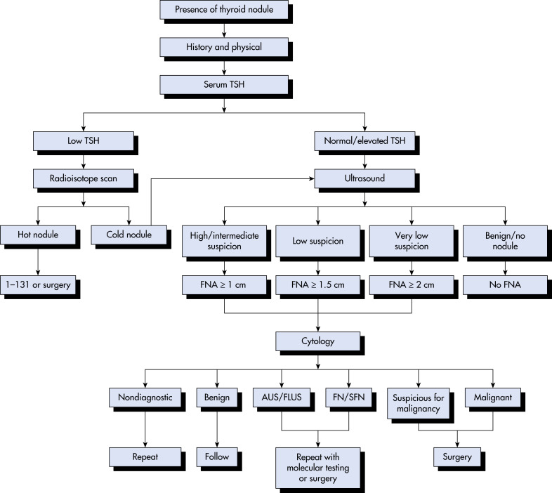
Workup (FIG. 1

Physical exam is helpful if there are overt signs of hyperthyroidism or malignancy, but these are uncommon. Diagnosis usually relies on laboratory tests, radiographic studies, and cytology.1

Figure 1 Workup of a thyroid nodule.

AUS, Atypia of undetermined significance; FLUS, follicular lesion of undetermined significance; FN, follicular neoplasm; FNA, fine-needle aspiration biopsy; SFN, suspicious for follicular neoplasm; TSH, thyroid-stimulating hormone.

Modified from Townsend CM et al: Sabiston textbook of surgery, ed 21, St Louis, 2022, Elsevier.

Laboratory Tests

  • Serum thyroid-stimulating hormone (TSH) required in all patients. If suppressed, obtain free T4 and free T3 and thyroid scan to screen for a “hot nodule,” indicative of a hyperfunctioning adenoma. If the TSH is normal or elevated, consider biopsy based on ultrasonographic characteristics.
  • Serum calcitonin is only recommended when suspecting medullary carcinoma (MTC), such as a patient with a family history of MEN II or familial MTC, or history of pheochromocytoma or hyperparathyroidism.
  • If the patient may have Hashimoto thyroiditis, check an antimicrosomal or antithyroid peroxidase antibody level to confirm (see “Thyroiditis” in Section I). These patients often have marked heterogeneity of the thyroid parenchyma on ultrasound, which can be mistaken for nodular disease.
  • Molecular analysis of thyroid tissue is now commercially available, and it may be useful when fine needle aspiration (FNA) biopsy results are indeterminate.2 Several companies offer the testing, and in general they are beneficial as rule-out tests. They can be used to identify a subpopulation of patients with a low likelihood of cancer, thereby avoiding unnecessary thyroid lobectomy in patients with indeterminate FNA. These tests have a high negative predictive value for cytologically indeterminate nodules (95% for an atypia or follicular lesion of undetermined significance, 94% for a follicular neoplasm). It is less common to find a gene that is highly predictive of cancer, such as BRAF, RET/PTC, and PAX8-PPAR gamma. The presence of RAS mutations is not as helpful, as it is commonly found in benign follicular adenomas, noninvasive follicular thyroid neoplasms with papillary-like nuclear features, and follicular cancers.
Biopsy

  • FNA biopsy is the best means of distinguishing benign from malignant nodules, but it requires the availability of an expert cytopathologist.
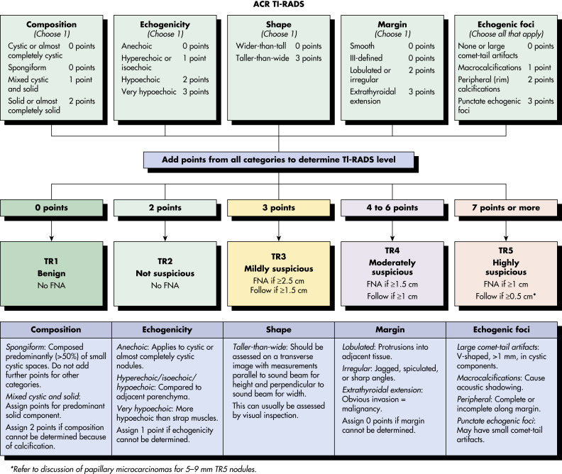
  • Decision to perform ultrasound-guided biopsy is based on size and ultrasound features. The American Thyroid Association guidelines for thyroid nodules tallies the number of benign or suspicious ultrasound features and then recommends biopsy or observation based on nodule size. The American College of Radiology has published a thyroid nodule scoring system called TI-RADS (thyroid imaging reporting and data system [Table 1]), which is used in a similar way. If the likelihood of malignancy is low, a 1-yr follow-up ultrasound should be performed.
  • FNA is discouraged for thyroid nodules <1 cm in diameter unless there are highly concerning features, such as concern for tracheal or nerve involvement, or suspicion of metastatic disease.
  • FNA biopsy is very low yield for thyroid cystic lesions because of a paucity of cellular material. Most asymptomatic simple cysts can be observed. Large compressive cysts, or those with a significant solid component, should be considered for resection.
  • Previous endocrine, surgery, and radiology algorithms recommended biopsy of all solid thyroid nodules over 1 cm. Over the last 5 yr, there has been a marked shift toward risk stratifying nodules by their ultrasound characteristics using the TI-RADS scoring system or the American Thyroid Association risk assessment. Lower-risk nodules can be followed with ultrasound unless they cross certain size thresholds. Higher-risk nodules still follow the 1-cm cutoff for FNA biopsy.
  • FNA biopsy results at most tertiary care institutions are now reported using the Bethesda classification system. The Bethesda Classification system (Table 2) estimates the probability of malignancy at histology based on FNA biopsy cytology and treatment recommendation.3

TABLE 2 Bethesda Classification System for Thyroid Nodules

Diagnostic Category Bethesda ClassificationRisk of MalignancyAction
I Nondiagnostic1-4Repeat biopsy after 4 wk; if very low risk, follow with ultrasound
II Benign1-31-yr follow-up ultrasound
III Atypia (follicular lesion) of undetermined significance (AUS/FLUS)5-15Repeat biopsy in 3 mo; consider genetic testing
IV Follicular neoplasm15-30Thyroid lobectomy; consider genetic testing if nodule appears low risk or patient wishes to avoid surgery
V Suspicious60-75Thyroid lobectomy or total thyroidectomy
VI Malignant97-99Thyroid lobectomy or total thyroidectomy

Adapted from Cibas ES, Ali SZ: The Bethesda system for reporting thyroid cytopathology, Am J Clin Pathol 132:658-665, 2009.

TABLE 1 American College of Radiology Thyroid Nodule Scoring System: TI-RADS

Scoring and ClassificationRecommendations
TR1: 0 pointsTR1: No FNA required (0.3% risk)
TR2: 2 pointsTR2: No FNA required (1.5% risk)
TR3: 3 pointsTR3: 1.5 cm follow-up at 1, 3, 5 yr, 2.5 cm FNA (4.8% risk)
TR4: 4-6 pointsTR4: 1.0 cm follow-up at 1, 2, 3, 5 yr 1.5 cm FNA (9.1% risk)
TR5: 7 pointsTR5: 0.5 cm follow-up each year, 1.0 cm FNA (35% risk)

ACR TI-RADS is a reporting system for thyroid nodules on ultrasound proposed by the American College of Radiology (ACR) and is based on composition, echogenicity, shape, margin, and echogenic foci.

From Tessler FN et al: ACR Thyroid Imaging, Reporting and Data System (TI-RADS): white paper of the ACR TI-RADS committee, J Am Coll Radiol 14:587-595, 2017.

Imaging Studies

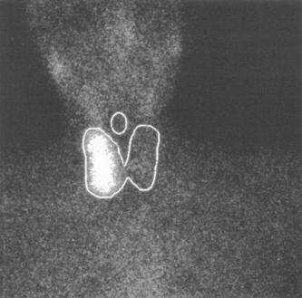
  • Iodine uptake scanning (Fig. 2) is only indicated in patients with suppressed TSH levels to determine if the nodule is toxic or not. If it is hot, biopsy is not indicated.
  • Ultrasonography is an inexpensive and effective modality to stratify malignancy risk. The American College of Radiology (ACR) Thyroid Imaging, Reporting and Data System (TI-RADS) is illustrated in Fig. 3.4
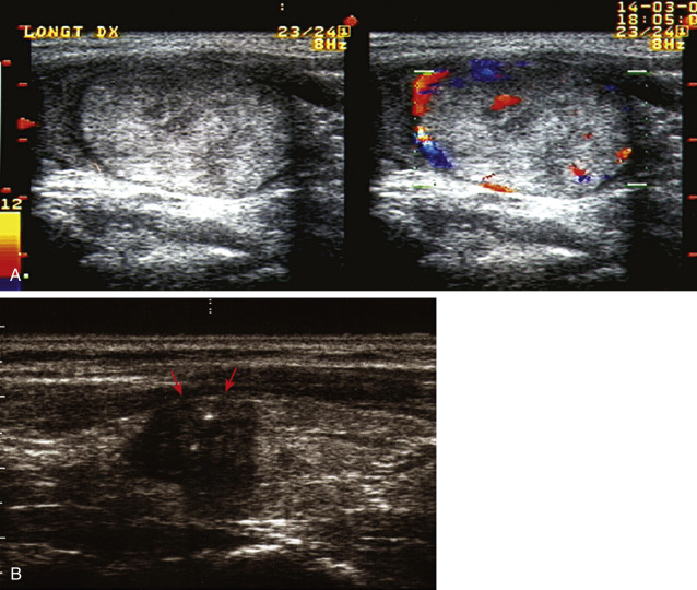
  • Ultrasound (Fig. E4) is useful to evaluate the size and number of nodules, as well as their characteristics. These include whether it is solid or cystic, echogenicity relative to normal thyroid tissue, irregular vs. smooth borders, presence of calcifications, shape, and vascularity. Table 3 summarizes ultrasound features of benign and malignant thyroid nodules. The three ultrasound characteristics most predictive of malignancy in solid thyroid nodules are shape taller than wide, microcalcifications, and hypoechogenicity, in that order. The positive predictive value for malignancy steadily rises as the number of suspicious ultrasound findings mounts. However, FNA biopsy remains necessary for a definitive diagnosis. Very low risk ultrasound features include simple cystic lesions and spongiform nodules (sponge-like in appearance with layers of solid and cystic contents). Ultrasound follow-up is reasonable in such cases without biopsy, although it is acceptable to biopsy spongiform nodules over 2 cm based on practitioner and patient preference.

TABLE 3 Ultrasound Features Suggestive of Benign and Malignant Thyroid Nodules

BenignSuspicious for Malignancy
Nodule characteristics
  • Cystic
  • Spongiform
  • Hyperechoic
  • “Comet tail” artefact (seen in colloid nodules; do not confuse with microcalcifications)
  • Hypoechoic
  • Very hypoechoic
  • Shape taller than wide
Nodule margin
  • Smooth, regular
  • Presence of halo
  • Irregular, lobulated, or poorly defined
  • Protrusion outside of thyroid capsule
Calcification
  • Absent
  • Coarse
  • Eggshell
  • Microcalcification
  • Broken eggshell
Metastatic spread
  • Invasion of the adjacent tissues
  • Enlarged ipsilateral or bilateral cervical lymph nodes; metastatic nodes are round, hyperechoic, and can contain microcalcifications
Figure 3 The American College of Radiology (Acr) Thyroid Imaging, Reporting and Data System (Ti-RADS) Lexicon, Tr Levels, and Criteria for Fine-Needle Aspiration Biopsy

From Tessler FN et al: ACR Thyroid Imaging, Reporting and Data System [TI-RADS]: white paper of the ACR TI-RADS Committee, J Am Coll Radiol 14:587-595, 2017.

Figure 2 131i Scan Demonstrating an Area of Increased Uptake in the Right Lobe of a 32-Yr-Old Woman with Increased Thyroid Function Test Values and a Palpable Nodule

This scan is consistent with a toxic or hyperfunctioning nodule.

From Townsend CM et al: Sabiston textbook of surgery, ed 21, St Louis, 2022, Elsevier.

Figure E4 Conventional and color flow Doppler ultrasonography.

A, Benign lesion. Sonogram shows well-defined, oval, hyperechoic nodule with perinodular and slight intranodular blood flow. B, Malignant lesion. Sonogram shows a nodule with inhomogeneous hypoechoic aspect, microcalcifications, irregular borders, and invasion of the thyroid capsule (arrows).

From Melmed S et al: Williams textbook of endocrinology, ed 12, Philadelphia, 2011, Elsevier.

Treatment

General Rx

  • Evaluation of results of FNA:
    1. Regardless of biopsy result, symptomatic compressive nodules, substernal nodules, and those causing tracheal deviation are all indications for thyroidectomy.
    2. Table 2 contains the guidelines for management of each of the possible results after FNA biopsy. Nondiagnostic biopsies contain insufficient cells and require repeat, unless they are obviously benign cystic lesions.
    3. Benign nodules carry a 5% false-negative biopsy rate and should be reassessed at 1 yr for growth. If they have grown more than 3 mm, repeat biopsy should be considered.
    4. AUS/FLUS carries a 15% risk of malignancy. Usually a repeat biopsy is performed at 3 mo in hopes of obtaining a definitive result. Genetic testing is a consideration.
    5. Follicular neoplasms are higher risk and warrant lobectomy or genetic testing. Ultrasound features should aid in the decision to perform genetic testing.
    6. Suspected malignancy and papillary thyroid cancer are treated similarly with lobectomy or total thyroidectomy.
Disposition

Variable with results of FNA biopsy. Once patients have had a benign biopsy and a 1-yr follow-up ultrasound shows no significant growth, they do not require annual thyroid ultrasound exams. They can be followed with an annual neck exam. If growth is detected or symptoms develop, repeat ultrasound is indicated.

Referral

Surgery or radiology referral for possible FNA biopsy if nodule is solid and over 1 cm

Pearls & Considerations

Comments

  • Many solid, benign nodules grow; therefore, an increase in nodule volume alone is not a reliable predictor of malignancy.
  • Thyroid nodules incidentally identified as FDG avid on fluorodeoxyglucose-PET (FDG-PET) scan done for other disorders have a higher malignancy rate (25%).
  • Highly suspicious nodules should be referred for surgical evaluation even if result of FNA is “benign.”
  • Most follicular neoplasms are benign, and patients should not be told they have a malignancy based on this cytology result.
Related Content

Thyroid Nodule (Patient Information)

Thyroiditis (Related Key Topic)

Thyroid Carcinoma (Related Key Topic)

Related Content

    1. Haugen B.R. : American Thyroid Association management guidelines for adult patients with thyroid nodules and differentiated thyroid cancer: the American Thyroid Association Guidelines Task Force on Thyroid Nodules and Differentiated Thyroid CancerThyroid. ;26(1):1-133, 2016.
    2. Livhits M.J. : Effectiveness of molecular testing techniques for diagnosis of indeterminate thyroid nodules: a randomized clinical trialJAMA Oncol. ;7(1):70-77, 2021.
    3. Cibas E.S., Ali S.Z. : The 2017 Bethesda system for reporting thyroid cytopathologyThyroid. ;27(11):1341-1346, 2017.
    4. Tessler F.N. : ACR Thyroid Imaging, Reporting and Data System (TI-RADS): white paper of the ACR TI-RADS CommitteeJ Am Coll Radiol. ;14(5):587-595, 2017.