section name header

Basic Information

AUTHOR: Maheswara Satya Gangadhara Rao Golla, MD

Definition

Angina pectoris is a term used to describe a clinical syndrome, typically characterized by chest, jaw, shoulder, back, or arm discomfort that is caused by myocardial ischemia. This is most commonly related to atheromatous plaque in one or more than one large epicardial coronary artery; however, myocardial ischemia may occur in the absence of obstructive coronary artery disease (CAD), such as uncontrolled hypertension, microvascular disease, valvular heart disease, hypertrophic cardiomyopathy, coronary spasm, or endothelial dysfunction. Any situation that causes an imbalance in myocardial oxygen supply and demand can cause an angina syndrome. Angina can be classified as follows:

  • Chronic stable angina, stable ischemic heart disease (SIHD), or chronic CAD:
    1. Predictable. Usually follows a precipitating event (e.g., climbing stairs, sexual intercourse, a heavy meal, emotional stress, cold weather)
    2. Generally, has the same severity as previous attacks; relieved by rest or by the customary dose of sublingual nitroglycerin
    3. Caused by a fixed coronary artery obstruction secondary to atherosclerosis. The presence of one or more obstructions in major coronary arteries is likely; the severity of stenosis is usually >70%
  • Unstable (rest, recent onset, crescendo angina; will be reviewed under “Acute Coronary Syndrome”):
    1. Rest angina: Angina occurring at rest and usually prolonged >20 min, occurring within 1 wk of presentation
    2. Recent onset: Angina of at least CCS Class III severity occurring less than 2 mo after the onset of the symptoms
    3. Crescendo angina: Previously diagnosed angina that is distinctly more frequent, longer in duration, or lower in threshold (i.e., increased by >1 CCS class within 2 mo of initial presentation to at least CCS Class III severity)
  • Prinzmetal variant:
    1. Occurs at rest, common after cold exposure
    2. Cyclical in nature
    3. ECG finding of episodic ST-segment elevations
    4. Caused by coronary artery spasm with or without superimposed CAD
    5. Patients are more likely to develop ventricular arrhythmias
  • Microvascular angina (syndrome X):
    1. Refers to patients with angina symptoms, positive exercise test, normal coronary angiograms, and no coronary spasm. Defective endothelium-dependent dilation in the coronary microcirculation contributes to the altered regulation of myocardial perfusion and the ischemic manifestations in these patients.
    2. Patients with chest pain and normal or nonobstructive coronary angiograms are predominantly women, and many have a prognosis that is not as benign as commonly thought (2% risk of death or myocardial infarction [MI] at 30 days of follow-up).
  • Refractory angina:
    1. Refers to patients who, despite optimal medical therapy with at least maximal doses, or as tolerated of two antianginal medications, in addition to aspirin, aggressive risk factor modification, such as smoking cessation, adequate control of hypertension, diabetes, and hyperlipidemia, still have both angina and objective evidence of ischemia
  • Other:
    1. Angina due to aortic stenosis and idiopathic hypertrophic subaortic stenosis, cocaine-induced coronary vasoconstriction
Functional Classification:

Stable angina should be classified using a grading system. The most commonly adopted is that of the Canadian Cardiovascular Society (CCS):

  • Class I: Ordinary physical activity, such as walking or climbing stairs, does not cause angina. Angina occurs with strenuous, rapid, or prolonged exertion at work or recreation.
  • Class II: Slight limitation of ordinary activity. Angina occurs on walking or climbing stairs rapidly; walking uphill; walking or stair climbing after meals, in cold, in wind, or under emotional stress; or only during the few hours after awakening. Angina occurs on walking more than two level blocks and climbing more than one flight of ordinary stairs at a normal pace and in normal conditions.
  • Class III: Marked limitations of ordinary physical activity. Angina occurs on walking one to two level blocks and climbing one flight of stairs in normal conditions and at a normal pace.
  • Class IV: Inability to carry on any physical activity without discomfort; anginal symptoms may be present at rest.
ICD-10CM CODES
I20.1Angina pectoris with documented spasm
I20.8Other forms of angina pectoris
I20.9Angina pectoris, unspecified
I25.110Atherosclerotic heart disease of native coronary artery with unstable angina pectoris
I25.111Atherosclerotic heart disease of native coronary artery with angina pectoris with documented spasm
I25.118Atherosclerotic heart disease of native coronary artery with other forms of angina pectoris
I25.119Atherosclerotic heart disease of native coronary artery with unspecified angina pectoris
I25.700Atherosclerosis of coronary artery bypass graft(s), unspecified, with unstable angina pectoris
I25.790Atherosclerosis of other coronary artery bypass graft(s) with unstable angina pectoris
I25.791Atherosclerosis of other coronary artery bypass graft(s) with angina pectoris with documented spasm
I25.798Atherosclerosis of other coronary artery bypass graft(s) with other forms of angina pectoris
I25.799Atherosclerosis of other coronary artery bypass graft(s) with unspecified angina pectoris
Epidemiology & Demographics

  • It is estimated that one in three adults in the U.S. (about 81 million) has some form of cardiovascular disease. Based on the NHANES survey 2007 to 2010, an estimated 15.4 million have coronary heart disease (CHD) of which 7.8 million have angina.
  • Angina is most common in middle-aged and elderly men. Among persons 60 to 79 yr of age, approximately 25% of men and 16% of women have CHD, and these figures rise to 37% and 23% among men and women >80 yr of age, respectively.
  • The incidence of CHD and angina in women after menopause is similar to that of men.
  • Although the survival rate has steadily improved over time, SIHD remains the number one cause of death in men and women (27% of deaths).
  • The initial manifestation of ischemic heart disease is angina pectoris in 50%, and about 50% of patients presenting to the hospital with acute coronary syndrome have preceding angina.
  • Two older population-based studies from Olmstead County, MN, and Framingham, MA, showed annual rate of MI in patients with symptomatic angina of 3% to 3.5%/yr.
  • Within 12 mo of initial diagnosis, 10% to 20% of patients with diagnosis of stable angina progress to MI or unstable angina.
Physical Findings & Clinical Presentation

  • The assessment of chest pain should include quality, location, severity, and duration of pain; radiation; associated symptoms; provocative factors; and alleviating factors. Anginal pain can be described as “squeezing,” “griplike,” “suffocating,” and “heavy,” but it is rarely sharp or stabbing and typically does not vary with position or respiration. The classic Levine sign is placing a clenched fist over the precordium to describe the pain. Many patients do not, however, describe angina as frank pain but as tightness, pressure, or discomfort. Other patients, in particular women and older adults, can present with atypical symptoms such as nausea, vomiting, midepigastric discomfort, sharp (atypical) chest pain, dizziness, or syncope.
  • Ischemic pain of more than 20 minutes’ duration should raise concern for possible acute coronary syndrome.
  • Women are more likely than men to report atypical chest pain or discomfort (65% reported on Women’s Ischemic Syndrome Evaluation [WISE] study).
  • Elderly and diabetics may report symptoms other than chest pain, such as dyspnea, fatigue, or diaphoresis.
Etiology
Risk Factors

  • Advanced age.
  • Male sex.
  • Genetic predisposition, family history of premature CAD in first-degree relatives (men <55 yr of age, and women <65 yr of age).
  • Smoking (risk of first MI is increased by near threefold).
  • Hypertension.
  • Hyperlipidemia.
  • Impaired glucose tolerance or diabetes mellitus.
  • History of stroke or peripheral arterial disease.
  • Chronic kidney disease (CKD).
  • Metabolic syndrome.
  • Physical inactivity.
  • Obesity (body mass index [BMI] >30% over ideal). A higher BMI during childhood is also associated with an increased risk of CHD in adulthood.
  • Entities that cause increased oxygen demand include hyperthermia (particularly if accompanied by volume contraction), hyperthyroidism, and cocaine or methamphetamine abuse.
  • Cocaine is used by >5 million Americans regularly and is responsible for >64,000 emergency department (ED) evaluations yearly to rule out myocardial ischemia. Cocaine causes sympathomimetic toxicity and not only increases myocardial oxygen demand but also induces coronary vasospasm and can cause infarction in young patients. Long-term cocaine use can cause premature development of SIHD.
  • Severe uncontrolled hypertension causes increased myocardial oxygen demand and decreased subendocardial perfusion that increases left ventricular (LV) wall tension. Hypertrophic cardiomyopathy and aortic stenosis can induce even more severe LV hypertrophy and resultant wall tension.
  • Other causes of increased myocardial oxygen demand are ventricular or supraventricular tachycardias. Ambulatory monitoring may be required to diagnose these.
  • Entities that limit myocardial oxygen supply such as anemia may cause angina when the hemoglobin drops to <9 g/dl, and ST-T-wave changes (depression or inversion) can occur at levels <7 g/dl.
  • Hypoxemia resulting from pulmonary disease (e.g., pneumonia, asthma, chronic obstructive pulmonary disease, pulmonary hypertension, interstitial fibrosis, or obstructive sleep apnea) can also precipitate angina.
  • Polycythemia, leukemia, thrombocytosis, and hypergammaglobulinemia.
  • Oral contraceptive and hormone replacement therapy use.
  • Coronary artery calcium is associated with an increased risk of MI.
  • Long-term use of NSAIDs.
  • Exposure to air pollution from traffic (dilute diesel exhaust) promotes myocardial ischemia and is associated with adverse cardiovascular events.
  • Low serum folate levels required for conversion of homocysteine to methionine are associated with an increased risk of fatal CHD. Hyperhomocysteinemia has a toxic effect on vascular endothelium and interferes with proliferation of arterial wall smooth muscle cells. Elevated plasma homocysteine level is a strong and independent risk factor for CHD events, especially in patients with type 2 diabetes mellitus.
  • Elevated levels of highly sensitive C-reactive protein (hs-CRP, cardio CRP). Diseases associated with systemic inflammation can lead to accelerated atherosclerosis.
  • Depression.
  • Vasculitis.
  • Elevated levels of lipoprotein-associated phospholipase A2.
  • Elevated fibrinogen levels.
  • Low level of red blood cell glutathione peroxidase-1 activity.
  • Radiation therapy.

Diagnosis

Differential Diagnosis

  • Nonischemic cardiovascular: Aortic dissection, pericarditis
  • Pulmonary: Pulmonary embolism, pneumothorax, pneumonia, pleuritis
  • Gastrointestinal: Esophageal, esophagitis, spasm, reflux, biliary colic, cholecystitis, choledocholithiasis, cholangitis, peptic ulcer, pancreatitis
  • Chest wall: Costochondritis, fibrositis, rib fracture, sternoclavicular arthritis, herpes zoster (before the rash)
  • Psychiatric: Anxiety disorders, hyperventilation, panic disorder, primary anxiety, affective disorders (i.e., depression), somatoform disorders, thought disorders (i.e., fixed delusions)
Workup

  • In patients with chest pain, the probability of CAD should be estimated on the basis of patient age, sex, cardiovascular risk factors, and pain characteristics.
  • The most important diagnostic element is the history. Chest pain or left arm pain or discomfort occurring with exertion and relieved by rest in a patient with cardiovascular risk factors is consistent with a high likelihood of CAD.
  • In assessing the likelihood of underlying SIHD it is helpful to classify the chest pain as typical angina, atypical angina, and/or noncardiac chest pain.
  • Typical angina, (definite) will have the following three features: (1) Substernal chest discomfort with a characteristic quality and duration, (2) provoked by exertion or emotional stress, and (3) relieved by rest and/or sublingual nitroglycerin.
  • Atypical angina, (probable) will have two of the above listed three features.
  • Noncardiac chest pain will have one or none of the previously listed features.
  • Physical examination may be completely normal in many patients; however, certain findings may be helpful in the assessment of the patient with suspected SIHD. Some findings may identify consequences of ischemia or possible causes of the anginal syndrome other than CAD. The presence of hypertension, arcus senilis, xanthelasma, carotid or peripheral bruits, and a prominent S4 are all physical signs that could raise concern for the presence of CAD. A murmur of mitral regurgitation may be a marker of an ischemic cardiomyopathy or transient ischemia. A murmur suggestive of hypertrophic cardiomyopathy or aortic stenosis may suggest a cause of angina other than CAD.
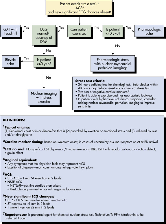
  • The ultimate goal for an SIHD patient’s evaluation is to identify high-risk CAD patients with minimal use of resources. In general, four common steps would help a clinician to assess high feature of SIHD. Those are (1) CAD risk assessment based on various classic risk factors, (2) functional capacity and stress test result, (3) LV and right ventricular (RV) function, and (4) coronary anatomy. Every patient does not need each of the modalities. One should classify stable angina without history of CAD patients to low, intermediate, and high pretest probability or likelihood for CAD after assessment of comorbidities and atherosclerotic vascular disease risk factors. Consider functional capacity and stress test (ETT [exercise tolerance test], ETT-MIBI [ETT-myocardial perfusion imaging (multiplex ion beam imaging with dipyridamole/technetium sestamibi)] or stress echo) for low and intermediate pretest probability patients. Exercise testing (Fig. 1) is preferred to pharmacologic stress tests when possible. Functional capacity and stress test results categorize SIHD patients based on their risk of annual mortality. Patients with an estimated annual mortality of <1% classify as low risk, 1% to 3% are considered as intermediate risk, and >3% annual mortality belongs to high-risk patient population (please see “Coronary Artery Disease” chapter).1,2

Figure 1 Stress test algorithm.

!!flowchart!!

ACS, Acute coronary syndrome; BBB, bundle branch block; DM, diabetes mellitus; ECG, electrocardiogram; echo, echocardiography; ED, emergency department; GTX, graded exercise test; LVH, left ventricular hypertrophy; NSTE, non-ST-segment elevation; NSTEMI, NSTE myocardial infarction; STE, ST-segment elevation; y/o, years old.

From Adams JG et al: Emergency medicine: clinical essentials, ed 2, Philadelphia, 2013, Saunders.

Laboratory Tests

  • Screen for hypertension, diabetes, and hyperlipidemia per routine guidelines.
  • Electrocardiogram should be obtained during pain and when the patient is free of any discomfort. A normal resting electrocardiogram is not unusual in patients with SIHD; in patients who present with chest pain, 1% to 6% who have an acute MI will have a normal or nondiagnostic electrocardiogram.
  • Chest x-ray PA and lateral if symptoms suggestive of heart failure, pericardial disease, aortic aneurysm/dissection.
  • Cardio-CRP (hs-CRP): Its elevation is a relatively moderate predictor of CHD, and it adds prognostic information to that conveyed by the Framingham risk score.
(See “coronary Artery Disease” Chapter, Fig. 1

  • Exercise testing is used for diagnosis as well as prognosis of SIHD. If the patient is physically capable to perform at least moderate physical exercise, exercise stress testing see (see Fig. 1) is useful because of the important prognostic information obtained from exercise performance and the hemodynamic response. Risk stratification based on noninvasive testing is summarized in Table 1. Patients who have an intermediate likelihood of CAD, as patients in a low-risk or high-likelihood category are more likely to have a false-positive or false-negative result, respectively. Risk assessment is also indicated in patients with SIHD who are being considered for revascularization of known coronary stenosis of unclear physiologic significance. The exercise treadmill test is contraindicated in patients with presence of ST depression >0.5 mm or Wolff-Parkinson-White syndrome, left bundle branch block, or pacemaker rhythm in baseline ECG.
  • Stress echocardiography or stress testing with myocardial perfusion imaging may be employed when baseline electrocardiographic abnormalities are present that render the electrocardiographic response to exercise uninterpretable. Stress echocardiography has the advantage of higher specificity and a lower cost. Stress radionuclide perfusion imaging has a higher sensitivity, particularly for single-vessel coronary disease, and has a higher technical success rate. When the patient is unable to exercise adequately, pharmacologic stress testing (i.e., dobutamine, adenosine, regadenoson) may be used with these imaging modalities.
  • A good predictor of risk for a patient with stable angina is the Duke treadmill score, which incorporates the patient’s functional status (METS or time in minutes during the Bruce protocol), ST-segment depression in millimeters, and an angina index (yes or no). Patients with favorable Duke scores (>5) have a 5-yr survival rate of >97%; this is independent of other factors such as coronary anatomy and LV function.
  • Echocardiography is indicated in patients with murmurs suggestive of aortic stenosis, hypertrophic cardiomyopathy, mitral regurgitation, mitral valve prolapse, previous MI, pathologic Q waves, complex ventricular arrhythmias, heart failure, hypertension, diabetes, and abnormal ECG.
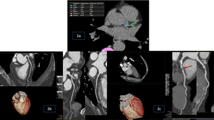
  • Cardiac computed tomography (CCTA; Fig. 2) is useful for the detection of subclinical CAD in asymptomatic patients with an intermediate Framingham 10-yr risk estimate of 10% to 20%. 2021 ACC/AHA/ASE chest pain guidelines recommend cardiac CT angiogram (CCTA) as a front-line imaging modality for stable angina. Trials comparing CCTA to invasive coronary angiography (ICA) in patients with stable angina have shown that an initial strategy of CCTA for anatomic imaging is associated with similar outcomes but fewer procedural complications and invasive angiograms.3 CCTA detects and quantifies coronary calcium and evaluates the lumen and wall of the coronary artery. CCTA can be useful as a first-line test for risk assessment in patients with SIHD who are unable to exercise to an adequate workload regardless of interpretability of ECG. Also can be used when a functional test has an indeterminate result and to assess bypass graft patency or patency of previous stents >3 mm diameter. CCTA CT cost and radiation exposure are limiting factors to recommending widespread routine use of this marker. CCTA has an excellent negative predictive value to rule out severe CAD in the setting of active chest pain evaluation in the emergency room (ROMICAT-II trial). In 2018 the SCOT-HEART trial showed coronary CTA resulted in a lower risk of nonfatal MI than standard care alone (ETT) in stable angina patients. Myocardial perfusion imaging and noninvasive fractional flow ratio would be possible with cardiac CTA as well.
  • Coronary artery calcium score is a strong predictor of incidence of CAD and provides predictive information in patients with low to intermediate pretest probability of CAD beyond that provided by standard risk factors. A score <100 indicates low risk and a score >400 high risk.
  • Cardiac magnetic resonance imaging, in addition to its use for diagnosis of arrhythmogenic right ventricular dysplasia, can also be used to assess myocardial perfusion and viability as well as function in patients unable to exercise. Additional studies are needed to determine the cost effectiveness of these studies in patients with ischemic cardiomyopathy.
  • Invasive coronary angiography remains the gold standard for the identification of clinically significant CAD. Angiography is performed to define the location and extent of coronary disease; indicated in selected patients who are candidates for coronary revascularization (either coronary artery bypass graft [CABG] surgery or angioplasty).
Figure 2 2a, Example of Coronary Artery Calcium Scoring in Which Calcified Foci are Identified Within the Left Anterior Descending (Green) and Left Main (Blue) Coronary Arteries; Table Shows Individual and Total Agatston Scores

2b, Cardiac CTA Showed Normal Coronary Arteries. 2c, Cardiac CTA Showed Proximal to Mid-Left Anterior Descending Artery Stenosis (80%) (Red Arrow).

Courtesy Maheswara Satya Gangadhara Rao Golla, MD.

TABLE 1 Risk Stratification Based on Noninvasive Testing

High Risk (>3% Annual Risk for Death or Myocardial Infarction)
  1. Severe resting left ventricular dysfunction (LVEF <35%) not readily explained by noncoronary causes
  1. Resting perfusion abnormalities involving 10% of the myocardium without previous known MI
  1. High-risk stress findings on the ECG, including:
    • 2-mm ST-segment depression at low workload or persisting into recovery
    • Exercise-induced ST-segment elevation
    • Exercise-induced VT/VF
  1. Severe stress-induced LV dysfunction (peak exercise LVEF <45% or drop in LVEF with stress 10%)
  1. Stress-induced perfusion abnormalities encumbering 10% of the myocardium or stress segmental scores indicating multiple vascular territories with abnormalities
  1. Stress-induced LV dilation
  1. Inducible wall motion abnormality (involving >2 segments or 2 coronary beds)
  1. Wall motion abnormality developing at a low dose of dobutamine (10 mg/kg/min) or at a low heart rate (<120 beats/min)
  1. Multivessel obstructive CAD (70% stenosis) or left main stenosis (50% stenosis) on CCTA
Intermediate Risk (1%-3% Annual Risk for Death or Myocardial Infarction)
  1. Mild to moderate resting LV dysfunction (LVEF of 35%-49%) not readily explained by noncoronary causes
  1. Resting perfusion abnormalities involving 5%-9.9% of the myocardium in patients without a history or previous evidence of MI
  1. 1-mm ST-segment depression occurring with exertional symptoms
  1. Stress-induced perfusion abnormalities encumbering 5%-9.9% of the myocardium or stress segmental scores indicating 1 vascular territory with abnormalities but without LV dilation
  1. Small wall motion abnormality involving 1-2 segments and only 1 coronary bed
  1. Single-vessel CAD with 70% stenosis or moderate CAD stenosis (50%-69% stenosis) in 2 arteries on CCTA
Low Risk (<1% Annual Risk for Death or Myocardial Infarction)
  1. Low-risk treadmill score (score 5) or no new ST-segment changes or exercise-induced chest pain symptoms when achieving maximal levels of exercise
  1. Normal or small myocardial perfusion defect at rest or with stress encumbering <5% of the myocardium
  1. Normal stress or no change in limited resting wall motion abnormalities during stress
  1. No coronary stenosis >50% on CCTA

CCTA, Cardiac computed tomography angiography; LVEF, left ventricular ejection fraction; VF, ventricular fibrillation; VT, ventricular tachycardia. Assessment of coronary artery calcium can also be used to contribute to risk assessment.

Although the published data are limited, patients with these findings will probably not be at low risk in the presence of either a high-risk treadmill score or severe resting LV dysfunction (LVEF <35%).

Modified from Fihn SD et al: ACCF/AHA/ACP/AATS/PCNA/SCAI/STS guideline for the diagnosis and management of patients with stable ischemic heart disease: a report of the American College of Cardiology Foundation/American Heart Association Task Force on Practice Guidelines, and the American College of Physicians, American Association for Thoracic Surgery, Preventive Cardiovascular Nurses Association, Society for Cardiovascular Angiography and Interventions, and Society of Thoracic Surgeons, Circulation 126:e354, 2012; in Zipes DP: Braunwald’s heart disease: a textbook of cardiovascular medicine, ed 11, Philadelphia, 2019, Elsevier.

Treatment

Five Fundamental Overlapping Strategies are Recommended

  • Patient education: Support active participation of patients in the decision-making process of their treatment.
  • Management of comorbid conditions that contribute to or worsen SIHD.
  • Aggressive modification of preventable risk factors such as smoking cessation, weight reduction in obese patients, regular aerobic exercise program (at least 30 to 60 min/day for 5 days a week), correction of folate deficiency, reduced intake of saturated fats (to <7% of total calories) and trans-fatty acids (to <1% of total calories), low-sodium diet (<2 g/day), and teaching importance of medication adherence. Whole grains as the main form of carbohydrates, an abundance of fruits and vegetables, and adequate omega-3 fatty acids are optimal for prevention of SIHD.
  • Evidence-based pharmacologic management to improve quality of life and survival.
  • Use appropriate revascularization procedures to improve survival and long-term outcomes in selected patients.
Pharmacologic Therapy (See “coronary Artery Disease”)

Treatment can be classified based on medications that prevent MI and death.

  • Aspirin reduces cardiovascular mortality and morbidity rates by 20% to 25% among patients with CAD. Appropriate dose is 75 to 162 mg/day in the absence of contraindications. It inhibits the enzyme cyclooxygenase and synthesis of thromboxane A2 and reduces the risk of adverse cardiovascular events by 33% in patients with unstable angina. Patients intolerant to aspirin can be treated with clopidogrel or can undergo aspirin desensitization. Clopidogrel irreversibly blocks the P2Y12 adenosine diphosphate receptor on the platelet surface, thereby interrupting platelet activation and aggregation. Clopidogrel can be combined with ASA in high-risk patients with SIHD with low risk for bleeding complications or can be given alone in patients that are aspirin intolerant. Dose is 75 mg/day.
  • Ticagrelor, in the PEGASUS-TIMI-54, reduced the risk of death, cardiovascular MI, or stroke in patients after 1 yr of MI. However, it is associated with an increased risk of bleeding when compared to placebo.
  • Dipyridamole is not recommended as an antiplatelet therapy for the treatment of patients with SIHD.
  • Beta-adrenergic blockers, which prevent MI and death, are first-line therapy in the management of angina pectoris. They achieve their major antianginal effect by decreasing myocardial oxygen demand in reducing heart rate and systolic blood pressure product, atrioventricular nodal conduction, and myocardial contractility, in this manner contributing to a reduction in angina onset, with improvement in the ischemic threshold during exercise and during the usual daily activities. Absent contraindications, they should be regarded as initial therapy for stable angina for all patients. Their dose should generally be adjusted to reduce the resting heart rate to 55 to 60 beats/min. Despite the difference among the available beta-blockers, they all seem to be equally efficacious in SIHD. Beta-blockers recommended for at least 2 to 3 yr after MI, and lifelong for patients with LV ejection fraction of <40% with heart failure or prior MI.
  • Nitrates cause venodilation and relaxation of vascular smooth muscle; the decreased venous return from venodilation decreases diastolic ventricular wall tension (preload) and thereby reduces mechanical activity (and myocardial oxygen consumption) during systole. Relaxation of vascular smooth muscle increases coronary blood flow and reduces systemic pressure. Dilation of the arterial wall will not be affected by plaque, but independent of an intact endothelium, leads to reduced resistance across the obstructed lumen. Nitroglycerin contributes to coronary blood flow redistribution by augmenting collateral flow and lowering ventricular diastolic pressure from areas of normal perfusion to ischemic zones. Nitroglycerin also has demonstrated antithrombotic and antiplatelet effects. Sublingual nitroglycerin or nitroglycerin spray should be prescribed to all patients with SIHD for immediate angina relief. Tolerance to nitrates can be minimized by avoiding sustained blood levels with a daily nitrate-free period (e.g., omission of bedtime dose of oral isosorbide dinitrate or 12 h on/12 h off transdermal nitroglycerin therapy). Nitrates are relatively contraindicated in patients with hypertrophic obstructive cardiomyopathy, and should also be avoided in patients with severe aortic stenosis. Nitrates should not be used within 24 h of sildenafil (Viagra) or vardenafil (Levitra) or within 48 h of tadalafil (Cialis) because of the potential for hypotension.
  • Calcium channel blockers are antiischemic medications that have no proven mortality benefit in SIHD. They improve myocardial oxygen supply by decreasing coronary vascular resistance and augmenting epicardial conduit vessel and systemic arterial blood flow. Myocardial demand is decreased by a reduction in myocardial contractility, systemic vascular resistance, and arterial pressure. They are first-line treatment when beta-blockers are contraindicated. They play a major role in preventing and terminating myocardial ischemia induced by coronary artery spasm. They are particularly effective in treating microvascular angina. All classes of calcium channel blockers reduce anginal episodes, increase exercise duration, and reduce use of sublingual nitroglycerin in patients with effort-induced angina. Short-acting calcium channel blockers should be avoided. Calcium channel blockers (particularly nondihydropyridine) should generally also be avoided in patients with CHF secondary to systolic dysfunction due to its negative inotropic effect.
  • Ranolazine, which has been tested in four different studies with a total of 1737 patients (MARISA, CARISA, RAN080, and ERICA), inhibits the late inward sodium current, indirectly reducing the sodium-dependent calcium current during ischemic conditions and leading to improvement in ventricular diastolic tension and oxygen consumption. It seems to increase the efficiency of energy production in the heart, maintaining cardiac function. Its antianginal and antiischemic effects do not depend on reductions in heart rate or blood pressure. It is indicated for treatment of chronic angina that is inadequately controlled with other antianginals. It represents a new class of drugs known as metabolic modulators and can be useful when prescribed as substitute for beta-blockers or in combination with them for relief of symptoms when initial treatment with beta-blockers is not successful or is contraindicated. Side effects include prolongation of QT interval. Low doses of diltiazem and verapamil should be used with ranolazine. The extended-release preparation reduces the frequency of angina, improves exercise performance, and delays the development of exercise-induced angina and ST-segment depression.
  • ACE inhibition through changes in the physiologic balance between angiotensin II and bradykinin could contribute to the reductions in LV and vascular hypertrophy, atherosclerosis progression, plaque rupture, and thrombosis; the favorable changes in cardiac hemodynamics; and the improved myocardial oxygen supply/demand. It has been shown to be effective in reducing cardiovascular death, MI, and stroke in patients who are at risk for or who had vascular disease. They are indicated in patients with hypertension, diabetes, LVEF <40%, and CKD. Angiotensin receptor blockers (ARBs) can be given to patients with SIHD who are intolerant to ACE inhibitors and qualify for them.
  • Use of high-intensity statin drugs is recommended in all patients with CAD. In late 2018 the American College of Cardiology (ACC) recommended an low density lipoprotein (LDL) goal of <70 mg/dl for secondary prevention of atherosclerotic cardiovascular disease. There is no LDL goal for primary prevention. The FDA approved PCSK9 (proprotein convertase subtilisin/kexin type 9) inhibitors (alirocumab and evolocumab) for heterozygous familial hypercholesterolemia in those who maximally tolerated statins. Newer 2018 lipid guidelines suggested starting PCSK9 inhibitor in very-high-risk atherosclerotic cardiovascular disease patients who did not meet the LDL goal on high-intensity statin and ezetimibe. One should always consider adding ezetimibe to high-intensity statin prior to initiation of PCSK9 inhibitor due to cost issues. Inclisiran, evinacumab, and bempedoic acid are reasonable nonstatin therapy alternatives in patients who are unable to tolerate statin and reach target LDL levels.
  • Influenza vaccine is recommended for patients with SIHD on annual basis to prevent all-cause mortality, morbidity, and hospitalization caused by the exacerbation of underlying medical conditions produced by influenza.
  • The EMPAREG OUTCOME, CANVAS, DECLARE-TIMI 58, and CREDENCE trial showed that SGLT2 inhibitors significantly reduce cardiovascular events in type 2 diabetic patients.
New Modalities for the Treatment of Chronic Stable Angina Pectoris:

  • Although a significant amount of progress has been made in the management of CAD with percutaneous coronary intervention (PCI) and CABG, many patients with the condition require additional therapeutic modalities for relief of symptoms and improvement in quality of life. This group of patients includes those with diffuse CAD who are not suitable for revascularization, patients with previous multiple PCIs or CABG limiting the chances for further revascularization, the lack of vascular conduits for CABG, severe LV systolic dysfunction in patients with previous CABG or PCI, and comorbidities that would render the patients at high risk for revascularization.
  • The following pharmacologic agents have been used for the management of stable angina in combination with the standard protocol of nitrates, beta-blockers, calcium channel blockers, and ranolazine: High-dose statin therapy, trimetazidine, perhexiline, nicorandil, allopurinol, ivabradine, fasudil, and testosterone.
  • Other, nonpharmacologic modalities that are highly experimental include stem cell therapy, therapeutic angiogenesis, and mechanical therapies such as external counterpulsation, spinal cord stimulation, transmyocardial laser revascularization, and coronary sinus reducing device.
  • In TACT (Trial to Assess Chelation Therapy), ethylenediaminetetraacetic acid (EDTA) intravenous infusion resulted in significant decrease in total mortality, recurrent MI, stroke, coronary revascularization, or hospitalization for angina. Thus chelation therapy was upgraded from Class III (not recommended) to Class IIb in the 2014 SIHD guidelines. Allopurinol, a xanthine oxidase inhibitor, was shown to reduce myocardial oxygen demand per unit of cardiac output in patients with heart failure in a small crossover study of 65 patients given 600 mg of allopurinol daily for 6 wk. Allopurinol increased the median time to ST depression from 232 sec at baseline to 393 sec. Further and larger studies are necessary to recommend allopurinol as an adjunctive therapy for stable angina.
  • Testosterone improves endothelial dysfunction and may be an effective antiangina agent. However, given the potential side effects, additional trials are necessary to recommend testosterone as an adjunctive drug for chronic angina.

The value of enhanced external counterpulsation (EECP) was assessed with the MUST-EECP trial, which randomly assigned 139 outpatients with angina, documented CAD, and a positive stress test to 35 h of active EECP. The results indicated the following regarding EECP: (1) Was well tolerated; (2) exercise duration increased in both groups; (3) active EECP patients had a significant increase in time to 1-mm ST-segment depression, whereas there was no change in the inactive group; (4) more patients undergoing active EECP had a decrease in angina episodes, and fewer had an increase in angina symptoms compared with the active group. These data corroborate similar data from multicenter registries. The American Heart Association, American College of Cardiology, Society for Cardiovascular Angiography and Interventions, American Thoracic Society, and Society of Thoracic Surgeons focused update states that EECP may be considered for relief of refractory angina.

The following treatments have NOT been shown to be beneficial in reducing cardiovascular risk or improving clinical outcomes: Estrogen therapy, vitamin C, vitamin E, and beta-carotene supplementation; treatment of elevated homocysteine with folate or vitamins B6 and B12; chelation therapy; garlic; coenzyme Q10; selenium; and chromium.

Referral

Revascularization:

  • Revascularization methods should be formulated taking into consideration improved survival or improved symptoms. Revascularization includes either PCI (balloon angioplasty and stenting) or CABG. However, note that although the role of PCI is unquestionable in the presence of an acute MI, its role is not so clear in stable CAD. The utilization of PCI for stable CAD was reduced by 51.7% from 2007 to 2011, and hospitals with higher volumes of PCI had the largest reduction of these procedures.
  • To improve survival:
    1. Perform CABG for patients with significant (>50% diameter stenosis) left main coronary artery stenosis, more than 70% diameter stenosis in proximal left anterior descending artery (LAD), or more than 70% diameter stenosis in three major epicardial vessels, >70% diameter stenosis in two major coronary arteries with severe or extensive myocardial ischemia, and in patients with mild to moderate LV systolic dysfunction (EF 35% to 50%) and significant multivessel CAD. Left internal mammary artery (LIMA) graft improves survival when used to bypass a proximal LAD artery stenosis. CABG is recommended in preference to PCI to improve survival in patients with multivessel CAD with Syntax score of >22 and multivessel CAD with diabetes, particularly if a LIMA graft to LAD is used.2
    2. PCI is reasonable as an alternative to CABG in selected stable patients (low or intermediate SYNTAX score and/or high STS score) with unprotected left main CAD, low risk of PCI procedural complications, and a high likelihood of good long-term outcome and clinical characteristics that predict a significantly increased risk of adverse surgical outcomes (e.g., STS-predicted risk of operative mortality >5).
  • To improve symptoms:

CABG or PCI to improve symptoms is beneficial in patients with one or more significant (>70% diameter) coronary artery stenosis amenable to revascularization and unacceptable angina despite maximal medical treatment, or in whom increasing medical therapy cannot be implemented because of medication contraindications, adverse effects, or patient preferences. In 2017, ORBITA trial showed no significant improvement in angina score after PCI for optimally treated stable angina patients. However, 85% of study patients in placebo group underwent PCI within 6 wk after ORBITA trial completed. In 2020, the Ischemia trial showed no benefit with routine invasive therapy compared to conservative treatment in moderate to severe ischemia patients. However, this study excludes patients with ACS, severe and frequent symptoms, EF <35%, and left main stenosis. Hybrid coronary revascularization: LIMA-to-LAD artery grafting and of >1 non-LAD coronary artery can be used in patients who have an unfavorable aorta, have poor target vessels for CABG, have unsuitable graft conduits, or have unfavorable LAD for PCI.

  • Compared with PCI, CABG is more effective in relieving angina and leads to fewer repeated revascularizations but has a higher risk for procedural stroke. Survival to 10 yr is similar for both procedures.
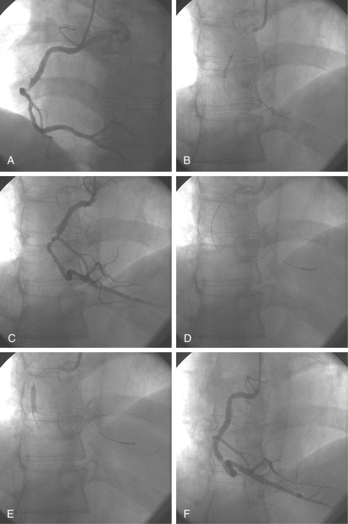
  • Angioplasty and coronary stents (Fig. E3).
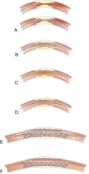
  • PCI has an established place in treating angina but is not superior to intensive medical therapy to prevent MI and death in symptomatic or asymptomatic patients. Patients selected for PCI should also be candidates for CABG. Approximately 80% of patients show immediate benefit after PCI. The development of coronary stents has increased the number of patients who can be treated in the cardiac laboratory. Cardiac stents (Fig. E4) are currently used in nearly 95% of all patients with PCI lesions. The rate of restenosis is reduced by placing a stent electively in primary atheromatous lesions. The major limitations of stenting are subacute thrombosis, restenosis within the stent, bleeding complications when antiplatelets are used after stenting, and higher cost. The combination of aspirin and P2Y12 antagonists is effective in preventing coronary stent thrombosis and the duration of therapy depends on whether bare metal stents (BMS) or drug-eluting stents (DES) are used. Duration of dual antiplatelet therapy can be as short as 4 wk for BMS, but 6 to 12 mo of therapy is generally required for DES. This difference in duration is due to the lack of endothelium proliferation in DES initially. New drug-eluting stents with thin struts releasing Limus-family analogs from durable polymers have lowered the risk of stent thrombosis compared with early-generation stents releasing sirolimus or paclitaxel. Current evidence supports the use of drug-eluting stents in most clinical settings without safety concerns (unless there are contraindications to use of dual antiplatelet therapy).2

Figure E3 Coronary angioplasty procedure.

A, Critical stenosis in midsegment of a right coronary artery. B, Inflation of a 3.5-mm-diameter angioplasty balloon. C, Angiographic control after balloon inflation. D, Placement of a 3.5-mm-diameter, 18-mm-long metal stent. E, Inflation of the balloon. F, Final result.

From Vincent JL et al: Textbook of critical care, ed 6, Philadelphia, 2011, Saunders.

Figure E4 Implantation of a coronary stent.

A, Placement of balloon catheter. B, Predilation with balloon catheter. C, Balloon is withdrawn. D, Placement of coronary stent, which has been crimped on a balloon catheter. E, Inflation of balloon and expansion of stent. F, Withdrawal of balloon catheter and final result.

From Vincent JL et al: Textbook of critical care, ed 6, Philadelphia, 2011, Saunders.

Pearls & Considerations

Comments

  • Although nitrate responsiveness is usually an integral part of a diagnostic strategy for SIHD, recent reports question its value and conclude that in a general population admitted for chest pain, relief of pain after nitroglycerin treatment does not predict active CAD and should not be used to guide diagnosis in the acute care setting.
  • CABG is associated with higher long-term survival rates and lower rates of repeat revascularization than PCI and stenting; however, patients often prefer stenting because it is less invasive, involves a shorter hospital stay, and has a lower in-hospital mortality rate.
Related Content

Angina (Patient Information)

Unstable Angina (Patient Information)

Acute Coronary Syndrome (Related Key Topic)

Coronary Artery Disease (Related Key Topic)

Myocardial Infarction (Related Key Topic)

Related Content

    1. ACCF/AHA/ACP/AATS/PCNA/SCAI/STS guideline for the diagnosis and management of patients with stable ischemic heart disease: a report of the American College of Cardiology Foundation/American Heart Association Task Force on Practice guidelines, and the American College of Physicians, American Association for Thoracic Surgery, Preventive Cardiovascular Nurses Association, Society for Cardiovascular Angiography and Interventions, and Society of Thoracic SurgeonsJ Am Coll Cardiol. ;60(24):e44-e164, 2012.
    2. 2021 ACC/AHA/SCAI guideline for coronary artery revascularization: a report of the American College of Cardiology/American Heart Association Joint Committee on Clinical Practice GuidelinesCirculation. ;145(3):e18-e114, 2022.
    3. Maurovich-Horvat P. : CT or invasive anigography in stable chest pain 10.1056 N Engl J Med. ;386:1591-1602, 2022.