section name header

Basic Information

AUTHORS: Sahar Mahani, MD and Craig L. Basman, MD, FACC, FSCAI

Definition

Coronary artery disease (CAD) is a clinical syndrome that suggests limitation of coronary blood flow to the myocardium as a result of atherosclerotic lesions. Atherosclerosis can be defined as the narrowing of the artery due to plaque formation in the setting of lipid accumulation inside the arterial walls. It is silent in the early stages and characterized by exertional symptoms in later stages as described in the following text.

This topic addresses only stable CAD. Acute coronary syndromes (ACSs), angina pectoris, and myocardial infarction (MI) are addressed as separate topics elsewhere.

Synonyms

Atherosclerotic heart disease

CAD

Chronic stable angina

Stable ischemic heart disease

Coronary arteriosclerosis

ICD-10CM CODES
I25.0Atherosclerotic cardiovascular disease
I25.1Atherosclerotic heart disease
I25.10Atherosclerotic heart disease of native coronary artery without angina pectoris
I25.110Atherosclerotic heart disease of native coronary artery with unstable angina pectoris
I25.111Atherosclerotic heart disease of native coronary artery with angina pectoris with documented spasm
I25.118Atherosclerotic heart disease of native coronary artery with other forms of angina pectoris
I25.119Atherosclerotic heart disease of native coronary artery with unspecified angina pectoris
Epidemiology & Demographics
Incidence

  • For persons who are 40 yr old, the lifetime risk of developing CAD is 49% in men and 32% in women. The risk increases with age in both men and women; however, the incidence in women lags behind that seen in men by approximately 10 yr.
  • The incidence in premenopausal women is relatively low, whereas the incidence increases significantly in postmenopausal women.
  • The initial presentation in men is more likely to be that of an MI, whereas women often present initially with angina.
Prevalence

The 2017 Heart Disease and Stroke Statistics update from the American Heart Association estimated that 16.5 million Americans 20 yr of age have CAD. There is a slight male predominance at 55%, with incidence increasing with age. CAD prevalence is 7.9% for men and 5.1% for women.1

Risk Factors

The most prevalent risk factors include tobacco use, diabetes mellitus, hypertension, hyperlipidemia, obesity, family history, age, male sex, and peripheral vascular disease. Coronary plaque, especially noncalcified plaque, is more prevalent and extensive in HIV-infected men, independent of CAD risk factors.2,3

Physical Findings & Clinical Presentation

  • Left anterior chest discomfort is often described as squeezing, heavy pressure, and burning. Associated symptoms include fatigue, dyspnea, weakness, lightheadedness, nausea, diaphoresis, altered mental status, and syncope.
  • Discomfort that radiates to the neck, jaw, shoulder, and arm (left more than right) is common.
  • Typical angina is exertional, resolves with rest after 3 to 5 min, and rarely lasts more than 30 min.
  • Women and diabetes may not present with classic symptoms but may manifest more frequently with dyspnea or GI complaints.
  • Discomfort is elicited by physical exertion, emotional stress, cold exposure, consumption of a large meal, and smoking.
  • Early coronary disease is asymptomatic.
  • Stigmata of atherosclerosis may include xanthelasma, tendon xanthomata, and evidence of peripheral vascular disease such as claudication and diminished peripheral pulses.
Etiology

The process of atherosclerosis begins when the endothelium is damaged or dysfunctional by a variety of different pathways/disease states: Hypertension, hyperlipidemia, trauma, toxins from drugs or tobacco use, or endothelial dysfunction sometime thought to be related to genetics. When the endothelium is dysfunctional, low-density lipoprotein (LDL) cholesterol circulating in the blood begins to accumulate within the intima. As the cholesterol accumulates within this plaque precursor, it begins to oxidize over time, which subsequently signals monocytes to migrate into the intima and convert to macrophages-this accumulation is known as a fatty streak. The macrophage cells enlarge and engulf cholesterol but can become overwhelmed and subsequently undergo apoptosis, leaving behind foam cells (the remnants of cholesterol-filled macrophages), as well as releasing inflammatory cytokines. Atherogenesis is further propagated by this resultant cellular necrosis within the forming plaque, promoting inflammatory mediator expression, intimal thickening, and migration of more macrophage cells.

Initially atheromatous plaques develop in an outward direction-positive remodeling, with enlargement of the external radius of the artery, thereby maintaining the inner luminal diameter and thus blood flow. Luminal obstruction and vascular calcification are both later stages of atherogenesis. As the process continues, a well-defined core of extracellular lipids form-the collection is, at this stage, called an atheroma or fibrous plaque. Signals from the apoptotic macrophages prompt smooth muscle cell migration from the media, accelerating plaque formation by multiple actions, including secretion of collagen and elastin forming a protein fibrous cap, deposition of calcium, and releasing signals for neovascularization. By this time in the formation of the atheroma, the internal elastin membrane has become dysfunctional, allowing the passage of smooth muscle cells and perforations of neovascularization. Due to the previously mentioned changes and thickening of the intimal layer, there can be luminal narrowing, which reduces flow and ultimately can lead to symptoms of ischemia. This process of atherosclerosis typically occurs over many years.4

Diagnosis

Differential Diagnosis of Acute Chest Pain

  • Cardiovascular causes
    1. Acute coronary syndrome
    2. Aortic dissection
    3. Pericarditis
    4. Coronary arterial vasospasm
  • Pulmonary causes
    1. Pulmonary embolism
    2. Pneumonia
    3. Pleuritis
    4. Pneumothorax
  • Gastrointestinal causes
    1. Esophageal spasm
    2. Esophagitis
    3. GERD
  • Chest wall causes
    1. Rib fracture
    2. Sternoclavicular arthritis
    3. Herpes zoster
    4. Costochondritis
  • Psychiatric causes
    1. Anxiety disorders
Workup

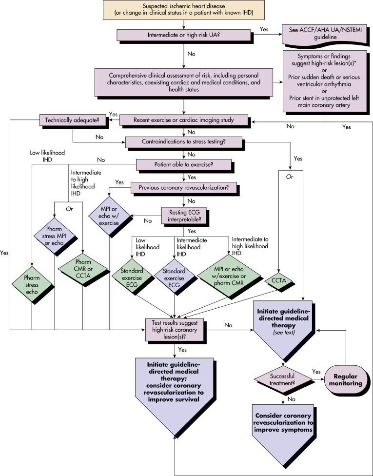
See Fig. 1. ACC/HFA guidelines for stress testing and advanced imaging are summarized in Tables 1 to 3 to .

TABLE 3 ACC/AHA Guidelines for Coronary Angiography to Assess Risk in Patients With Known or Suspected Stable Ischemic Heart Disease

ClassIndicationLevel of Evidence
I (indicated)
  1. Patients with SIHD who have survived sudden cardiac death or potentially life-threatening ventricular arrhythmia should undergo coronary angiography to assess cardiac risk.
B
  1. Patients with SIHD in whom symptoms and signs of heart failure develop should be evaluated to determine whether coronary angiography should be performed for risk assessment.
B
  1. Coronary arteriography is recommended for patients with SIHD whose clinical characteristics and results of noninvasive testing indicate a high likelihood of severe IHD and when the benefits are deemed to exceed risk.
C
IIa (good supportive evidence)
  1. Coronary angiography is reasonable to further assess risk in patients with SIHD who have depressed LV function (EF <50%) and moderate-risk criteria on noninvasive testing with demonstrable ischemia.
C
  1. Coronary angiography is reasonable to further assess risk in patients with SIHD and inconclusive prognostic information after noninvasive testing or in patients for whom noninvasive testing is contraindicated or inadequate.
C
  1. Coronary angiography for risk assessment is reasonable for patients with SIHD who have unsatisfactory quality of life because of angina, have preserved LV function (EF >50%), and have intermediate-risk criteria on noninvasive testing.
C
III (no benefit)
  1. Coronary angiography for risk assessment is not recommended in patients with SIHD who elect not to undergo revascularization or who are not candidates for revascularization because of comorbid conditions or individual preferences.
B
  1. Coronary angiography is not recommended to further assess risk in patients with SIHD who have preserved LV function (EF >50%) and low-risk criteria on noninvasive testing.
B
  1. Coronary angiography is not recommended to assess risk in patients who are at low risk according to clinical criteria and who have not undergone noninvasive risk testing.
C
  1. Coronary angiography is not recommended to assess risk in asymptomatic patients with no evidence of ischemia on noninvasive testing.
C

ACC/AHA, American College of Cardiology/American Heart Association; EF, ejection fraction; IHD, ischemic heart disease; LV, left ventricle; SIHD, stable ischemic heart disease.

From Zipes DP: Braunwald’s heart disease: a textbook of cardiovascular medicine, ed 11, Philadelphia, 2019, Elsevier.

TABLE 2 ACC/AHA Guidelines for Stress Testing and Advanced Imaging for Patients With Known Stable Ischemic Heart Disease Who Require Noninvasive Testing for Risk Assessment

TestExercise StatusECG InterpretableAdditional ConsiderationsRecommendationLevel of Evidence
AbleUnableYesNo
Patients Able to Exercise
Exercise ECGXXIB
Exercise ECG with MPI or echoXXAbnormalities other than LBBB or ventricular pacingIB
Exercise ECG with MPI or echoXXIIaB
Pharmacologic stress CMRXXIIaB
CCTAXXIIbB
Pharmacologic stress imaging or CCTAXXIIIC
Patients Unable to Exercise
Pharmacologic stress with nuclear MPI or echoXEitherIB
Pharmacologic stress CMRXEitherIIaB
CCTAXEitherWithout previous stress testIIaC
Regardless of Ability to Exercise
Pharmacologic stress with nuclear MPI or echoEitherXLBBB presentIB
Exercise or pharmacologic stress with nuclear MPI, echo, or CMREitherEitherKnown coronary stenosis being considered for revascularizationIB
CCTAEitherEitherIndeterminate result of functional testingIIaC
EitherEitherUnable to undergo stress imagingIIbC
EitherEitherAlternative to invasive coronary angiography when functional testing indicates moderate to high riskIIbC
Multiple stress tests or cardiac imaging at the same timeEitherEitherIII

ACC/AHA, American College of Cardiology/American Heart Association; CCTA, coronary computed tomography angiography; CMR, cardiac magnetic resonance; ECG, electrocardiography; echo, echocardiography; LBBB, left bundle branch block; MPI, myocardial perfusion imaging.

From Zipes DP: Braunwald’s heart disease: a textbook of cardiovascular medicine, ed 11, Philadelphia, 2019, Elsevier.

TABLE 1 ACC/AHA Guidelines for Stress Testing and Advanced Imaging for Initial Diagnosis in Patients With Suspected Stable Ischemic Heart Disease Who Require Noninvasive Testing

TestExercise StatusECG InterpretablePretest Probability of Ischemic Heart DiseaseRecommendationLevel of Evidence
AbleUnableYesNoLowIntermediateHigh
Patients Able to Exercise
Exercise ECGXXXIA
Exercise ECG with MPI or echoXXXXIB
Exercise ECGXXXIIaC
Exercise ECG with MPI or echoXXXXIIaB
Pharmacologic stress CMRXXXXIIaB
CCTAXEitherXIIbB
Exercise echoXXXIIbC
Pharmacologic stress with nuclear MPI, echo, or CMRXXAnyIIIC
Exercise stress with MPIXXXIIIC
Patients Unable to Exercise
Pharmacologic stress with nuclear MPI or echoXEitherXXIB
Pharmacologic stress echoXEitherXIIaC
CCTAXEitherXXIIaB
Pharmacologic stress CMRXEitherXXIIaB
Exercise ECGXXAnyIIIC
Other Reasons for Cardiac Computed Tomography Angiography
Continued symptoms after normal test results
Inconclusive stress test results
Unable to undergo stress test
EitherEitherXIIaC
CACEitherEitherXIIbC

ACC/AHA, American College of Cardiology/American Heart Association; CAC, coronary artery calcium (imaging); CCTA, coronary computed tomography angiography; CMR, cardiac magnetic resonance; ECG, electrocardiography; echo, echocardiography; MPI, myocardial perfusion imaging.

From Zipes DP: Braunwald’s heart disease: a textbook of cardiovascular medicine, ed 11, Philadelphia, 2019, Elsevier.

Figure 1 Diagnosis of patients with suspected ischemic heart disease.

!!flowchart!!

CCTA is reasonable only for patients with intermediate probability of IHD. ACC/AHA, American College of Cardiology Foundation/American Heart Association; CCTA, computed coronary tomography angiography; CMR, cardiac magnetic resonance; ECG, electrocardiogram; echo, echocardiography; IHD, ischemic heart disease; MPI, myocardial perfusion imaging; Pharm, pharmacologic; UA, unstable angina; UA/NSTEMI, unstable angina/non-ST-elevation myocardial infarction.

From 2012 ACC/AHA/ACP/AATS/PCNA/SCAI/STS Guideline for the Diagnosis and Management of Patients With Stable Ischemic Heart Disease: a report of the American College of Cardiology/American Heart Association Task Force on Practice Guidelines, and the American College of Physicians, American Association for Thoracic Surgery, Preventive Cardiovascular Nurses Association, Society for Cardiovascular Angiography and Interventions, and Society of Thoracic Surgeons, J Am Coll Cardiol 60:e44-e164, 2012.

Laboratory Tests

  • Focused metabolic studies to rule out noncardiac causes (basic metabolic panel, liver function tests).
  • CBC count can help rule out anemia-related chest discomfort.
  • Fasting cholesterol panel is important in assessment of lipid-related risk factors.
  • Hemoglobin A1c is important to follow glycemic control.
  • High-sensitivity C-reactive protein.
Imaging Studies

  • When choosing the appropriate imaging study, one must keep in mind the patient’s baseline activity levels and baseline ECG findings. In general, an exercise-based stress test yields a better idea of the patient’s ischemic burden. Furthermore, each imaging modality focuses on a different element within the ischemia cascade. The ischemia cascade suggests that, with coronary artery luminal narrowing, there is a progression of findings as the demand for oxygen-carrying hemoglobin overwhelms the delivered blood supply. The ischemia cascade: Perfusion defect >diastolic dysfunction >systolic dysfunction >ECG changes and finally chest pain. See Tables 1 and 2 for further guidance on appropriate stress testing.
  • Electrocardiography (ECG) is important for assessing ischemia or prior infarct.
  • Echocardiogram to assess left ventricular ejection fraction (LVEF) and the presence of wall motion abnormalities.
  • Exercise treadmill test (ETT) is the gold standard to assess physiologic cardiac stress response in patient with low or intermediate risk if the patient is able to exercise and has an “interpretable” baseline ECG (normal, right bundle branch block [RBBB], left ventricular hypertrophy [LVH] without repolarization abnormalities, <1 mm ST depression at rest). ETT provides high sensitivity but poor specificity. Therefore, if ETT is positive, further imaging studies may be obtained.
  • Stress tests combined with imaging (exercise stress echo or exercise myocardial perfusion study) are the next best options if ETT cannot be performed because of physical limitation, if baseline ECG is uninterpretable or if ETT is positive.
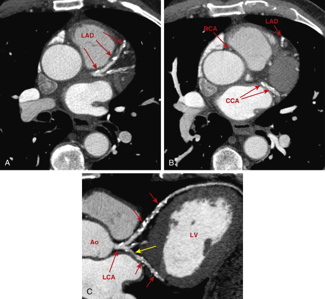
  • Although not used for angina or in an acute setting, a coronary artery calcium score can be used to improve CVD risk classification. The coronary artery calcium (CAC) is estimated from a noncontrast CT (Fig. E2) scan. Normal coronary arteries do not have plaques or calcium, and the normal score is 0. A CAC score of 300 or higher, or 75th percentile or higher for age, sex, and ethnicity is considered a high risk. According to the recent ACC/AHA guidelines, CAC is most appropriate among adults with an estimated 10-yr ASCVD risk <7.5% in whom questions remain about whether statin therapy is indicated. A CAC score >75th percentile for age, sex, and ethnicity is considered high risk and would justify revising a patient’s risk upward.5
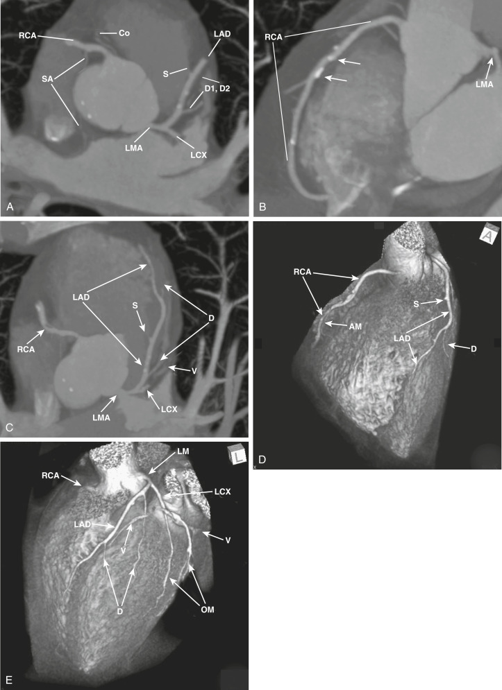
  • Cardiac computed tomography angiography (CCTA, Fig. E3) is an emerging imaging modality that can be considered as an alternative to stress testing. CCTA has excellent negative predictive value for obstructive CAD. CCTA may also identify plaque, which may change management (i.e., addition of a statin).
  • Those with high-risk features (Box 1) should undergo invasive coronary angiography to assess coronary anatomy for revascularization.
  • Coronary angiography is useful for both diagnostic and therapeutic interventions if there is evidence of significant ischemia by noninvasive assessment or progressive symptoms despite optimal medical therapy (as in the following).6,7

BOX 1 Noninvasive Risk Stratification

High risk (>3% annual mortality rate)
  1. Severe resting left ventricular dysfunction (LVEF <35%)
  2. High-risk treadmill score (–11)
  3. Severe exercise left ventricular dysfunction (exercise LVEF <35%)
  4. Stress-induced large perfusion defect (particularly if anterior)
  5. Stress-induced multiple perfusion defects of moderate size
  6. Large, fixed perfusion defect with LV dilation or increased lung uptake (thallium-201)
  7. Stress-induced moderate perfusion defect with LV dilation or increased lung uptake (thallium-201)
  8. Echocardiographic wall motion abnormality (involving >2 segments) developing at low dose of dobutamine (10 mg/kg/min) or at a low heart rate (<120 beats/min)
  9. Stress echocardiographic evidence of extensive ischemia
Intermediate risk (1%-3% annual mortality rate)
  1. Mild/moderate resting left ventricular dysfunction (LVEF 35%-49%)
  2. Intermediate-risk treadmill score (score between –11 and <5)
  3. Stress-induced moderate perfusion defect without LV dilation or increased lung intake (thallium-201)
  4. Limited stress echocardiographic ischemia with a wall motion abnormality only at doses of dobutamine involving 2 segments
Low risk (<1% annual mortality rate)
  1. Low-risk treadmill score (5)
  2. Normal or small myocardial perfusion defect at rest or with stress
  3. Normal stress echocardiographic wall motion or no change of limited resting wall motion abnormalities during stress

Figure E2 Coronary artery calcification and computed tomography angiography.

A, Electrocardiogram-gated contrast-enhanced computed tomography shows calcification of the left anterior descending (LAD) coronary artery and a branch. B, At a lower level, calcification (arrows) of the LAD artery and circumflex coronary artery (CCA) is visible. The right coronary artery (RCA) is noncalcified. C, A curved reconstruction showing the left coronary artery (LCA) and the LAD artery and CCA along their axes. Multiple calcifications are visible (red arrows). A noncalcified stenosis of the CCA (yellow arrow) is also shown. Ao, Aorta; LV, left ventricle.

From Webb WR et al: Fundamentals of body CT, ed 4, Philadelphia, 2015, Saunders.

FIG E3 Normal Coronary Artery Anatomy, as Visualized by Computed Tomography Angiography

A, Axial maximum intensity projection (MIP) slab image showing the courses of the proximal coronary arteries. B, An oblique coronal MIP reformatted image of the right coronary artery (RCA). Focal hyperdensities within the RCA (arrows) are foci of calcific atherosclerosis. The proximal left main coronary artery (LMA) is also visualized. C, An oblique axial MIP reformatted image of the left anterior descending (LAD) artery. A small septal perforator (S) and diagonal branches (D) arise from the LAD. Nearby coronary veins (V) are also visualized. D and E, Volume-rendered reformatted images of the coronary arteries. AM, Acute marginal; Co, conus branch; LCX, left circumflex artery; OM, obtuse marginal; Sa, SA (sinoatrial) nodal branch.

From Soto JA, Lucey BC: Emergency Radiology: the requisites, ed 2, Philadelphia, 2017, Elsevier.

Treatment

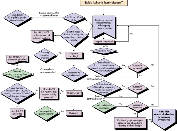
Figure 4 Algorithm for Guideline-Directed Medical Therapy for Patients with Stable Ischemic Heart Disease

The algorithms do not represent a comprehensive list of recommendations (see text for all recommendations).The use of bile acid sequestrant is relatively contraindicated when triglycerides are 200 mg/dl and is contraindicated when triglycerides are 500 mg/dl.Dietary supplement niacin must not be used as a substitute for prescription niacin. ACC, American College of Cardiology; ACEI, angiotensin-converting enzyme inhibitor; AHA, American Heart Association; ARB, angiotensin-receptor blocker; ASA, aspirin; ATP III, Adult Treatment Panel 3; BP, blood pressure; CCB, calcium channel blocker; CKD, chronic kidney disease; JNC VII, Seventh Report of the Joint National Committee on Prevention, Detection, Evaluation, and Treatment of High Blood Pressure; LV, left ventricular; MI, myocardial infarction; NHLBI, National Heart, Lung, and Blood Institute; NTG, nitroglycerin; SIHD, stable ischemic heart disease.

From Fihn SD et al: 2012 ACC/AHA/ACP/AATS/PCNA/SCAI/STS Guideline for the diagnosis and management of patients with stable ischemic heart disease: a report of the American College of Cardiology Foundation/American Heart Association Task Force on Practice Guidelines, and the American College of Physicians, American Association for Thoracic Surgery, Preventive Cardiovascular Nurses Association, Society for Cardiovascular Angiography and Interventions, and Society of Thoracic Surgeons, J Am Coll Cardiol 60:e44-e164, 2012.

TABLE 4 ACCF/AHA Guidelines for Risk Factor Modification

ClassIndicationLevel of Evidence
Lipid Management
I (indicated)
  1. Lifestyle modifications, including daily physical activity and weight management, are strongly recommended for all patients with SIHD.
B
  1. Dietary therapy for all patients should include reduced intake of saturated fats (to <7% of total calories), trans fatty acids (to <1% of total calories), and cholesterol (to <200 mg/day).
B
  1. In addition to therapeutic lifestyle changes, a moderate or high dose of a statin should be prescribed in the absence of contraindications or documented adverse effects.
A
IIa (good supportive evidence)For patients who do not tolerate statins, LDL cholesterol-lowering therapy with bile acid sequestrants, niacin, or both is reasonable.B
Blood Pressure Management
I (indicated)
  1. All patients should be counseled about the need for lifestyle modification: Weight control; increased physical activity; alcohol moderation; sodium reduction; and emphasis on increased consumption of fresh fruits, vegetables, and low-fat dairy products.
B
  1. In patients with SIHD and a BP of 140/90 mm Hg or higher, antihypertensive drug therapy should be instituted in addition to or after a trial of lifestyle modifications.
A
  1. The specific medications used for the treatment of high BP should be based on specific patient characteristics and may include ACE inhibitors and/or beta-blocking agents, as well as the addition of other drugs such as thiazide diuretics or calcium channel blocking agents if needed to achieve a goal BP of less than 140/90 mm Hg.
B
Diabetes Management
IIa (good supportive evidence)
  1. For selected individual patients, such as those with a short duration of diabetes mellitus and a long life expectancy, a goal hemoglobin A1c (HbA1c) of 7% or less is reasonable.
B
  1. A goal HbA1c between 7% and 9% is reasonable for certain patients according to age, history of hypoglycemia, presence of microvascular or macrovascular complications, or presence of coexisting medical conditions.
C
IIb (weak supportive evidence)Initiation of pharmacotherapy interventions to achieve a target HbA1c might be reasonable.A
III (not indicated)Therapy with rosiglitazone should not be initiated in patients with SIHD.C
Physical Activity
I (indicated)
  1. For all patients, clinicians should encourage 30-60 min of moderate-intensity aerobic activity at least 5 days and preferably 7 days/wk, supplemented by an increase in daily lifestyle activities (e.g., walking breaks at work, gardening, household work) to improve cardiorespiratory fitness and move patients out of the least-fit, least-active, high-risk cohort (bottom 20%).
B
  1. For all patients, risk assessment with a physical activity history and/or an exercise test is recommended to guide prognosis and prescription.
B
  1. Medically supervised programs (cardiac rehabilitation) and physician-directed, home-based programs are recommended for at-risk patients at first diagnosis.
A
IIa (good supportive evidence)It is reasonable for clinicians to recommend complementary resistance training at least 2 days/wk.C
Weight Management
I (indicated)
  1. BMI and/or waist circumference should be assessed at every visit, and clinicians should consistently encourage weight maintenance or reduction through an appropriate balance of lifestyle physical activity, structured exercise, caloric intake, and formal behavioral programs when indicated to maintain or achieve a BMI of between 18.5 and 24.9 kg/m2 and a waist circumference of less than 102 cm (40 inches) in men and less than 88 cm (35 inches) in women (less for certain racial groups).
B
  1. The initial goal of weight loss therapy should be to reduce body weight by approximately 5%-10% from baseline. With success, further weight loss can be attempted if indicated.
C
Smoking Cessation
I (indicated)Smoking cessation and avoidance of exposure to environmental tobacco smoke at work and home should be encouraged for all patients with SIHD. Follow-up, referral to special programs, and pharmacotherapy are recommended, as is a stepwise strategy for smoking cessation (Ask, Advise, Assess, Assist, Arrange, Avoid).B
Management of Psychologic Factors
IIa (good supportive evidence)It is reasonable to consider screening patients with SIHD for depression and to refer or treat when indicated.B
IIb (weak supportive evidence)Treatment of depression has not been shown to improve cardiovascular disease outcomes but might be reasonable for its other clinical benefits.C
Alcohol Consumption
IIb (weak supportive evidence)In patients with SIHD who drink alcohol, it might be reasonable for nonpregnant women to have 1 drink (4 oz of wine, 12 oz of beer, or 1 oz of spirits) a day and for men to have 1 or 2 drinks a day unless alcohol is contraindicated (such as in patients with a history of alcohol abuse or dependence or those with liver disease).C
Exposure to Air Pollution
IIa (good supportive evidence)It is reasonable for patients with SIHD to avoid exposure to increased air pollution to reduce their risk for cardiovascular events.C

ACE, Angiotensin-converting enzyme; ACC/AHA, American College of Cardiology/American Heart Association; BMI, body mass index; BP, blood pressure; LDL, low-density lipoprotein; SIHD, stable ischemic heart disease.

From Zipes DP: Braunwald’s heart disease: a textbook of cardiovascular medicine, ed 11, Philadelphia, 2019, Elsevier.

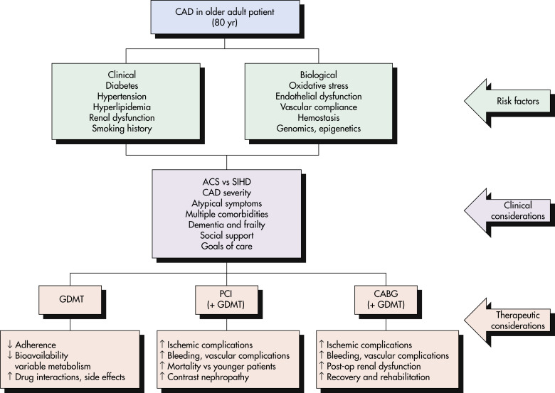
Figure E5 Risk Factors, Clinical Influences, and Treatment Considerations in Older Patients with Coronary Artery Disease (CAD)

ACS, Acute coronary syndrome; CABG, coronary artery bypass grafting; GDMT, Goal directed medical therapy; PCI, percutaneous coronary intervention; SIHD, stable ischemic heart disease.

From Madhavan MV et al: Coronary artery disease in patients 80 yr of age, J Am Coll Cardiol 71:2015-2040, 2018. With permission. In Warshaw G et al: Ham’s primary care geriatrics, ed 7, Philadelphia, 2022, Elsevier.

Nonpharmacologic Therapy
Lifestyle Modification

  • 150 min/wk of moderate-intensity activity or 75 min/wk of vigorous-intensity activity or a combination thereof (for adults) as one of the seven components of ideal cardiovascular health.
  • A Mediterranean diet with a focus on vegetables, fruit, fish, whole grains, and olive oil has proven to reduce cardiovascular events to a degree greater than low-fat diets and equal to or greater than the benefit observed in statin trials.8
  • Smoking cessation.
  • Weight reduction can reduce various risk factors with improved lipid, blood pressure, and glycemic control.
Therapeutic Interventions

Coronary angiography with percutaneous coronary intervention (PCI) or coronary artery bypass graft (CABG) for patients on optimal medical therapy with persistent symptoms.

Acute General Rx for Stable Angina

  • Rest
  • Sublingual nitroglycerin if rest does not provide adequate relief
Chronic Rx

  • Antianginal therapy:
    1. Nitrates (isosorbide mononitrate and isosorbide dinitrate). These medications treat ischemia by venodilation to decrease preload, dilate epicardial coronary arteries, and recruit coronary collaterals. Furthermore, they attenuate platelet aggregation. Although they do not influence survival or decrease cardiovascular death in patients with chronic CAD, they do lower the rate of angina frequency and increase time to ischemic ECG findings on treadmill testing.
    2. Beta-adrenergic antagonists (metoprolol, atenolol, carvedilol, or any other beta-blockers with the exception of those with intrinsic sympathomimetic activity). These medications work to relieve angina by decreasing myocardial oxygen demand by reducing heart rate, blood pressure, and contractility.
    3. Calcium channel blockers (CCBs; amlodipine or verapamil). The antianginal efficacy of CCBs is comparable to beta-blockers.
    4. Ranolazine. This is a selective inhibitor of late sodium influx into myocytes, which leads to decreased myocardial contractility. It can be used in combination with beta-blockers and significantly reduces frequency of angina and increases exercise duration and time to onset of angina. Although it rarely may cause QT prolongation, it has not been linked to any clinically important arrhythmias.9
    5. May benefit from combination therapy of the above.
  • Antiplatelet therapy:
    1. Aspirin therapy (81 mg/day).10 In regard to pulmonary prevention the 2021 U.S. Preventive Services Task Force (USPSTF) recommendation statement on the use of low-dose aspirin for middle-age people (ages 40 to 59) with a 10-year cardiovascular risk 10% states that the decision should be an individual one and respect professional judgment and patient preference. For age 60 the USPSTF recommends against low-dose aspirin for primary prevention.11
    2. Clopidogrel for those intolerant to aspirin therapy.
    3. Combination of aspirin and clopidogrel does not reduce cardiovascular events in stable CAD.12
    4. Antiplatelet drugs such as prasugrel and ticagrelor are used for ACS and do not have a recommended role in patients with stable CAD.
  • Statins:
    1. HMG-CoA reductase inhibitors (statins) with high-intensity therapy for a target LDL reduction of >50% if safely achieved in high-risk patients; if not a candidate for high-intensity therapy, patient should receive at least moderate-intensity statin therapy that lowers LDL by 30% to 50%.
  • PCSK9 inhibitors:
    1. This is a novel class of monoclonal antibodies that inhibit proprotein convertase subtilisin/kexin type 9. They cause dramatic reductions in LDL cholesterol and have been shown to reduce cardiovascular events. They are generally reserved for patients with familial hypercholesterolemia and patients with established vascular disease with LDL above goal despite maximal tolerated dose of statin.13
  • Colchicine: In a randomized trial involving patients with chronic coronary disease, the risk of cardiovascular events was 31% lower among those who received 0.5 mg of colchicine once daily than among those who received placebo.14,15
  • Glucagon-like peptide 1 receptor agonists (GLP-1RAs) and sodium-glucose co-transporter-2 (SGLT2) inhibitors reduce cardiovascular mortality. GLP-1RAs reduce stroke, and SGLT2 inhibitors reduce heart failure hospitalization and are preferred agents in patients with diabetes who also have, or are at high risk for, CV disease.15a,15b
Coronary Artery Revascularization

  • Patients with symptoms refractory to optimal medical therapy as above or those with high clinical stress testing or an angiographic risk profile and suitable coronary anatomy may benefit from revascularization with either PCI or CABG surgery.16 ACC/AHA guidelines for revascularization are summarized in Tables E5 and 6.
  • ACC/AHA class I indications for bypass surgery in chronic CAD patients include the following:
    1. High grade (>50%) left main CAD
    2. Left main CAD-equivalent anatomy including >70% luminal stenosis in the left anterior descending artery and left circumflex arteries. CABG is also recommended over PCI when high complexity CAD is present. PCI is reasonable in selected patients if equivalent revascularization is possible
    3. Three-vessel disease with LVEF <50%
    4. Single- or two-vessel CAD with a large area of viable myocardium at risk
    5. Severe angina despite medical therapy if CABG can be performed with acceptable risk
  • PCI has not been shown to reduce long-term rates of MI and death in patients with stable chronic CAD and therefore has no class I indications in this group. PCI is suitable in patients with suitable anatomy with refractory or lifestyle-limiting angina who have failed optimal medical therapy. Trials such as COURAGE, ORBITA, and ISCHEMIA have demonstrated no significant difference between optimal medical therapy and PCI in overall survival, MI, and ACS in patients with stable CAD. However, there is 30% crossover to PCI in patients who are managed with optimal medical therapy alone.7,17
  • The SYNTAX trial compared PCI to CABG in patients with stable angina. The trial used a numerical score based on qualitative plaque features on angiography with a primary composite endpoint of stroke, mortality, and MI (MACCE). In patients with low burden of CAD (SYNTAX <22) there was no difference between PCI and CABG up to 5 yr. In patients with intermediate burden (SYNTAX 23-32) there was no difference in MACCE at 5 yr (but a decrease in MI and/or revascularization). In patients with high burden of CAD (SYNTAX >33) it showed that surgical revascularization was associated with a lesser risk MACCE.18
  • Patients with diabetes and multivessel CAD involving the left anterior descending artery should undergo CABG instead of PCI.19
  • Trials continue to support CABG as superior to PCI in diabetic patients (BARI 2D, FREEDOM) with multivessel CAD and should remain the revascularization strategy of choice in this patient population.

TABLE 6 ACC/AHA Guidelines for Revascularization to Improve Symptoms in Patients With Significant Anatomic (>50% Left Main or >70% Nonleft Main Coronary Artery Disease) or Physiologic (Fractional Flow Reserve <0.80) Coronary Artery Stenoses

Clinical SettingRecommendationLevel of Evidence
1 significant stenosis amenable to revascularization and unacceptable angina despite GDMTICABG or PCIA
1 significant stenoses and unacceptable angina in whom GDMT cannot be implemented because of medication contraindications, adverse effects, or patient preferencesIIaCABG or PCIC
Previous CABG with 1 significant stenosis associated with ischemia and unacceptable angina despite GDMTIIaPCIC
IIbCABGC
Complex 3-vessel CAD (e.g., SYNTAX score 22) with or without involvement of the proximal LAD artery and a good candidate for CABGIIaCABG preferred over PCIB
Viable ischemic myocardium that is perfused by coronary arteries that are not amenable to graftingIIbTMR as an adjunct to CABGB
No anatomic or physiologic criteria for revascularizationIIICABG or PCIC

ACC/AHA, American College of Cardiology/American Heart Association; CABG, coronary artery bypass graft; GDMT, guideline-directed medical therapy; LAD, left anterior descending; PCI, percutaneous coronary intervention; SYNTAX, synergy between PCI with taxus and cardiac surgery; TMR, transmyocardial revascularization.

From Zipes DP: Braunwald’s heart disease: a textbook of cardiovascular medicine, ed 11, Philadelphia, 2019, Elsevier.

TABLE E5 ACC/AHA Guidelines for Revascularization to Improve Survival Versus Medical Therapy in Patients With Stable Ischemic Heart Disease

Anatomic SettingClassRecommendationLevel of Evidence
Unprotected Left Main or Complex Coronary Artery Disease
CABG and PCIIHeart team approachC
CABG and PCIIIaCalculation of STS and SYNTAX scoresB
Unprotected Left Main
CABGIB
PCIIIaFor SIHD when both of the following are present:
1. Anatomic conditions associated with a low risk for PCI procedural complications and a high likelihood of a good long-term outcome
2. Clinical characteristics that predict a significantly increased risk for adverse surgical outcomes
B
IIbFor SIHD when both of the following are present:
1. Anatomic conditions associated with a low to intermediate risk for PCI procedural complications and an intermediate to high likelihood of a good long-term outcome
2. Clinical characteristics that predict increased risk for adverse surgical outcomes (e.g., STS-predicted operative mortality >2%)
B
IIIFor SIHD in patients (versus performing CABG) with unfavorable anatomy for PCI and who are good candidates for CABGB
Three-Vessel Coronary Artery Disease with or Without Proximal Left Anterior Descending Coronary Artery Disease
CABGIB
IIaIt is reasonable to choose CABG over PCI in patients with complex 3-vessel CAD (e.g., SYNTAX score 22) who are good candidates for CABGB
PCIIIbB
Two-Vessel Coronary Artery Disease With Proximal Left Anterior Descending Coronary Artery Disease
CABGIB
PCIIIbB
Two-Vessel Coronary Artery Disease Without Proximal Left Anterior Descending Coronary Artery Disease
CABGIIaWith extensive ischemiaB
IIbWithout extensive ischemiaC
PCIIIbB
One-Vessel Proximal Left Anterior Descending Coronary Artery Disease
CABGIIaWith LIMAB
PCIIIbB
One-Vessel Coronary Artery Disease Without Proximal Left Anterior Descending Coronary Artery Disease
CABGIIIHarmB
PCIIIIHarmB
Left Ventricular Dysfunction
CABGIIaEF of 35%-50%B
CABGIIbEF <35% without significant left main diseaseB
PCIN/AInsufficient data
Survivors of Sudden Cardiac Death With Presumed Ischemia-Mediated Ventricular Tachycardia
CABGIB
PCIIC
No Anatomic or Physiologic Criteria for Revascularization
CABGIIIHarmB
PCIIIIHarmB

ACC/AHA, American College of Cardiology/American Heart Association; CABG, coronary artery bypass graft; CAD, coronary artery disease; EF, ejection fraction; LIMA, left internal mammary artery; N/A, not applicable; PCI, percutaneous coronary intervention; SIHD, stable ischemic heart disease; STS, Society of Thoracic Surgery; SYNTAX, synergy between PCI with taxus and cardiac surgery.

From Zipes DP: Braunwald’s heart disease: a textbook of cardiovascular medicine, ed 11, Philadelphia, 2019, Elsevier.

Disposition

  • Risk estimators for cardiovascular events in patients without known coronary artery disease are summarized in Table 7.
  • CAD is a common chronic condition with which many patients can live for years with good symptom control on optimal medical therapy.

TABLE 7 Risk Estimators for Cardiovascular Events in Patients Without Known Coronary Artery Disease

Clinical Risk EstimatorPurpose of Risk EstimatorLimitationsWeb or Mobile App Available
ASCVD Risk Estimator10-yr risk of ASCVD event (CV death, MI, stroke)Not validated for patients 80 yr old, excludes family historyYes
Framingham Risk ScoreEstimates risk for CV death, MI, stable and unstable anginaExcludes family historyYes
MESA Risk Score10-yr risk for CV death, MI, cardiac arrest, coronary revascularizationExcludes family historyYes
Reynolds CVD Risk ScoreEstimate risk for CV death, MI, stroke, revascularizationDoes not account for treatment of hypertensionYes
Adult Treatment Panel (ATP) IIIEstimate risk for death or MIExcludes DM or family historyYes

ASCVD, Atherosclerotic cardiovascular disease; CV, cardiovascular; DM, diabetes mellitus; MESA, Multi-Ethnic Study of Atherosclerosis; MI, myocardial infarction.

From Warshaw G et al: Ham’s primary care geriatrics, ed 7, Philadelphia, 2022, Elsevier.

Referral

To a cardiovascular disease specialist

Pearls & Considerations

Comments

  • The transition from stable CAD to unstable angina must be carefully monitored. Symptoms of concern include more frequent episodes of chest pain, exertional dyspnea, chest pain that is less responsive to nitroglycerin, or first episode of chest pain. Regarding unstable angina, please refer to section on acute coronary syndromes.
  • ACC/AHA guidelines for follow-up noninvasive testing are summarized in Tables 8 and 9.

TABLE 8 ACC/AHA Guidelines for Follow-Up Noninvasive Testing in Patients With Known Stable Ischemic Heart Disease: New, Recurrent, or Worsening Symptoms (Not Consistent With Unstable Angina)

TestExercise StatusECG InterpretableAdditional ConsiderationsRecommendationLevel of Evidence
AbleUnableYesNo
Patients Able to Exercise
Exercise ECGXXIB
Exercise ECG with MPI or echoXXIB
Exercise ECG with MPI or echoXEitherPrevious requirement for imaging or known to be at high risk for multivessel CADIIaB
Pharmacologic stress MPI, echo, or CMRXXIIIC
Patients Unable to Exercise
Pharmacologic stress with nuclear MPI or echoXEitherIB
Pharmacologic stress CMRXEitherIIaB
Exercise ECGXXIIIC
Regardless of Ability to Exercise
CCTAEitherEitherTo assess patency of coronary stent or bypass graft 3 mm in diameterIIbC
EitherEitherIn absence of known moderate or severe calcification and to assess coronary stent <3 mm in diameterIIbC
EitherEitherKnown moderate or severe calcification or assessment of stent <3 mm in diameterIIIC

ACC/AHA, American College of Cardiology/American Heart Association; CAD, coronary artery disease; CCTA, cardiac computed tomography angiography; CMR, cardiac magnetic resonance; ECG, electrocardiography; echo, echocardiography; MPI, myocardial perfusion imaging.

From Zipes DP: Braunwald’s heart disease: a textbook of cardiovascular medicine, ed 11, Philadelphia, 2019, Elsevier.

TABLE 9 ACC/AHA Guidelines for Follow-Up Noninvasive Testing in Patients With Known Stable Ischemic Heart Disease: Asymptomatic or Stable Symptoms

TestExercise StatusECG InterpretablePretest Probability of IschemiaAdditional ConsiderationsRecommendationLevel of Evidence
AbleUnableYesNo
Exercise or pharmacologic stress with MPI, echo, or CMR at 2-yr intervalsXXPrevious evidence of silent ischemia or at high risk for recurrent eventUnable to exercise, uninterpretable ECG, or incomplete revascularizationIIaC
Exercise ECG at 1-yr intervalsXXPrevious silent ischemia or at high risk for recurrent eventIIbC
Exercise ECGXXNo previous silent ischemia and not at high risk for recurrent eventsIIbC
Exercise or pharmacologic stress imaging or CCTAEitherEither<5-yr intervals after CABG or <2 yr intervals after PCIIIIC

ACC/AHA, American College of Cardiology/American Heart Association; CABG, coronary artery bypass graft; CCTA, cardiac computed tomography angiography; CMR, cardiac magnetic resonance; ECG, electrocardiography; echo, echocardiography; MPI, myocardial perfusion imaging; PCI, percutaneous coronary intervention.

From Zipes DP: Braunwald’s heart disease: a textbook of cardiovascular medicine, ed 11, Philadelphia, 2019, Elsevier.

Related Content

Coronary Artery Disease (Patient Information)

Acute Coronary Syndrome (Related Key Topic)

Angina Pectoris (Related Key Topic)

Hypercholesterolemia (Related Key Topic)

Hyperlipoproteinemia, Primary (Related Key Topic)

Myocardial Infarction (Related Key Topic)

Related Content

    1. Benjamin E.J. : Heart disease and stroke statistics-2017 update: a report from the American Heart AssociationCirculation. ;135(10), 2017.
    2. Goff D.C. : ACC/AHA guideline on the assessment of cardiovascular risk: a report of the American College of Cardiology/American Heart Association task force on practice guidelinesCirculation. ;129(24 Suppl):S49-S73, 2014.
    3. Post W.S. : Associations between HIV infection and subclinical coronary atherosclerosisAnn Intern Med. ;160:458-467, 2014.
    4. Vergallo R., Crea F. : Atherosclerotic plaque healingN Engl J Med. ;383:846-857, 2020.
    5. Polonsky T.S. : Coronary artery calcium scoreJ Am Med Assoc. ;312:837-838, 2014.
    6. Wolk M.J. : ACC/AHA/ASE/ASNC/HFSA/HRS/SCAI/SCCT/SCMR/STS 2013 multimodality appropriate use criteria for the detection of stable ischemic heart diseaseJ Am Coll Cardiol. ;63:380-406, 2014.
    7. Fihn S.D. : ACCF/AHA/ACP/AATS/PCNA/SCAI/STS guideline for the diagnosis and management of patients with stable ischemic heart disease: executive summaryJ Am Coll Cardiol. ;60:2564-2603, 2012.
    8. Estruch R. : Primary prevention of cardiovascular disease with a Mediterranean dietN Engl J Med. ;368:1279-1290, 2013.
    9. Banon D. : The usefulness of ranolazine for the treatment of refractory chronic stable angina pectoris as determined from a systematic review of randomized controlled trialsAm J Cardiol. ;113:1075-1082, 2014.
    10. Nudy M. : Aspirin for primary atherosclerotic cardiovascular disease prevention as baseline risk increases: a meta-regression analysisAm J Med. ;133:1056-1064, 2020.
    11. aspirin use to prevent cardiovascular disease: preventive medication, USPSTF, 2021.https://www.uspreventiveservicestaskforce.org/uspstf/draft-recommendation/aspirin-use-to-prevent-cardiovascular-disease-preventive-medication
    12. Bhatt D.L. : Clopidogrel and aspirin versus aspirin alone for the prevention of atherothrombotic events-CHARISMA trialN Engl J Med. ;354(16):1706-1717, 2006.
    13. Sabatine M.S. : Efficacy and safety of evolocumab in reducing lipids and cardiovascular eventsN Engl J Med. ;372:1500-1509, 2015.
    14. Nidorf S.M. : Colchicine in patients with chronic coronary diseaseN Engl J Med. ;383:1838-1847, 2020.
    15. Tong D.C. : Colchicine in patients with acute coronary syndrome: the Australian COPS randomized clinical trialCirculation. ;142(20):1890-1900, 2020.
    16. Kanie T. : Dipeptidyl peptidase-4 inhibitors, glucagon-like peptide 1 receptor agonists and sodium-glucose co-transporter-2 inhibitors for people with cardiovascular disease: a network meta-analysisCochrane Database Syst Rev. ;10(10), 2021.
    17. Braunwald E. : Gliflozins in the management of cardiovascular diseaseN Engl J Med. ;386(21):2024-2034, 2024.
    18. Stergiopoulos K. : Percutaneous coronary intervention outcomes in patients with stable obstructive coronary artery disease and myocardial ischemia: a collaborative meta-analysis of contemporary randomized clinical trialsJAMA Intern Med. ;174:232-240, 2014.
    19. Boden W.E. : Optimal medical therapy with or without PCI for stable coronary diseaseN Engl J Med. ;356:1503-1516, 2007.
    20. Serruys P.W. : Percutaneous coronary intervention versus coronary-artery bypass grafting for severe coronary artery diseaseN Engl J Med. ;360(10):961-972, 2009.
    21. Lawton J.S. : ACC/AHA/SCAI guideline for coronary artery revascularization: executive summary: a report of the American College of Cardiology/American Heart Association Joint Committee on clinical practice guidelinesCirculation. ;145(3):e4-e17, 2022.