section name header

Basic Information

AUTHORS: Michael J. Farias, BS and John D. Milner, MD and Manuel F. DaSilva, MD

Definition

Rickets is a result of deficient mineralization of osteoid matrix before closure of the epiphyseal plate causing softening and weakening of bones in infants and children. The mineralization impairment may be secondary to abnormal calcium, phosphorus, or vitamin D metabolism leading to accumulation of osteoid before epiphyseal closure, compromising bone stability at sites of rapid bone growth.1 When this occurs in adulthood after epiphyseal closure, it is referred to as osteomalacia. Renal osteodystrophy is a term used to describe a similar condition in patients with chronic kidney disease. Certain forms of the disorder may respond only to high doses of vitamin D and are referred to as vitamin D-resistant rickets (VDRR).2

ICD-10CM CODES
E55.0Rickets, active
E83.30Disorder of phosphorus metabolism, unspecified
M83.9Adult osteomalacia, unspecified
N25.0Renal osteodystrophy
Epidemiology & Demographics
Risk Factors

  • Children ages 6 to 24 mo
  • Premature infants
  • Residents of northern latitudes with inadequate sunlight exposure
  • Solely breastfed infants
  • Darker skin pigmentation
  • Use of anticonvulsants3
Physical Findings & Clinical Presentation

The classic clinical presentation of children with rickets includes:

  • Apathy, muscle weakness, muscle cramps, delayed growth and short stature
  • Skeletal pain and swollen joints (Figs. E1 and E2)
  • Protuberant abdomen due to muscle hypotonia4
  • Widened sutures, delayed closure of fontanelles, and frontal bossing of head1
  • Craniotabes (softening of skull bones)4
  • Delayed eruption of teeth and poorly mineralized teeth4
  • Bowing deformity of lower extremities, specifically femur and tibia (Fig. E3)
  • Table E1 summarizes clinical features of rickets
  • Rachitic rosary (enlargement and prominence of the rib head at the costochondral junctions)1
  • Harrison groove (indentation of the lower ribs)1
  • Breastbone that is pushed forward (pigeon chest)5
  • Pathologic fractures
  • Scoliosis or kyphosis

The less pronounced clinical presentation of adults with osteomalacia includes:4

  • Skeletal pain and bone tenderness
  • Muscle hypotonia and proximal muscle weakness
  • Pathologic fractures and decreased bone density
  • Gait disturbances

Figure E1 Rachitic “rosary” in a child with rickets.

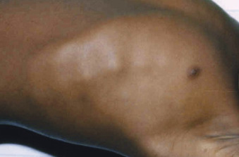
Courtesy Dr. Thomas D. Thacher, Rochester, MN. In Kliegman RM: Nelson textbook of pediatrics, ed 21, Philadelphia, 2020, Elsevier.

Figure E2 Hands and Forearms of a Young Child with Rickets Show Prominence above the Wrist, Resulting from Flaring and Poor Mineralization of the Lower End of the Radius and Ulna

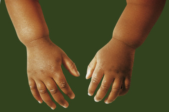
From Bullough PG: Orthopaedic pathology, ed 5, St Louis, 2010, Mosby, Fig 8-31. In Kliegman RM: Nelson textbook of pediatrics, ed 21, Philadelphia, 2020, Elsevier.

Figure E3 (A) A Typical Example of Rickets

Note the Bowing of the Femurs and Tibiae, Which May Result from Vitamin D Deficiency, Phosphate Deficiency, or Other Causes. (B) A Skeletal Radiograph of a Child with Rickets. Note that the Weight-Bearing Bones of the Lower Extremities are Bowed and that the Epiphyses are Open, Mottled, and Overgrown. (C) Looser Zones or Pseudofractures that are Characteristic of Osteomalacia or Rickets. Because the Epiphyses are Closed, the Patient is an Adult. This Radiograph is Diagnostic of Osteomalacia.

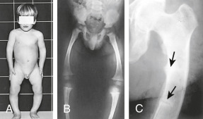
From Stewart A: Metabolic bone diseases. In Andreoli TE et al [eds]: Cecil essentials of medicine, ed 8, Philadelphia, 2010, Saunders.

TABLE E1 Clinical Features of Rickets

General
  • Failure to thrive (malnutrition)
  • Listlessness
  • Protruding abdomen
  • Muscle weakness (especially proximal)
  • Hypocalcemic dilated cardiomyopathy
  • Fractures (pathologic, minimal trauma)
  • Increased intracranial pressure
Head
  • Craniotabes
  • Frontal bossing
  • Delayed fontanel closure (usually closed by 2 yr)
  • Delayed dentition
    • No incisors by age 10 mo
    • No molars by age 18 mo
  • Caries
  • Craniosynostosis
Chest
  • Rachitic rosary
  • Harrison groove
  • Respiratory infections and atelectasis
Back
  • Scoliosis
  • Kyphosis
  • Lordosis
Extremities
  • Enlargement of wrists and ankles
  • Valgus or varus deformities
  • Windswept deformity (valgus deformity of one leg with varus deformity of other leg)
  • Anterior bowing of tibia and femur
  • Coxa vara
  • Leg pain
Hypocalcemic Symptoms
  • Tetany
  • Seizures
  • Stridor caused by laryngeal spasm

These features are associated most frequently with vitamin D deficiency disorders.

These symptoms develop only in children with disorders that produce hypocalcemia.

Etiology

The most common cause of rickets and osteomalacia is vitamin D deficiency. Insufficient calcium and phosphorus intake can also lead to rickets. Vitamin D and parathyroid hormone (PTH) play an important role in calcium homeostasis. Vitamin D is required for adequate calcium absorption in the gastrointestinal tract. Low vitamin D leads to reduced intestinal absorption of calcium, causing a compensatory increase in PTH and resorption of bone, leading to increased alkaline phosphatase levels.3 This may arise from various conditions including inadequate dietary intake, malabsorption, chronic parenteral nutrition, and the additional risk factors listed previously. Malabsorption that causes inefficient mineralization is observed in diseases such as cystic fibrosis, intestinal disorders such as celiac disease, and in many chronic illnesses, including severe liver disease.3

Chronic renal failure can produce bone mineralization deficiency due to electrolyte abnormalities causing hypocalcemia and renal osteodystrophy (renal rickets). This results in decreased excretion of phosphate, therefore elevating serum phosphorus, along with elevated PTH and low levels of 1,25-OH vitamin D.1

Other causes specifically include vitamin D-dependent rickets (VDDR type I and II) and VDRR, also known as hereditary hypophosphatemic rickets.4 (Table E2)

  • VDDR type I (VDDR-1) results from mutations in the vitamin D 1-alpha hydroxylase enzyme that converts inactive vitamin D to the active metabolite of 1,25(OH)2D.
  • VDDR type II (VDDR-II), also known as hereditary vitamin-D resistant rickets (HVDRR), results from defective vitamin D receptors due to a mutation in the vitamin D receptor gene (Table E3).

TABLE E2 Causes of Vitamin D-Deficiency Rickets or Osteomalacia, or Both

Extrinsic
Inadequate dietary intake of vitamin D
Decreased exposure or avoidance of sunlight
Use of sunscreens (especially a solar protective factor >8)
Excess clothing (veil/hijab)
Increased dark skin pigmentation
Intrinsic
Advancing age with decreased cutaneous production of vitamin D
Malabsorption due to various gastrointestinal disorders
  • Gastrectomy (partial, total, or bypass procedure)
  • Small intestinal disease, resection, or bypass
  • Gluten enteropathy (celiac sprue)
  • Biliary cirrhosis (uncommon)
  • Pancreatic insufficiency including cystic fibrosis (uncommon)
Impaired or genetically defective vitamin D 25-hydroxylase enzyme
  • Immaturity
  • Neonatal hepatitis
  • Cirrhosis of the liver
  • Genetic defect (vitamin D-dependent rickets type 1B)
Impaired or genetically defective 25-hydroxyvitamin D 1α-hydroxylase enzyme
  • Genetic defect (vitamin D-dependent rickets type 1A)
  • Chronic renal failure
“Acquired” vitamin D deficiency
  • Increased catabolism due to microsomal enzyme induction
  • Anticonvulsants
  • Calcium deficiency with secondary hyperparathyroidism

From Melmed S et al: Williams textbook of endocrinology, ed 14, 2019, Elsevier.

TABLE E3 Causes of Hypophosphatemic Rickets or Osteomalacia, or Both

Genetic
Autosomal dominant rickets
Autosomal recessive rickets
X-linked dominant rickets (X-linked hypophosphatemia)
X-linked recessive
Hypophosphatemic hypercalciuric hereditary rickets (or Dent disease)
Neurofibromatosis
Fibrous dysplasia
Genetic Fanconi syndrome (may have renal failure)
Acquired
Tumor-induced osteomalacia (most common acquired cause)
Renal tubular damage or nonfamilial Fanconi syndrome
  • Paraproteinemia
  • Wilson disease
  • Galactosemia
  • Tyrosinemia
  • Glycogenosis
Drug induced
  • Tenofovir and adefovir (second most common acquired cause)
  • Phosphate-binding antacids
  • Cadmium poisoning
  • Use of outdated tetracycline
  • Amphotericin

From Melmed S et al: Williams textbook of endocrinology, ed 14, 2019, Elsevier.

Diagnosis

Differential Diagnosis

  • Osteoporosis (Table E4 contrasts features of four metabolic bone diseases)
  • Hyperparathyroidism
  • Hyperthyroidism
  • Hypophosphatasia
  • Skeletal dysplasia

TABLE E4 Contrasting Features of Four Major Metabolic Bone Diseases

VariableRickets and OsteomalaciaOsteoporosisOsteitis FibrosaOsteitis Deformans
Basic abnormalityDefective mineralization of cartilage and boneInsufficient replacement of normal lamellar boneReplacement by woven bone and fibrous tissueAbnormal woven bone
PrevalenceSecond most commonMost commonUncommonNot uncommon
Serum calciumNormal/lowNormalHigh/very highNormal
Serum phosphateNormal/low/very lowNormalFrequently lowNormal
Alkaline phosphataseHighNormal/highHigh/very highHigh
Parathyroid hormoneHighNormal/highHigh/very highNormal
25-Hydroxyvitamin DLow/normalNormal/lowFrequently lowNormal/low
Cortical thinningYes, except in X-linked hypophosphatemiaSometimesYesNo
Vertebral deformitiesBiconcave or cod fishlikeWedged/compressedVariableEnlarged size
Long bone deformitiesBowing and pseudofracturesNone, except fragility fracturesBrown tumorsBowing and stress fractures
Workup

  • Rickets is a clinical diagnosis. It is often challenging to determine the exact etiology; however, clinical, biochemical, and radiologic findings can be useful.
  • A thorough dietary and medication history for children is important. Bone biopsy is generally not performed; however, it is the diagnostic gold standard.6
  • Fig. E4 illustrates a clinical algorithm based on biochemical abnormalities in evaluating patients suspected of having rickets or osteomalacia.

Figure E4 Suggested Clinical Algorithm Based on Biochemical Abnormalities in Evaluating Patients Suspected of Having Rickets, Osteomalacia, or Both

!!flowchart!!

Analytes: Ca, Serum calcium; P, serum phosphate; 25-D, serum 25-hydroxyvitamin D; 1,25-D, 1,25-dihydroxyvitamin D; PTH, serum parathyroid hormone; TmP/GFR, tubular maximum for phosphate reabsorption/glomerular filtration rate; HVO I, hypovitaminosis D osteopathy stage I; TRP, tubular reabsorption of phosphate; AA, urine amino acid analysis; HCO3, urine bicarbonate excretion; Glu, urine glucose. Conditions: VDD, vitamin D deficiency; VDDR, vitamin D-dependent rickets; XLH, X-linked hypophosphatemic rickets and osteomalacia; TIO, tumor-induced osteomalacia; HHRH, hereditary hypophosphatemic rickets with hypercalciuria.

From Melmed S et al: Williams textbook of endocrinology, ed 14, 2019, Elsevier.

Laboratory Tests

Blood testing (Table E5) should include serum calcium, inorganic phosphorus (Pi), alkaline phosphate, PTH, 25-OH vitamin D, creatinine, and liver enzymes. Tables E6 and E7 summarize laboratory findings in various disorders that cause rickets and osteomalacia. Children may present solely with an elevated alkaline phosphatase level and characteristic physical findings.4

TABLE E5 Evaluation of Osteomalacia

In all patients: Calcium, phosphorus, alkaline phosphatase, urinary calcium levels; 25-hydroxyvitamin D and intact parathyroid hormone levels
In selected patients:
  • 1,25-Dihydroxyvitamin D levels (e.g., renal insufficiency and vitamin D-resistant osteomalacia or rickets)
  • Vitamin D absorption test: Obtain 25-hydroxyvitamin D levels at 0, 4, and 8 hr (e.g., some cases of malabsorption)
  • Tubular reabsorption of phosphate (e.g., vitamin D-resistant osteomalacia or rickets)
  • Bone biopsy with double tetracycline labels

From Firestein GS et al: Firestein & Kelley’s textbook of rheumatology, ed 11, Philadelphia, 2021, Elsevier.

TABLE E6 Relevant Abnormalities in Various Types of Vitamin D-Related Rickets and Osteomalacia

VariableVitamin D DeficiencyVitamin D Dependency Type 1BVitamin D Dependency Type 1AVitamin D Dependency Type 2Hypophosphatemic Vitamin D Resistanta
Basic defectNutritional/malabsorption25-Hydroxylase defect1α-Hydroxylase defectVDR defectExcess FGF23
Gene locusNot applicableChromosome 11p15.2Chromosome 12q13.1Chromosome 12q12-q14Xp22.11
Enzyme defectNot applicableCYP2R1CYP27B1Receptor defectPHEX gene defect
Serum calciumLow/normalLowLowLowNormal/high
Serum phosphateNormal/lowNormal/lowNormal/lowNormal/lowVery low
Alkaline phosphataseHighHighHighHighHigh
Parathyroid hormoneHighHighHighHighNormal/high
25-Hydroxyvitamin DLowLow/very lowNormalNormalNormal
1,25-Dihydroxyvitamin DVariableLow/low normalLowHighLow
Urine calciumLowLowLowLowNormal/high
Vitamin D dose required to heal/cure ricketsb1,000-2,000 IU daily
Few weeks to months
10,000 IU daily
Lifelong
10,000 IU daily
Lifelong
100,000 units daily
Lifelong
100,000 units daily
Lifelong
Calcitriol dose required to heal/cure rickets0.04 μg/kg per day
Few weeks to months
0.04 μg/kg per day
Lifelong
0.04 μg/kg per day
Lifelong
1-2 μg/day
Lifelong
1-2 μg/day and oral phosphate
Lifelong

CYP, Cytochrome P-450; FGF23, fibroblast growth factor 23; PHEX, phosphate-regulating endopeptidase homolog, X-linked; VDR, vitamin D receptor.

a Previously referred to as vitamin D-resistant rickets because of the large doses of vitamin D required to heal rickets; the current name is X-linked hypophosphatemic rickets and osteomalacia.

b Shown only to illustrate comparative effective doses. Both calcitriol and alfacalcidol are now widely available and used to treat all vitamin D-dependent rickets and osteomalacia.

TABLE E7 Key Abnormalities in Various Types of Rickets and Osteomalacia

VariableVitamin D DeficiencyVitamin D-Resistant HypophosphatemicTumor-Induced OsteomalaciaDrug-Induced Osteomalacia
Basic defectNutritional/malabsorptionPHEX gene defect
Defective catabolism of FGF23
Ectopic production of FGF23Renal tubular damage
Direct effect on bone mineralization
FGF23 levelsNot applicableUsually highAlmost always highHigh/variable
Serum calciumLow/normalNormal/highNormalVariable
Serum phosphateNormal/lowVery lowLow/very lowVariable
Alkaline phosphataseHighHighHighVariable
Parathyroid hormoneHighNormal/highNormalVariable
25-Hydroxyvitamin DLowNormalNormalLow/normal
1,25-Dihydroxyvitamin DVariableLowLowLow/variable
Bone mineral densityLowNormal/highOften lowOften low
Therapeutic strategyVitamin D + calciumCalcitriol and phosphate
Burosumab
Resection of the tumor
Other therapies (see text)
Discontinuation of offending drug

FGF23, Fibroblast growth factor 23; PHEX, phosphate-regulating endopeptidase homolog, X-linked.

From Melmed S et al: Williams textbook of endocrinology, ed 14, 2019, Elsevier.

Imaging Studies

  • In rickets, characteristic radiologic findings include irregular epiphyseal-metaphyseal junctions with widening and flaring of the epiphyses of long bones, causing bowing.1,4
  • Pseudofractures (Looser zones) and narrow radiolucent lines may be seen as a result of microfractures at high stress points or at the location of entry of blood vessels into the bone and are observed in severe rickets and osteomalacia.4

Treatment

Referral

  • Because of the complex nature of many of these disorders, a qualified endocrinologist and nephrologist should be consulted for treatment.
  • Orthopedic consultation may be required for some cases of bowlegs or spinal deformities, which may require specific bracing for long-term growth of the bones. Corrective surgery may be necessary if the deformity is very severe.
  • Surgical care is also indicated for slipped capital femoral epiphysis, which is common in renal rickets.
Related Content

Neurofibromatosis (Related Key Topic)

Osteoporosis (Related Key Topic)

Vitamin D Deficiency (Patient Information)

Vitamins and Their Functions (Patient Information)

Related Content

  1. Creo A.L. : Nutritional rickets around the world: an updatePaediatr Int Child Health. ;37(2):84-98, 2017.doi:10.1080/20469047.2016.1248170
  2. Vahed L.K. : The frequency of clinical manifestations of hypophosphatemic rickets in patients with therapeutic strategiesClin Pract. ;8(2), 2018.doi:10.4081/cp.2018.1072
  3. Munns C.F. : Global consensus recommendations on prevention and management of nutritional ricketsJ Clin Endocrinol Metab. ;101(2):394-415, 2016.doi:10.1210/jc.2015-2175
  4. Lambert A.S., Linglart A. : Hypocalcaemic and hypophosphatemic ricketsBest Pract Res Clin Endocrinol Metab. ;32(4):455-476, 2018.doi:10.1016/j.beem.2018.05.009
  5. Nagaratnam S. : Debilitating pain and fractures: a rare case of hypophosphatemic osteomalacia with concomitant vitamin D deficiency in neurofibromatosis type 1J ASEAN Fed Endocr Soc. ;35(1):105-108, 2020.doi:10.15605/jafes.035.01.17
  6. Bhan A. : Bone histomorphometry in the evaluation of osteomalaciaBone Rep. ;8:125-134, 2018.doi:10.1016/j.bonr.2018.03.005