section name header

Basic Information

AUTHORS: Natasha Choudhury, MD and Corey Elam Goldsmith, MD, FAAN

Definition

Multiple sclerosis (MS) is a chronic, predominantly autoimmune demyelinating disease of the central nervous system (CNS), characterized by subacute neurologic deficits correlating with CNS lesions (typical for MS in location, shape, and orientation) separated in time (typically at least 1 mo) and space, and excluding other possible disease.1-3

An MS relapse is defined as an acute to subacute (peaking over hours to days) onset of neurologic dysfunction (typically focal) lasting at least 24 h, and caused by inflammatory CNS demyelination. Relapses can be symptomatic or asymptomatic, the latter of which are represented by new enhancing MRI lesions without correlating symptoms.

Active subtypes include:

  • Relapsing-remitting MS (RRMS) (85%): Relapses followed by complete or near-complete recovery over weeks to months (rarely beyond 6 mo), 50% to 85% of which later evolve to secondary progressive MS.2-3
  • Primary progressive MS (PPMS) (10% to 15%): Progressive worsening of neurologic disability from the onset, with rare distinct relapses.2-3
  • Secondary progressive MS (SPMS): Progressive worsening of neurologic disability over at least 1 yr with few or no distinct relapses, from a prior course consistent with RRMS.
    • Progressive-relapsing or relapsing-progressive courses can be incorporated into definitions of PPMS or SPMS, respectively.
  • Clinically isolated syndrome (CIS): An initial, isolated clinical event lasting at least 24 h and typical for MS relapse, but not yet meeting criteria for dissemination in time and space. Correlating demyelinating lesions on MRI are associated with 60% to 80% risk of developing a second relapse (and therefore, MS) within several years, vs. 20% without the presence of typical MS lesions on MRI.2-3 The definition for RRMS is met when another distinct relapse occurs or when MRI demonstrates new lesions typical for MS.2,4
  • Radiologically isolated syndrome (RIS): The presence of CNS lesions on MRI that meet the diagnostic imaging criteria for MS, without correlating symptoms or symptoms typical for MS. Approximately 50% develop MS within 10 years.1,2
  • Solitary sclerosis: Characterized by isolated or minimal CNS demyelinating lesion(s) with associated progressive neurologic morbidity similar to that in progressive MS, without any clinical or radiologic evidence of new MS-type lesions.2

Rare MS variants include:

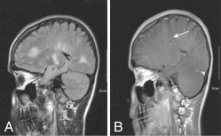
  • Marburg variant: Characterized by acute onset and a fulminant, often malignant course (severe disability or death can occur within a year of initial symptoms). MRI reveals tumefactive (tumor-like) demyelinating lesion(s) with extensive edema. Pathology shows severe inflammation with extensive necrosis. May also involve the peripheral nerves.5
  • Baló’s concentric sclerosis: Has a monophasic and rapidly progressive course. Neuroimaging and pathology show alternating rings of high- and low-signal intensity (on MRI) representing demyelination and intact myelination, resembling an onion bulb.5 More common in those of Chinese and Filipino descents.
  • Schilder’s disease (myelinoclastic diffuse sclerosis): Onset is typically in childhood with 1 to 2 large, confluent lesions. Usually progresses to involve widespread, bilateral regions of the CNS, and has variable course and prognosis.6-7 Etiology is unclear but may have a possible association with preceding infectious illness.
Synonyms

MS

Disseminated sclerosis

ICD-10CM CODE
G35Multiple sclerosis
Epidemiology & Demographics
Prevalence

More common in people raised in northern latitudes and in certain genetic clusters.1 Global prevalence of MS is estimated to be around 36 per 100,000 people (3 million people).8 There are currently nearly 1 million people living with MS in the United States.9

Predominant Sex & Age

Female:male ratio is approximately 3:1.1

Peak Incidence

Most common permanently disabling disorder of the central nervous system in young adults.1 Two thirds of patients have incidence between 20 and 40 yr; mean age of onset is 30 yr. Ranges from infancy to 70 yr.10

Race

Although more common in White people of Northern European descent, MS has been demonstrated to occur across most races and ethnic backgrounds. Recent studies suggest up to 47% higher risk of MS in Black vs. White women in the United States. Hispanic, Asian, and Native American populations have demonstrated lower incidence of MS thus far, compared to White and Black populations. Features of MS that have been more commonly associated with Black patients (compared to White patients) include transverse myelitis, vision loss from optic neuritis, earlier progression to disability, and higher lesion burden. Hispanic patients may also more frequently have optic neuritis and transverse myelitis, as well as younger age of disease onset and more severe disease course. Asian patients may have higher rates of optic nerve and spinal cord involvement.11

Genetics

Frequency of MS in dizygotic twins and siblings is 3% to 5%, and 30% to 50% in monozygotic twins. 200 genes have been identified that contribute to the risk of developing MS.1 Most common associations include human leukocyte antigen classes I and II (DRB11501, DQA10102, DQB10602), (DRB10405-DQA10301-DQB10302 in the Mediterranean population). A notable epigenetic interaction between vitamin D and the main MS-linked HLA-DRB11501 allele has been elucidated. Conversely, HLA-A02 has been associated with a reduced odds of developing MS.12

Physical Findings & Clinical Presentation

Findings depend on the location of the CNS lesion(s) and may include the following:

  • Common: Nonspecific complaints such as fatigue (most common, with 80% lifetime prevalence), blurred vision, diplopia, vertigo, falls, hemiparesis, paraparesis, monoparesis, numbness, paresthesias, ataxia, cognitive impairment, depression, anxiety, pseudobulbar affect (involuntary crying or laughing out of context), sexual dysfunction, and bowel/bladder dysfunction
  • Visual abnormalities: Horizontal nystagmus, visual field deficits, Marcus Gunn pupil (i.e., relative afferent papillary defect-normal consensual light reflex; however, when swinging a flashlight from the unaffected eye to the affected eye, direct light causes paradoxical pupillary dilation in the affected eye), sixth nerve palsy, internuclear ophthalmoplegia (paresis of the adducting eye on conjugate lateral gaze with simultaneous horizontal nystagmus of the abducting eye) (Fig. 1)
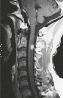
  • Corticospinal tract(s) involvement: Transverse myelitis, upper motor neuron signs such as spasticity (particularly leg spasms at night or after prolonged immobility), hyperreflexia, clonus, extensor plantar responses, tonic spasms, upper motor neuron pattern of weakness
  • Sensory involvement: May include partial or full dermatomal loss of pain and temperature, loss of vibration (common) and position sense, temperature dysregulation, thoracic band of sensory loss, paresthesias, trigeminal neuralgia
  • Ataxia: Intention tremor, dysmetria, dysdiadochokinesis, titubation, inability to tandem gait
  • Bladder dysfunction: Detrusor hyperreflexia (urge incontinence), urinary frequency, flaccidity (neurogenic bladder), and dyssynergia (bladder contracts against a closed sphincter)
  • Lhermitte sign: Flexion of the neck elicits an electrical sensation extending down the spine and occasionally into the extremities, due to involvement of the posterior cervical spinal cord2,13
  • Uhthoff phenomenon: Transient recurrence or worsening of preexisting neurologic deficits with small elevations in core body temperature (e.g., during exercise or warm bathing)2

Figure 1 Internuclear ophthalmoplegia.

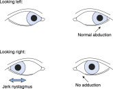
When the patient in the figure looks to the left (top row), both eyes move normally, but when the patient looks to the right (bottom row), the left eye fails to adduct (“weak” medial rectus) and the contralateral eye develops a jerk nystagmus. The finding is named for the side with weak adduction (i.e., in this example, a left internuclear ophthalmoplegia), and the lesion is in the ipsilateral medial longitudinal fasciculus (i.e., left medial longitudinal fasciculus in this example). See the text.

From McGee S: Evidence-based physical diagnosis, ed 4, Philadelphia, 2018, Elsevier.

Etiology

Likely multifactorial, with evidence for autoimmunity (autoreactive T and B lymphocytes), environmental factors (low sunlight exposure, vitamin D deficiency, smoking, obesity, shift work), and genetics (Mendelian and epigenetic). Environmental risk factors during childhood include exposure to certain viruses (e.g., Epstein-Barr virus and human herpes virus 6), low ultraviolet light exposure, and month of birth (higher in spring).1,14

Diagnosis

TABLE 1 Summary of Revised 2017 McDonald Criteria for Diagnosis of Multiple Sclerosis

RRMS/Clinical AttacksClinical LesionsParaclinical Testing Needed
22None
21MRI dissemination in space or a second clinical attack at a different CNS site
12MRI dissemination in time or CSF-specific oligoclonal bands
11
  1. Additional clinical attack at a different CNS site or MRI dissemination in space
  2. MRI dissemination in time or CSF-specific oligoclonal bands

Evidence of clinical lesions by physical examination or evoked potentials. CNS, Central nervous system; CSF, cerebrospinal fluid; MRI dissemination in space, 1 T2 lesions in 2 of the 4 typical areas for MS lesions-periventricular, juxtacortical, infratentorial, or spinal cord; MRI dissemination in time, a new lesion at follow-up MRI at any time, or presence of both an enhancing and nonenhancing lesion at any time; RRMS, relapsing-remitting multiple sclerosis.

Differential Diagnosis15TABLE 2

  • Autoimmune: Acute disseminated encephalomyelitis (ADEM), postvaccination encephalomyelitis, neuromyelitis optica spectrum disorder (NMOSD), myelin oligodendrocyte glycoprotein antibody disease (MOGAD), antiphospholipid antibody syndrome, autoimmune glial fibrillary acidic protein (GFAP) astrocytopathy
  • Degenerative: Amyotrophic lateral sclerosis, primary lateral sclerosis, cerebral autosomal dominant arteriopathy with subcortical infarcts and leukoencephalopathy (CADASIL), multisystem atrophy
  • Genetic: Fabry disease, Wilson disease, spinocerebellar ataxia, hereditary spastic paraparesis
  • Hematologic: Lymphoma, histiocytosis, thrombotic thrombocytopenic purpura
  • Infectious: Lyme disease, neurosyphilis, HIV, tropical spastic paraparesis (human T-lymphotropic virus type 1 or HTLV-1), progressive multifocal leukoencephalopathy (PML, caused by the JC virus), Listeria, Whipple disease (Tropheryma whipplei), acute flaccid myelitis, chronic meningitis, CNS tuberculosis, or fungal disease
  • Inflammatory: Systemic lupus erythematosus, vasculitis, neurosarcoidosis, Sjögren syndrome, Guillain-Barré syndrome, Behçet disease, celiac disease
  • Toxic/Nutritional/Metabolic: Vitamin B12 deficiency, copper deficiency, nitrous oxide toxicity, inherited leukodystrophies, central pontine myelinolysis
  • Mitochondrial: Leber hereditary optic neuropathy; mitochondrial encephalopathy, lactic acidosis, and stroke-like episodes (MELAS)
  • Neoplasms: CNS lymphoma, metastases, paraneoplastic disease, gliomatosis cerebri
  • Vascular: Susac syndrome, subcortical infarcts, Binswanger disease, amyloid angiopathy, cavernous or arteriovenous malformation, cerebral venous sinus thrombosis

TABLE 2 Conditions That Can Be Mistaken for Multiple Sclerosis and Other Diseases of Myelin

Vascular Disease
Small-vessel cerebrovascular disease
Vasculitis
CADASIL
Antiphospholipid antibody syndrome
Structural Lesions
Craniocervical junction, posterior fossa, or spinal tumors
Cervical spondylosis or disc herniation
Chiari malformation or syrinx
Degenerative Diseases
Hereditary myelopathy
Spinocerebellar degeneration
Infections
HTLV-1 infection
HIV myelopathy or HIV-related cerebritis
Neuroborreliosis (e.g., Lyme disease)
John Cunningham (JC) JC virus/progressive multifocal leukoencephalopathy
Neurosyphilis
Other Inflammatory Conditions
Systemic lupus erythematosus
Sjögren syndrome
Sarcoidosis
Monofocal or Monophasic Demyelinating Syndromes
Neuromyelitis optica spectrum disorder
Acute disseminated encephalomyelitis
Other Conditions
Hashimoto thyroiditis with or without encephalopathy
Nonspecific MRI abnormalities related to migraine, aging, or trauma

CADASIL, Cerebral autosomal dominant arteriopathy with subcortical infarcts and leukoencephalopathy; HIV, human immunodeficiency virus; HTLV, human T-cell lymphotropic virus; MRI, magnetic resonance imaging.

From Goldman L, Schafer AI: Goldman’s Cecil medicine, ed 24, Philadelphia, 2012, Saunders.

Diagnostic Studies (TABLE 3

  • Lumbar puncture to evaluate for presence of CSF-specific OCBs (Fig. 2), for cases that are atypical, and to evaluate for mimics of MS. Typical CSF abnormalities (Table 4) may include elevated protein (>100 mg/dl), mild pleocytosis, and elevated CSF-specific immunoglobulin G (IgG) synthesis rate. 70% of clinically definite MS (CDMS) demonstrate elevated CSF-specific IgG synthesis and 90% demonstrate CSF-specific OCBs2 (serum protein electrophoresis should be sent to the lab simultaneously with CSF for both of these tests). False-positive results for IgG synthesis, and rarely for positive OCBs, can be seen in CNS infections, parainfectious processes, vasculitis, and CNS lymphoma.
  • Serum: CBC with differential, comprehensive metabolic panel, liver function tests (LFTs), vitamin B12, copper, 25-OH vitamin D3.
  • Consider in the proper clinical setting: AQP4-IgG (to diagnose neuromyelitis optica), MOG-IgG, antinuclear antibody (ANA), ACE (a test with low specificity and sensitivity for sarcoidosis), antineutrophil cytoplasmic antibodies, antiphospholipid and anticardiolipin antibodies, Sjögren antibodies, thyroid stimulating hormone (TSH), free T4, peripheral blood smear, very-long-chain fatty acids, arylsulfatase A, lactate, pyruvate, urine organic acids, plasma amino acids.
  • Consider optical coherence tomography (OCT) or evoked potentials (visual, somatosensory, and brain stem auditory evoked response).

TABLE 3 Comparison of Sensitivity of Laboratory Testing in Multiple Sclerosis

VERBAERSSEPOCBMRI
Clinically definite multiple sclerosis80%-85%50%-65%65%-80%85%-95%90%-97%

Numbers show the percentage of patients with abnormal study results.

BAER, Brain stem auditory evoked response; MRI, magnetic resonance imaging; OCB, oligoclonal band; SSEP, somatosensory evoked potential; VER, visual evoked response.From Jankovic J et al: Bradley and Daroff’s neurology in clinical practice, ed 8, Philadelphia, 2022, Elsevier.

TABLE 4 Cerebrospinal Fluid Abnormalities in Multiple Sclerosis

AlbuminIgG/TPIgG/AlbuminIgG IndexOligoclonal Banding of Ig
Clinically definite multiple sclerosis23%67%60%-73%70%-90%85%-95%
Normal controls3%-36%3%7%

Other neurologic disease.

IgG/TP, Immunoglobulin G value/total protein.From Jankovic J et al: Bradley and Daroff’s neurology in clinical practice, ed 8, Philadelphia, 2022, Elsevier.

Figure 2 Electrophoresis of CSF of a Patient with Multiple Sclerosis (Left), Compared to the CSF of Someone with No Central Nervous System Inflammatory Disease (Right), Shows Three Distinct, Horizontal Oligoclonal Bands

From Kaufman DM et al: Kaufman’s clinical neurology for psychiatrists, ed 9, Philadelphia, 2023, Elsevier.

Imaging

MRI of the brain with and without gadolinium contrast Fig. 3 is recommended in all cases. MRI with and without contrast of the cervical (Fig. E4) and thoracic spines should also be performed, if possible, to assess for spinal cord lesions. MRI assesses for both acute and chronic lesions, as well as for atrophy, and should be repeated at least annually for disease surveillance and monitoring of disease-modifying therapy (DMT) efficacy. A normal MRI of the brain does not definitively exclude early MS but makes it extremely unlikely.4

Figure 3 Multiple sclerosis.

(A) Sagittal fluid-attenuated inversion recovery image magnetic resonance scan shows multiple lesions in corpus callosum, “Dawson fingers” (periventricular fingerlike lesions oriented toward the ventricles), along with ovoid and punctuate lesions in the deep white matter. (B) Gadolinium-enhanced scan shows an enhancing lesion (arrow).

Figure E4 This Magnetic Resonance Image of a Patient with Multiple Sclerosis Reveals a Plaque-the Hyperintense Lesion-in the High Cervical Spinal Cord

From Kaufman DM et al: Kaufman’s clinical neurology for psychiatrists, ed 9, Philadelphia, 2023, Elsevier.

Treatment

Nonpharmacologic Therapy

Patient education regarding disease characteristics, treatment options, risks and benefits of treatment, and prognosis. Often, patients need to incorporate intermittent rest periods on a daily basis and when physically active (for energy conservation), and avoid exposure to heat, which typically worsens symptoms (but not the disease).

Recommend physical therapy for new or worsening weakness, incoordination, or spasticity.

Acute General Rx (TABLE 5

For relapses: High-dose intravenous (IV) or oral methylprednisolone (1 g/day for 3 to 5 days).16 A proton pump inhibitor and insulin sliding scale should be simultaneously administered to prevent gastrointestinal ulcers and treat steroid-induced hyperglycemia.

TABLE 5 Approved Medications for Multiple Sclerosis

Generic (Brand) Name and Mode of AdministrationIndications(s)Side EffectsChemical Structure
Glatiramer acetate, Copaxone
Daily SQ injection
To reduce relapse frequency in patients with relapsing-remitting multiple sclerosis (MS) and patients who have experienced a first clinical episode and have MRI features consistent with MSInjection site reactions, lipoatrophy with prolonged useSynthetic polymer
IM IFN-β-1a, Avonex
Weekly IM injection
To slow accumulation of physical disability and decrease frequency of clinical exacerbations in patients with relapsing forms of MS and patients who have experienced a first clinical episode and have MRI features consistent with MSFlulike symptoms, injection site reactions, neutropenia, anemia, liver function test abnormalitiesInterferon beta-1 alpha
SC IFN-β-1a, Rebif
Three times per wk SQ injections
To slow accumulation of physical disability and decrease frequency of clinical exacerbations in relapsing forms of MSFlulike symptoms, injection site reactions, neutropenia, anemia, liver function test abnormalitiesInterferon beta-1-alpha
IFN-β-1b, Betaseron
Every other day SQ injections
To reduce the frequency of clinical exacerbations in patients with relapsing forms of MS and patients who have experienced a first clinical episode and have MRI features consistent with MSFlulike symptoms, injection site reactions, neutropenia, anemia, liver function test abnormalitiesInterferon beta-1-beta
Fingolimod, Gilenya
Daily oral tablet
To reduce frequency of clinical exacerbations and delay accumulation of physical disability in patients with relapsing forms of MSRisk of herpes virus infections, bradycardia, macular edema, and changes on pulmonary function test.Sphingosine-1-phosphate receptor blocker
Natalizumab, Tysabri
Monthly IV infusions
As monotherapy for relapsing forms of MS; to delay accumulation of physical disability and reduce frequency of clinical exacerbationsPML brain infection (risk 1:1,000)α4-integrin adhesion molecule blocker
Mitoxantrone, Novantrone
IV chemotherapy
To reduce neurologic disability and/or the frequency of clinical relapses in secondary (chronic) progressive, progressive relapsing or worsening relapsing-remitting MSCardiomyopathy, increased risk of secondary lymphoid malignanciesCytotoxic chemotherapy; synthetic antineoplastic anthracenedione
Teriflunomide, Aubagio
Daily oral tablet
To reduce frequency of relapses in relapsing-remitting MSHepatotoxicity, bone marrow suppression, peripheral neuropathyPyrimidine synthesis inhibitor
Dimethyl fumarate, Tecfidera
Twice daily capsule
To reduce frequency of relapses in relapsing-remitting MSFlushing, gastrointestinal side effects, lymphopeniaDimethyl fumarate
Ocrelizumab, Ocrevus
Every-6-mo infusion
To reduce frequency of relapses in relapsing-remitting MS; slows progression in primary progressive MSInfusion reactions; risk of infectionB-cell antibody
Cladribine
2 wk of oral pills/yr
To reduce frequency of relapses in relapsing-remitting MSPotential cancer risk; risk of infectionPurine synthesis modulator

IV, Intravenous; MRI, magnetic resonance imaging; MS, multiple sclerosis.

From Jankovic J et al: Bradley and Daroff’s neurology in clinical practice, ed 8, Philadelphia, 2022, Elsevier.

The Optic Neuritis Treatment Trial showed worse outcomes in optic neuritis after oral methylprednisolone compared with intravenous.17 However, the COPOUSEP trial published in 2015 demonstrated nearly equivalent efficacy in improvement of symptoms and/or full recovery between 1000 mg daily for 3 days of oral and IV methylprednisolone in all types of relapses. High-dose corticosteroids do not alter long-term outcomes of the disease, but instead accelerate recovery from symptoms of the current relapse.17-18

Plasma exchange can be considered for steroid-refractory cases.19

Chronic Rx

  • Most FDA-approved disease-modifying therapies (DMTs) are approved for use only in relapsing forms of MS (which include CIS, RRMS, and active SPMS-or SPMS with relapses). Ocrelizumab and mitoxantrone are the first FDA-approved DMTs for use in PPMS and SPMS, respectively.
  • Injectable DMTs: These medications include the first generation of MS drugs (interferons and glatiramer acetate), which have a low risk of side effects but modest efficacy in preventing future relapses. Ofatumumab is a newer injectable DMT with high efficacy but higher risk of side effects than the older injectable DMTs.
    1. Interferons: Include interferon beta-1a (intramuscular injection [IM Avonex, dose of 30 mcg once per wk; IM Plegridy, starting dose of 63 mcg uptitrated over 1 mo to 125 mcg every 2 wk; subcutaneous (SC) Rebif, dose of 22 or 44 mcg three times per wk]) and interferon beta-1b (SC Betaseron, SC Extavia; doses of 0.25 mg every other day). Involve a complex and multifactorial mechanism of action, including increased expression of antiinflammatory mediators and downregulated expression of proinflammatory cytokines. Side effects can include fatigue, flulike symptoms, injection site reactions such as skin necrosis, depression, hepatotoxicity, seizures, anemia, leukopenia, thrombocytopenia, and thrombotic microangiopathy. CBC with differential, LFTs, and thyroid function tests should be obtained before initiating and regularly monitored during treatment (initially every 3 mo for 6 mo, then annually).
    2. Glatiramer acetate (SC Copaxone, SC Glatopa; doses of 20 mg daily or 40 mg three times per wk): Synthetic protein that simulates myelin basic protein and shifts T cells from the proinflammatory Th1 to regulatory Th2 profile. Common side effects include injection site reactions (such as lipoatrophy and skin necrosis) and brief postinjection reactions such as flushing, chest tightness, tachycardia, and dyspnea; no laboratory monitoring is needed.
    3. Ofatumumab (SC Kesimpta; dose of 20 mg weekly for 3 wk, no injection for 1 wk, then 20 mg per mo): Anti-CD20 monoclonal antibody injectable. Side effects can include injection site reactions, headache, hepatitis B virus (HBV) reactivation, low immunoglobulins, and increased risk of infections (especially upper respiratory tract infections [URIs]). Before initiating and during treatment, obtain CBC with differential (including lymphocyte subsets), LFTs, immunoglobulins, and HBV and JC virus antibodies. Live or live-attenuated vaccinations should be administered at least 1 mo before starting treatment.
  • Oral DMTs: These medications are newer and more efficacious than the initial injectable DMTs in preventing MS relapses, but carry a higher risk of serious side effects and infections. Review the patient’s general risk of infections before starting any oral DMT.
    1. Fingolimod (Gilenya; dose of 0.5 mg daily), siponimod (Mayzent; starting dose of 0.25 mg daily, titrated up to 1 to 2 mg daily over 4 to 5 days), ozanimod (Zeposia; starting dose of 0.23 mg uptitrated over 1 wk to 0.92 mg daily), or ponesimod (Ponvory; starting dose of 2 mg uptitrated over 2 wk to 20 mg daily): Mechanism of action involves sphingosine-1-phosphate receptor modulation and lymphocyte sequestration. Side effects can include diarrhea, headache, back pain, abdominal pain, cough, hepatotoxicity, bradycardia with the first dose (requiring cardiac monitoring and ECG for at least 8 h after administration), arrhythmia, orthostatic hypotension, hypertension, pancytopenia, macular edema (requiring ophthalmologic exam at baseline, at 3 mo, and annually thereafter for those with history of diabetes or uveitis), increased risk of infections (including PML, cryptococcal meningitis, herpes simplex virus [HSV] encephalitis, disseminated VZV), posterior reversible encephalopathy syndrome (PRES), increased risk of skin cancers (requiring regular skin examinations), and reduced pulmonary function. Before initiating treatment, obtain baseline HSV, JC virus, and VZV serologies, CBC with differential, LFTs, macular OCT, and possibly CYP2C9 genotype (if considering siponimod); monitor CBC with differential and LFTs every 3 to 6 mo during treatment. Live or live-attenuated vaccinations should be administered at least 1 mo before starting treatment.
    2. Teriflunomide (Aubagio; dose of 14 mg daily): Reversible inhibitor of pyrimidine synthesis (enzyme dihydroorotate dehydrogenase). Side effects can include headache, nausea, diarrhea, hypertension, alopecia, peripheral neuropathy, paresthesias, severe hepatotoxicity, hypersensitivity reactions (anaphylaxis, angioedema, Stevens-Johnson syndrome, toxic epidermal necrolysis, drug reaction with eosinophilia and systemic symptoms [DRESS]), and interstitial lung disease. Patients should avoid becoming pregnant while on teriflunomide (pregnancy category X). Serum levels can be measured, and the drug can be eliminated by a course of activated charcoal or cholestyramine. Before initiating treatment, check baseline blood pressure, CBC with differential, LFTs, and tuberculosis (TB) test; monitor monthly LFTs for the first 6 mo, and CBC and LFTs every 3 to 6 mo during treatment.
    3. Dimethyl fumarate (Tecfidera; starting dose of 120 mg twice daily for 2 wk, then 240 mg twice daily), diroximel fumarate (Vumerity; starting dose of 231 mg twice daily for 1 wk, then 462 mg twice daily), or monomethyl fumarate (Bafiertam; starting dose of 95 mg twice daily for 1 wk, then 190 mg twice daily): Inhibit transcription of nuclear factor-κB (NF-κB; in the nuclear factor erythroid 2-related factor 2 [Nrf-2] pathway). Side effects can include nausea, abdominal discomfort (can be reduced with use of H2 blockers or proton pump inhibitors), pruritus, and flushing (can be reduced with concurrent use of aspirin and taking with food)-especially during the first month-and rarely, lymphopenia, with increased risk of infections such as PML. Before initiating treatment, check CBC with differential, LFTs, VZV and JC virus antibodies; monitor CBC with differential and LFTs every 3 to 6 mo during treatment.
    4. Cladribine (Mavenclad; total dose of 3.5 mg/kg administered orally in two short courses 1 yr apart): A purine antimetabolite that inhibits DNA synthesis and depletes both T and B lymphocytes. FDA-approved for use in relapsing forms of MS, except for CIS. Side effects can include hematologic toxicity, alopecia, increased risk of infections, increased risk of malignancy, weight loss, and hepatic injury. Pregnancy should be avoided during treatment and for at least 6 mo after the last dose, and breastfeeding is contraindicated during treatment days and for 10 days after the last dose. It is also contraindicated in patients who are immunocompromised, have active chronic infections, or have cancer. CBC with differential, TB test, and viral serologies (e.g., HIV, VZV, HSV, hepatitis B and C, JC virus) should be checked before initiating treatment and monitored regularly during treatment. Live or live-attenuated vaccinations should be administered at least 1 mo before starting treatment.
  • IV DMTs: These are generally the newest and most efficacious for preventing MS relapses. They consequently also carry a higher risk of serious side effects and infections. Live or live-attenuated vaccinations should be administered at least 1 mo before starting treatment with any of these.
    1. Natalizumab (Tysabri; dose of 300 mg monthly): A humanized monoclonal antibody that binds to the lymphocyte surface protein α4-integrin, which inhibits binding to VCAM-1 and, therefore, movement of lymphocytes across the bloodstream. It has been associated with a higher risk of PML than other immunomodulatory therapies. Testing for JC virus antibody (along with CBC with differential and LFTs) should be done every 6 mo because conversion to a positive antibody occurs at increased frequency while on this drug. If positive, PML is still rare, but the risk increases with increased length of therapy (>2 yr), history of chemotherapy or prior immunosuppression, and high level of JC virus antibodies. Other potential serious side effects can include infusion reactions, HSV and other infections, hepatotoxicity, and thrombocytopenia.
    2. Ocrelizumab (Ocrevus; dose of 600 mg every 6 mo, with the first dose divided as 300 mg given 2 wk apart): Humanized monoclonal antibody to CD20+ B cells. FDA-approved for use in PPMS as well as all relapsing forms of MS. Side effects can include leukopenia, low immunoglobulins, infusion reactions, increased risk of infections (upper and lower respiratory tract infections, UTI, HSV, skin infections), HBV reactivation, and possibly an increased risk of breast cancer. Before starting treatment, obtain baseline CBC with differential, immunoglobulins, TB test, and viral serologies, including HBV, HCV, VZV, HSV, HIV, and JC virus. Monitor CBC with differential (including lymphocyte subsets), immunoglobulins, HBV, and JC virus antibodies 1 to 2 times per yr. This medication is contraindicated in patients with history of active HBV infection.
    3. Alemtuzumab (Lemtrada; dose of 12 mg daily for 5 days, then 12 mg daily for 3 days 1 yr later): Anti-CD52 humanized monoclonal antibody and second-line therapy for patients who have failed at least two FDA-approved MS therapies. FDA-approved for use in relapsing forms of MS, except for CIS. Side effects can include the development of autoimmune conditions (34% developed autoimmune thyroid disorders), cytopenias, hepatotoxicity, infusion reactions, stroke, arterial dissection, hemophagocytic lymphohistiocytosis (HLH), bleeding or clotting disorders such as immune thrombocytopenic purpura (ITP) and TTP, pneumonitis, Goodpasture disease, increased risk of certain cancers, and increased risk of infections including PML. Only available through a restricted access program. It is contraindicated in patients who are immunocompromised or have chronic active infections. Requires monthly monitoring of CBC with differential (including lymphocyte subsets), LFTs, TSH, renal function tests, and urinalysis until at least 4 yr after the last course, and HSV prophylaxis for at least 2 mo after each dose.
    4. Mitoxantrone (Novantrone; 12 mg/m2 every 3 mo for 2 yr): An antineoplastic anthracenedione (DNA-reactive agent) that inhibits B-cell, T-cell, and macrophage proliferation and impairs antigen presentation. FDA-approved for use in SPMS, progressive-relapsing MS, and worsening RRMS; however, now used very infrequently. Potential serious side effects include secondary acute myeloid leukemia, neutropenia, and cardiotoxicity (CHF). Is contraindicated if baseline neutrophil count <1,500 cells/mm3 and hepatic impairment (due to reduced clearance). Requires initial evaluation and regular monitoring (prior to each dose) of CBC with differential, LFTs, ECG, echocardiogram (for left ventricular ejection fraction), and pregnancy test if relevant (pregnancy category D).
  • Symptomatic therapy:
    1. Gait dysfunction: Initial management should be referral to physical therapy, use of ankle-foot orthoses (for footdrop), and/or mobility aids. Dalfampridine (Ampyra) is a potassium channel blocker that is FDA-approved to improve walking speed (objectively measured by a timed 25-ft walk test) in patients with MS; its use is contraindicated in patients with epilepsy.
    2. Spasticity: Initial management should be twice-daily stretching exercises and referral to physical therapy. Can next consider baclofen (starting dose of 10 mg twice daily) or tizanidine (starting dose of 2 mg nightly; requires monitoring of liver enzymes). For severe spasticity not responsive to these medications, dantrolene (starting dose of 25 mg daily; requires monitoring of liver enzymes) or diazepam (average daily dose of 15 mg) may be tried. Onabotulinum toxin type A injection can be used for focal intractable spasticity. Intrathecal baclofen pump can be used for generalized intractable spasticity. Acute worsening of spasticity may be caused by infections such as UTI, injury, recent surgery, or colder temperatures.20
    3. Urge incontinence and retention: Obtain urinalysis and post-void residual bladder ultrasound. Recommend fluid restriction at night, scheduled voiding, and avoidance of caffeine and alcohol. Incontinence can be initially treated with anticholinergic/muscarinic therapy such as oxybutynin, trospium, tolterodine, or solifenacin. Mirabegron is a beta-3 agonist indicated for treatment of neurogenic detrusor overactivity (overactive or neurogenic bladder). Urinary retention can be treated with tamsulosin. In both cases, UTI or bladder infection should be ruled out. Intermittent catheterization, intradetrusor onabotulinum toxin A injections, and neuromodulation (tibial nerve stimulation) are more advanced options for severe neurogenic bladder symptoms and require management by a urologist.21
    4. Dysesthesias: Can treat with carbamazepine (200 mg twice daily), oxcarbazepine (starting dose of 300 mg daily), gabapentin (starting dose of 300 mg daily), or pregabalin (starting dose of 50 mg daily).
    5. Fatigue: Initial management should involve medication review, depression screening, evaluating for underlying sleep disorder, energy conservation strategies, exercise program, and weight loss for obesity. Can next consider medical management with amantadine (100 mg twice daily), modafinil (starting dose of 100 mg every morning), or a stimulant such as methylphenidate.
    6. Tremor: Can treat with clonazepam (starting dose of 0.5 mg daily), propranolol (starting dose of 20 to 40 mg twice daily), or gabapentin (starting dose of 300 mg daily).
    7. Cognitive impairment: Can objectively screen with the Symbol Digit Modalities Test, if available. Assess for other causes of cognitive impairment, such as neurodegenerative disease, depression, fatigue, disordered sleep, and polypharmacy. Recommend conservative strategies, such as the use of checklists, reminders, diaries, and calendars, and regular physical exercise and social activity.
    8. Depression and anxiety: Refer for cognitive-behavioral therapy. Consider starting a selective serotonin reuptake inhibitor (SSRI) or serotonin norepinephrine reuptake inhibitor (SNRI). Distinguish between depression and pseudobulbar affect, which is treated with dextromethorphan/quinidine (Nuedexta).
Disposition

Most patients have complete or near-complete recovery weeks to months after a relapse, even without acute treatment with high-dose steroids. Typically, two relapses occur in RRMS patient per year (75% will have >1 relapse). Although the rate of disease progression is highly variable, there is higher risk of greater long-term disability with higher relapse rate during the first 2 to 5 yr, poor recovery from initial relapses, older age of onset, involvement of multiple systems, male sex, African American, and primary progressive disease.

DMTs have significantly improved since their introduction, and the current treatment goal is no relapses and no disease progression on imaging.

Referral

  • Referral to a neurologist is highly recommended. Referral to an MS specialist should be considered in cases of poor response to initial therapy and/or if there is concern about complications of therapies.
  • Referrals for physical, speech, and occupational therapy for motor, sensory, and speech symptoms.
  • Referral to a mental health provider and/or psychiatrist for depression/anxiety.
  • Referral to neuropsychology for cognitive impairment symptoms.
  • Referral to urology if bladder-sphincter dyssynergia is possible, if symptoms are not responsive to first-line medications, or in case of complications such as recurrent UTI, hematuria, renal impairment, hydronephrosis, or stress incontinence.

Pearls & Considerations

Related Content

Multiple Sclerosis (Patient Information)

Neuromyelitis Optica Spectrum Disorder (Related Key Topic)

Optic Neuritis (Related Key Topic)

Related Content

    1. Reich D.S. : Multiple sclerosisN Engl J Med. ;378(2):169-180, 2018.doi:10.1056/NEJMra1401483
    2. Thompson A.J. : Diagnosis of multiple sclerosis: 2017 revisions of the McDonald criteriaLancet Neurol. ;17(2):162-173, 2018.doi:10.1016/S1474-4422(17)30470-2
    3. Polman C.H. : Diagnostic criteria for multiple sclerosis: 2010 revisions to the McDonald criteriaAnn Neurol. ;69(2):292-302, 2011.doi:10.1002/ana.22366
    4. Filippi M. : MRI criteria for the diagnosis of multiple sclerosis: MAGNIMS consensus guidelinesLancet Neurol. ;15(3):292-303, 2016.doi:10.1016/S1474-4422(15)00393-2
    5. Capello E., Mancardi G.L. : Marburg type and Balò’s concentric sclerosis: rare and acute variants of multiple sclerosisNeurol Sci. ;25(4):s361-s363, 2004.doi:10.1007/s10072-004-0341-1
    6. Poser C.M. : Schilder's myelinoclastic diffuse sclerosis [published correction appears in Pediatrics 78(1):138, 1986]Pediatrics. ;77(1):107-112, 1986.
    7. Mara Genç H. : Long-term clinical and radiologic follow-up of Schilder's diseaseMult Scler Relat Disord. ;13:47-51, 2017.doi:10.1016/j.msard.2017.02.007
    8. Walton C. : Rising prevalence of multiple sclerosis worldwide: insights from the Atlas of MS, third editionMult Scler. ;26(14):1816-1821, 2020.doi:10.1177/1352458520970841
    9. Wallin M.T. : The prevalence of MS in the United States: a population-based estimate using health claims data [published correction appears in Neurology 93(15):688, 2019]Neurology. ;92(10):e1029-e1040, 2019.doi:10.1212/WNL.0000000000007035
    10. Multiple sclerosis. National Institute of Neurological Disorders and Stroke. Available at https://www.ninds.nih.gov/health-information/disorders/multiple-sclerosis.
    11. Ward M., Goldman M.D. : Epidemiology and pathophysiology of multiple sclerosisContinuum (Minneap Minn). ;28(4):988-1005, 2022.doi:10.1212/CON.0000000000001136
    12. Moutsianas L. : Class II HLA interactions modulate genetic risk for multiple sclerosisNat Genet. ;47(10):1107-1113, 2015.doi:10.1038/ng.3395
    13. Gutrecht J.A. : Anatomic-radiologic basis of Lhermitte's sign in multiple sclerosisArch Neurol. ;50(8):849-851, 1993.doi:10.1001/archneur.1993.00540080056014
    14. Nourbakhsh B., Mowry E.M. : Multiple sclerosis risk factors and pathogenesisContinuum (Minneap Minn). ;25(3):596-610, 2019.doi:10.1212/CON.0000000000000725
    15. Miller D.H. : Differential diagnosis of suspected multiple sclerosis: a consensus approachMult Scler. ;14(9):1157-1174, 2008.doi:10.1177/1352458508096878
    16. Le Page E. : Oral versus intravenous high-dose methylprednisolone for treatment of relapses in patients with multiple sclerosis (COPOUSEP): a randomised, controlled, double-blind, non-inferiority trialLancet. ;386(9997):974-981, 2015.doi:10.1016/S0140-6736(15)61137-0
    17. Newman N.J. : The optic neuritis treatment trialOphthalmology. ;127(4S):S172-S173, 2020.doi:10.1016/j.ophtha.2019.09.046
    18. Perumal J.S. : Oral prednisone taper following intravenous steroids fails to improve disability or recovery from relapses in multiple sclerosisEur J Neurol. ;15(7):677-680, 2008.doi:10.1111/j.1468-1331.2008.02146.x
    19. Weinshenker B.G. : A randomized trial of plasma exchange in acute central nervous system inflammatory demyelinating disease doi:10.1002/1531-8249(199912)46:6<878::AID-ANA10>3.0.CO;2-Q Ann Neurol. ;46(6):878-886, 1999.
    20. Otero-Romero S. : Pharmacological management of spasticity in multiple sclerosis: systematic review and consensus paperMult Scler. ;22(11):1386-1396, 2016.doi:10.1177/1352458516643600
    21. Tornic J., Panicker J.N. : The management of lower urinary tract dysfunction in multiple sclerosisCurr Neurol Neurosci Rep. ;18(8), 2018.doi:10.1007/s11910-018-0857-z