AUTHORS: Vasken Keleshian, MD and Pranav M. Patel, MD, FACC, FAHA, FSCAI

Carotid artery aneurysm is a localized enlargement of the carotid artery caused by weakening of the artery wall and is categorized as either a true or false aneurysm. True aneurysm involves the full thickness of the arterial wall and has >50% increase in vessel diameter compared to the expected normal. False aneurysm, also known as pseudoaneurysm, forms due to disruption or injury to the artery wall that results in a contained hematoma connecting with the inner artery lumen.
Aneurysm of common carotid artery
Aneurysm of external carotid artery
Aneurysm of internal carotid artery
| ||||||||||||
Aneurysms of the carotid artery are rare. It is estimated that 0.1% to 2% of all carotid procedures are performed for correction of aneurysmal disease.1 It accounts for 0.4% to 4% of all aneurysms in the peripheral arteries.2
Overall, carotid artery aneurysm accounts for <1% of all arterial aneurysms and up to 4% of aneurysms in the peripheral arteries.3
Most patients are male, and most are diagnosed in the sixth or seventh decade of life.3
Genetic conditions such as fibromuscular dysplasia, connective tissue disorders, inflammatory diseases, and congenital defects can cause distortion of the arterial wall and lead to aneurysm.4 If a genetic condition is suspected, further investigation, including obtaining detailed family history, obtaining comprehensive imaging of other vascular beds, and referring for genetic testing, should be considered.
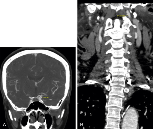
The majority of carotid artery aneurysms are found incidentally on computed tomography (CT) scan or MRI studies. Larger aneurysms can cause cranial nerve deficit, tracheal obstruction, dysphagia, tinnitus, hoarseness, and dizziness.5 Physical examination includes palpation of a pulsatile mass in the neck and auscultation of a carotid artery bruit and can produce an abnormal neurologic examination if a cranial nerve is affected.
The most common cause is related to atherosclerosis and infection. Primary infections of the artery wall can lead to true aneurysm, whereas infections related to carotid artery intervention cause secondary infection which predisposes to pseudoaneurysm. Injury from blunt or penetrating trauma can lead to either type of aneurysm depending on the mechanism of arterial wall injury.6 Carotid artery aneurysm can occur as a result of spontaneous dissection, which can occur in setting of radiation exposure (Table E1).7
TABLE E1 Type and Localization of Carotid Artery Aneurysm
| Type 1 | Isolated and short aneurysm of the internal carotid artery above the carotid bulb | ||
| Type 2 | Long aneurysm of the carotid artery extending below the carotid bulb | ||
| Type 3 | Aneurysm of the proximal internal carotid artery and the carotid bifurcation | ||
| Type 4 | Combined aneurysm of the internal and common carotid artery | ||
| Type 5 | Isolated common carotid artery aneurysm |