section name header

Basic Information ⬇

Author: Arielle Katcher, MD

Definition

Cancers of the uterus include tumors that originate from epithelial or mesenchymal tissue. Tumors that arise from the epithelium are referred to as adenocarcinomas, whereas tumors that arise from mesenchymal tissue (i.e., connective, muscular, vascular) are referred to as sarcomas. These tumors may be found in various locations of the uterus including the endometrium or myometrium.

Historically, endometrial adenocarcinomas were divided into two categories: Type 1 and Type 2. Type 1 endometrial carcinomas refer to endometrioid subtypes, whereas Type 2 endometrial carcinomas refer to serous, clear cell, and mixed Mullerian subtypes (see "Endometrial Cancer"). Recent molecular studies have shown carcinosarcomas (MMMT) to be closely related to epithelial tumors compared to sarcomas.

This chapter will focus on sarcomas of the uterus arising from the endometrial stroma or myometrium.

Synonyms

  • Sarcomas of the uterus
  • Leiomyosarcomas
  • Endometrial stromal sarcoma
  • Adenosarcomas
  • Undifferentiated sarcoma
ICD-10CM CODES
C54.1Malignant neoplasm of endometrium
C54.2Malignant neoplasm of the myometrium
C54.0Malignant neoplasm of isthmus uteri
C54.8Malignant neoplasm of overlapping sites of corpus uteri
Epidemiology & Demographics
Incidence & Prevalence:

Incidence of all uterine cancer is 28.1 per 100,000 women per year with 5.2 deaths per 100,000 women as of 2024. In the most recent update from Surveillance, Epidemiology, and End Results (SEER) Annual Report to the Nation, uterine cancer showed the highest increase frequency in death of all cancers among U.S. women. Endometrial cancer remains the most common uterine malignancy in the U.S.; however, sarcomas of the uterus are rare. Sarcomas account for approximately 3% to 8% of all cancers of the uterine corpus. Sarcomas are associated with a poor prognosis compared to endometrial cancer.

Predominant Age:

Mean age at diagnosis: 50 yr old

Risk Factors:

Box E1 describes risk factors for uterine sarcoma.

BOX E1 Risk Factors for Uterine Sarcoma

  • •Nulliparity
  • •Obesity
  • •History of pelvic radiation
  • •Exposure to tamoxifen

From Fielding JR et al: Gynecologic imaging, Philadelphia, 2011, Saunders.

Physical Findings & Clinical Presentation

  • •Abnormal vaginal bleeding is the most common symptom (90% of women with diagnosis)
  • •Vaginal discharge also may be a presenting symptom (10% of these patients have non-bloody discharge)
  • •May also present as pelvic pain or pressure and pelvic mass on examination (10% of women with uterine sarcoma)
  • •Urinary symptoms
  • •Abdominal pain or distention
  • •Weight loss
Etiology

  • •Endometrial cancers are thought to derive from an excess of unopposed estrogen causing a proliferation of disorganized dysplastic endometrium.
  • •A minority of patients may have a genetic predisposition for endometrial cancer (Lynch syndrome).
  • •The exact etiology for sarcomas is largely unknown.

Diagnosis ⬆ ⬇

Differential Diagnosis

  • •Endometrial hyperplasia
  • •Endometrial polyp
  • •Leiomyoma
Workup

Diagnosis can be made histologically by biopsy for abnormal bleeding. Workup includes biopsy (in the office or operating room, in conjunction with hysteroscopy) and imaging (see "Imaging Studies"). Surgical removal of the uterus is the most common way to diagnose a uterine sarcoma. Histologic criteria include mitotic index, cellular atypia, loss of polarity, and necrosis.

Laboratory Tests

  • •CBC
  • •Comprehensive metabolic panel (CMP)
  • •CA-125 (high levels can be a sign of metastasis, not diagnostic, not always elevated)
Imaging Studies

  • •Pelvic ultrasound is a lower-cost method of detecting uterine corpus mass or thickened endometrial lining. Features concerning for uterine sarcoma: Heterogeneous texture, central necrosis, irregular vessel distribution, and rapid growth of the uterus.
  • •Chest x-ray examination should be considered for preoperative testing with suspicion of uterine sarcoma.
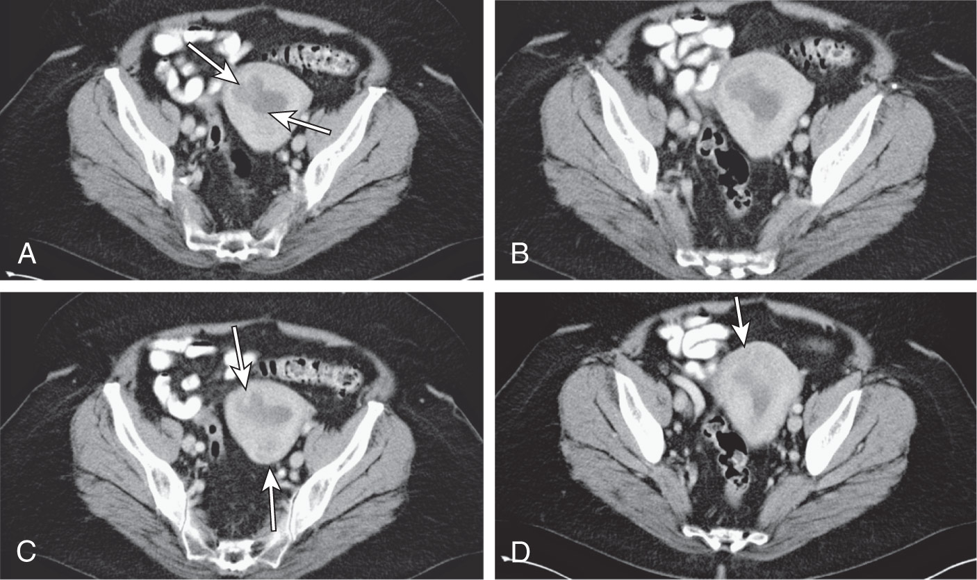
  • •Computed tomography (CT) scans (Fig. E1), MRI, and PET may be useful for assessing tumor spread once diagnosis is made or characterizing a uterine mass.

Figure E1 A 50-yr-old patient with uterine sarcoma.

A, Axial contrast-enhanced computed tomographic (CT) image. Low-attenuation lobulated and infiltrating soft tissue fills the endometrial canal (arrows), extending into the myometrium. B, Axial contrast-enhanced CT image showing same as A. Low-attenuation, lobulated, and infiltrating soft tissue fills the endometrial canal, extending into the myometrium. C, Axial contrast-enhanced CT image. Low-attenuation lobulated and infiltrating soft tissue fills the endometrial canal. Subtle myometrial invasion is seen anterior and posterior (arrows). D, Axial contrast-enhanced CT image. Low-attenuation lobulated and infiltrating soft tissue fills the endometrial canal (arrow), showing same. Subtle myometrial invasion is seen fundally.

(From Fielding JR et al: Gynecologic imaging, Philadelphia, 2011, Saunders.)

Staging

Staging of endometrial carcinoma and uterine adenosarcoma is summarized in Tables E1 and E2.

TABLE E1 International Federation of Gynecology and Obstetrics 2023 Staging for Endometrial Carcinoma (Including Carcinosarcoma)

Stage
IConfined to the uterine corpus and ovary
IADisease limited to the endometrium OR nonaggressive histologic type, i.e., low-grade endometrioid, with invasion of less than half of myometrium with no or focal lymphovascular space involvement (LVSI) OR good prognosis disease
IA1Nonaggressive histologic type limited to an endometrial polyp OR confined to the endometrium
IA2Nonaggressive histologic types involving less than half of the myometrium with no or focal LVSI
IA3Low-grade endometrioid carcinomas limited to the uterus and ovary
IBNonaggressive histologic types with invasion of half or more of the myometrium, and with no or focal LVSI
ICAggressive histologic types limited to a polyp or confined to the endometrium
IIInvasion of cervical stroma without extrauterine extension OR with substantial LVSI OR aggressive histologic types with myometrial invasion
IIAInvasion of the cervical stroma of nonaggressive histologic types
IIBSubstantial LVSI of nonaggressive histologic types
IICAggressive histologic types with any myometrial involvement
IIILocal and/or regional spread of the tumor of any histologic subtype
IIIAInvasion of uterine serosa, adnexa, or both by direct extension or metastasis
IIIA1Spread to ovary or fallopian tube (except when meeting stage IA3 criteria)
IIIA2Involvement of uterine subserosa or spread through the uterine serosa
IIIBMetastasis or direct spread to the vagina and/or to the parametria or pelvic peritoneum
IIIB1Metastasis or direct spread to the vagina and/or the parametria
IIIB2Metastasis to the pelvic peritoneum
IIICMetastasis to the pelvic or paraaortic lymph nodes or both
IIIC1Metastasis to the pelvic lymph nodes
IIIC1iMicrometastasis
IIIC1iiMacrometastasis
IIIC2Metastasis to paraaortic lymph nodes up to the renal vessels, with or without metastasis to the pelvic lymph nodes
IIIC2iMicrometastasis
IIIC2iiMacrometastasis
IVSpread to the bladder mucosa and/or intestinal mucosa and/or distance metastasis
IVAInvasion of the bladder mucosa and/or the intestinal/bowel mucosa
IVBAbdominal peritoneal metastasis beyond the pelvis
IVcDistant metastasis, including metastasis to any extraabdominal or intraabdominal lymph nodes above the renal vessels, lungs, liver, brain, or bone

Gaffney D et al: 2023 FIGO staging system for endometrial cancer: the evolution of the revolution, Gynecol Oncol 184:245-253, 2024. doi:10.1016/j.ygyno.2024.02.002.

TABLE E2 International Federation of Gynecology and Obstetrics 2009 Staging for Uterine Sarcoma (Including Leiomyosarcoma and Endometrial Stromal Sarcoma)

Stage
ITumor limited to the uterus
IATumor 5 cm or less in greatest dimension
IBTumor more than 5 cm
IITumor extends beyond the uterus, within the pelvis
IIAAdnexal involvement
IIBExtrauterine pelvic tissue involvement
IIIInvolvement of abdominal tissues
IIIA1 site
IIIB>1 site
IIICRegional lymph node metastasis
IV
IVATumor invades bladder or rectum
IVBDistant metastases

From Niederhuber JE: Abeloff’s clinical oncology, ed 6, Philadelphia, 2020, Elsevier.

Treatment ⬆

Disposition

  • •Survival varies with each type of sarcoma but is generally very poor. Box E2 describes uterine prognostic factors.
  • •5-yr survival for grade I endometrial stromal sarcoma is 91% and drops to 42% for grade III.
  • •5-yr survival for leiomyosarcoma ranges from 76% for stage 1 to 29% for stage 4.
  • •5-yr survival for undifferentiated sarcoma ranges from 70% for stage 1 to 23% for stage 4.

BOX E2 Uterine Sarcoma Prognostic Factors

  • •Tumor stage
  • •Tumor grade
  • •Tumor size
  • •Patient age
  • •Vascular space involvement
  • •Mitotic count
  • •Residual disease at surgery or uterine morcellation
  • •Adjuvant chemotherapy

From Fielding JR et al: Gynecologic imaging, Philadelphia, 2011, Saunders.

Referral

  • •A gynecologic oncologist should manage uterine sarcoma.
  • •Key points in the management of uterine sarcoma are described in Box E3.

BOX E3 Uterine Sarcoma:

Key Points

  • •The disease mainly affects women aged 40-60 yr old depending on the type of sarcoma.
  • •Patients may present with abnormal uterine bleeding, abdominal distention, enlarging pelvic mass, pelvic pain/pressure, or may be asymptomatic.
  • •The primary treatment is hysterectomy and bilateral salpingo-oophorectomy.
  • •Adjuvant radiotherapy to the pelvis and/or systemic chemotherapy may be considered if > stage 1 sarcoma.

From Greer IA et al: Mosby’s color atlas and text of obstetrics and gynecology, London, 2001, Harcourt.

Related Content