Author: Bethany Dus, MD, MSc
Endometrial cancer, also called endometrial carcinoma (EC), is cancer of the endometrium, which is the lining of the uterus. Traditionally, EC was divided into two types: Type 1, which is estrogen-driven, and type 2, which is not estrogen driven. However, EC is now more commonly subdivided into different types based on histology-how the cells appear under the microscope (Box 1).
BOX 1 Endometrial Primary Adenocarcinomas
From Gershenson DM et al: Comprehensive gynecology, ed 8, Philadelphia, 2022, Elsevier.
Most endometrial cancers, >87%, are adenocarcinomas with endometrioid cancer being the most common type of adenocarcinoma (Table 1).
TABLE 1 Pathogenetic Subsets of Endometrial Carcinoma
| Parameter | Type I | Type II |
| Age | 50s-60s | 60s-70s |
| Obesity | Common | Uncommon |
| Estrogenic stimuli | Common | Uncommon |
| Endometrium | Anovulatory | Atrophic |
| Precursor | Endometrial intraepithelial neoplasia | Presumed EmGD |
| Transition | Slow | Unknown |
| Type | Endometrioid | Papillary serous or mixed |
| Molecular genetics | MSI, PTEN mutation; loss of PAX2 | p53 mutation, 1p deletions; loss of PAX2 |
| Familial | Hereditary nonpolyposis colonic cancer syndrome | |
| Spread | Lymph nodes | Peritoneum |
| Concurrent ovarian | Common | Uncommon |
| Prognosis | Good | Poor |
EmGD, Endometrial glandular dysplasia; MSI, microsatellite instability.
From Crum CP et al: Diagnostic gynecologic and obstetric pathology, ed 3, Philadelphia, 2018, Elsevier.
| ICD-10CM CODES | |||
| C54.1 | Malignant neoplasm of endometrium | ||
| C54.9 | Malignant neoplasm of corpus uteri, unspecified | ||
| C55 | Malignant neoplasm of uterus, part unspecified | ||
TABLE 2 Risk Factors for Endometrial Cancer
| Factor | Relative Risk | ||
| Overweight (lbs): | |||
| 3.0 | |||
| 10.0 | |||
| Nulliparous: | |||
| 2.0 | |||
| 5.0 | |||
| Late menopause (>52 vs. 49 yr) | 2.4 | ||
| Diabetes mellitus | 2.7 | ||
| Unopposed estrogen therapy | 6.0 | ||
| Tamoxifen therapy | 2.0 | ||
| Sequential oral contraceptives | 7.0 | ||
| Combination oral contraceptives | 0.5 | ||
| Cowden syndrome (PTEN mutation) | Three- to fivefold increased risk | ||
| Hereditary nonpolyposis colonic cancer syndrome | 40%-60% lifetime risk | ||
| Family member with endometrial cancer | 3.4 |
From Crum CP et al: Diagnostic gynecologic and obstetric pathology, ed 3, Philadelphia, 2018, Elsevier.
TABLE 3 Differential Diagnosis of Endometrial Carcinoma (Curettings)
| Parameter | Mimicking | Differential Diagnosis |
| Gland architecture | Cancer | Telescoping artifact; stromal collapse breakdown; sectioning artifacts |
| Benign | Microglandular mucinous carcinoma; surface endometrioid carcinoma | |
| Nuclear atypia | Cancer | Surface or glandular repair; Arias-Stella changes (hormonal therapy); radiation effect |
| Papillary changes | Cancer | Exfoliation artifact; stromal breakdown with papillary changes; papillary syncytial changes |
| Benign | Papillary mucinous carcinoma |
From Crum CP et al: Diagnostic gynecologic and obstetric pathology, ed 3, Philadelphia, 2018, Elsevier.
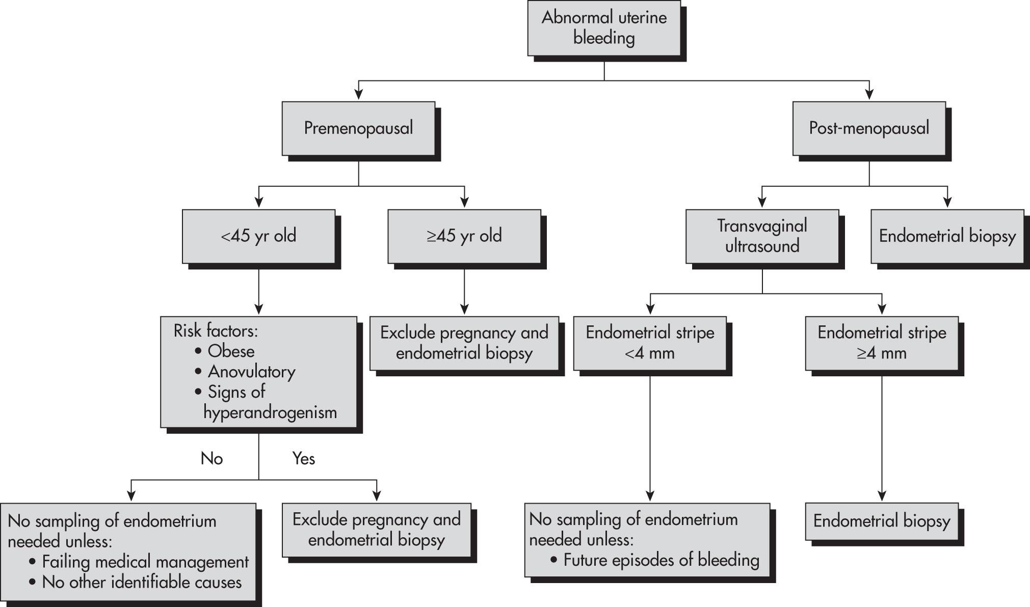
TABLE 4 National Comprehensive Cancer Network Treatment Guidelines for Endometrial Carcinoma After Comprehensive Surgical Staging
| Stage IA | |||
| Grade 1 without ARF | Observe | ||
| Grade 1 with ARF | Observe or VBT | ||
| Grade 2 or 3 without ARF | Observe or VBT | ||
| Grade 2 or 3 with ARF | Observe or VBT and/or pelvic RT | ||
| Stage IB | |||
| Grade 1 without ARF | Observe | ||
| Grade 1 with ARF | Observe or VBT | ||
| Grade 2 without ARF | Observe or VBT | ||
| Grade 2 with ARF | Observe or VBT and/or pelvic RT | ||
| Grade 3 without ARF | Observe or VBT and/or pelvic RT | ||
| Grade 3 with ARF | Observe or VBT and/or pelvic RT ± chemotherapy | ||
| Stage II | |||
| Grade 1 | VBT and/or pelvic RT | ||
| Grade 2 | Pelvic RT and VBT | ||
| Grade 3 | Pelvic RT and VBT ± chemotherapy | ||
| Stage IIIA | Chemotherapy ± pelvic RT or tumor-directed RT ± chemotherapy or pelvic RT ± VBT | ||
| Stage IIIB-IIIC | Chemotherapy and/or tumor-directed RT | ||
| Stage IVA-IVB | Chemotherapy ± RT |
ARF, Adverse risk factors (age, positive lymphovascular space invasion, tumor size, lower uterine or cervical involvement); RT, radiation therapy; VBT, vaginal brachytherapy.
From Niederhuber JE: Abeloffs clinical oncology, ed 6, Philadelphia, 2020, Elsevier.
TABLE 5 FIGO Staging for Endometrial Cancer (2023)2
| Stages* | Characteristic | ||
| IA | Nonaggressive histologic type of endometrial carcinoma limited to a polyp or confined to the endometrium | ||
| IA2 | Nonaggressive histologic types of endometria involving <50% of the myometrium with no or focal lymphovascular space invasion (LVSI) as defined by WHO criteria | ||
| IA3 | Low-grade endometrioid carcinomas limited to the uterus with low-grade endometrioid ovarian involvement | ||
| IB | Nonaggressive histologic types involving 50% or more of the myometrium with no LVSI or focal LVSI | ||
| IC | Aggressive histologic types, i.e., serous, high-grade endometrioid, clear cell, carcinosarcomas, undifferentiated, mixed, and other unusual types without any myometrial invasion | ||
| IIA | Nonaggressive histologic types that infiltrate the cervical stroma | ||
| IIB | Nonaggressive histologic types that have substantial LVSI | ||
| IIC | Aggressive histologic types with any myometrial invasion | ||
| IIIA | Differentiating between adnexal vs. uterine serosa infiltration | ||
| IIIB | Infiltration of vagina/parametria and pelvic peritoneal metastasis | ||
| IIIC | Refinements for lymph node metastasis to pelvic and paraaortic lymph nodes, including micrometastasis and macrometastasis | ||
| IVA | Locally advanced disease infiltrating the bladder or rectal mucosa | ||
| IVB | Extrapelvic peritoneal metastasis | ||
| IVC | Distant metastasis |
FIGO, Fédération Internationale de Gynécologie et dObstétrique (International Federation of Gynecology and Obstetrics).
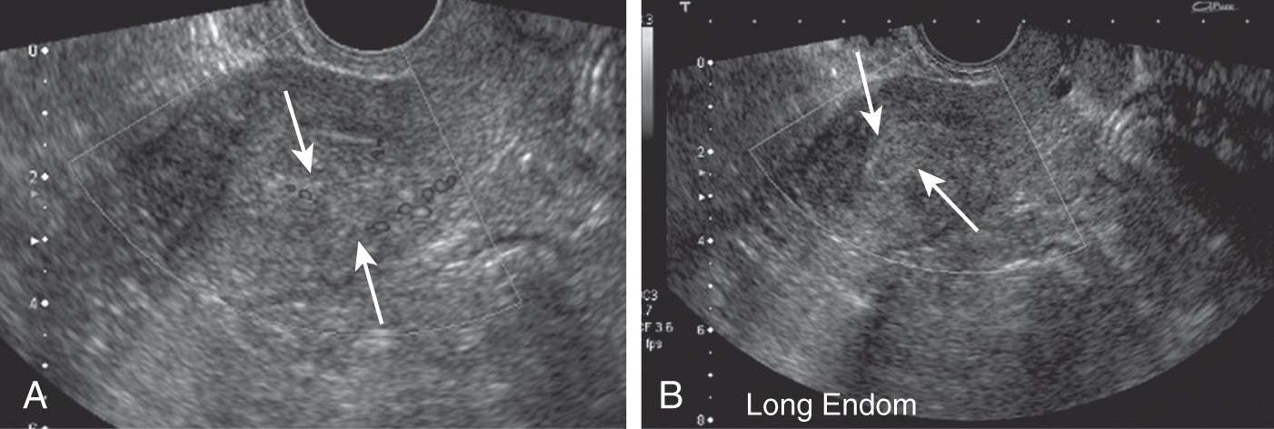
Figure E2 A 48-yr-old woman with endometrial carcinoma.

A, Endovaginal ultrasound (US) showing thickened, heterogeneous, cystic, and vascular hyperechoic tissue filling the endometrial cavity (arrows). B, Second sagittal US image showing the same (arrows).
(From Fielding JR et al: Gynecologic imaging, Philadelphia, 2011, Saunders.)
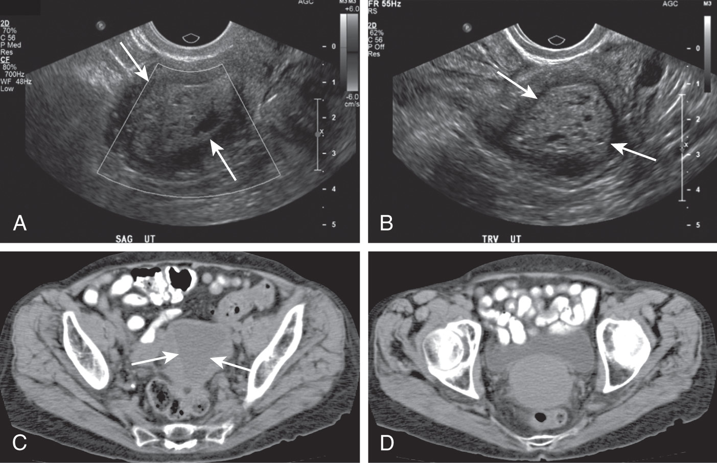
Figure E3 A 56-yr-old woman with endometrial carcinoma.

A, Sagittal ultrasound (US) image showing thickened cystic echogenic soft tissue filling the endometrial cavity (arrows). B, Axial US image showing thickened cystic echogenic soft tissue filling the endometrial cavity (arrows). C, Noncontrast-enhanced axial computed tomographic (CT) image showing low-attenuation tissue filling the endometrial canal (arrows) in a postmenopausal patient. Note fundal thinning. D, Noncontrast-enhanced axial CT image showing cervical soft tissue fullness.
(From Fielding JR et al: Gynecologic imaging, Philadelphia, 2011, Saunders.)
Any woman with postmenopausal bleeding or abnormal uterine bleeding with risk factors for endometrial cancer needs evaluation by a gynecologist and either endometrial biopsy and/or pelvic ultrasound. When endometrial cancer is diagnosed, the patient should be cared for by a gynecologic oncologist and undergo surgical staging in a minimally invasive procedure when possible.