WorkupFig. E1 describes an algorithm for the diagnosis of intrahepatic cholangiocarcinoma. The approach to the diagnosis of perihilar cholangiocarcinoma is summarized in Fig. E2. The Bismuth-Corlette classification of cholangiocarcinoma based on location is illustrated in Fig. E3. Staging systems for cholangiocarcinoma are described in Tables E1 to E3.
TABLE E1 TNM and American Joint Committee on Cancer (AJCC)/International Union Against Cancer (UICC) Staging Systems for Intrahepatic Cholangiocarcinoma
| TNM Stage | Criteria |
| Tx | Primary tumor cannot be assessed |
| T0 | No evidence of primary tumor |
| Tis | Carcinoma in situ (intraductal tumor) |
| T1a | Solitary tumor ≤5 cm without vascular invasion |
| T1b | Solitary tumor >5 cm without vascular invasion |
| T2 | Solitary tumor with vascular invasion OR multiple tumors, with or without vascular invasion |
| T3 | Tumor perforating the visceral peritoneum |
| T4 | Tumor involving the local extrahepatic structures by direct invasion |
| Nx | Regional lymph nodes cannot be assessed |
| N0 | No regional lymph node metastases |
| N1 | Regional lymph node metastases present |
| M0 | No distant metastases |
| M1 | Distant metastases |
| AJCC/UICC Stage | Tumor | Node | Metastasis |
| 0 | Tis | N0 | M0 |
| IA | T1a | N0 | M0 |
| IB | T1b | N0 | M0 |
| II | T2 | N0 | M0 |
| IIIA | T3 | N0 | M0 |
| IIIB | T4 | N0 | M0 |
| Any T | N1 | M0 |
| IV | Any T | Any N | M1 |
TNM, Tumor, node, metastasis.
From Feldman M et al: Sleisenger and Fordtran’s gastrointestinal and liver disease, ed 11, Philadelphia, 2021, Elsevier.
TABLE E2 MSKCC Staging System for Perihilar Cholangiocarcinoma
| Stage | Criteria |
| T1 | Tumor involving biliary confluence ± unilateral extension to secondary radicles |
| T2 | - Tumor involving biliary confluence ± unilateral extension to secondary radicles
- AND ipsilateral portal vein involvement ± ipsilateral hepatic lobe atrophy
|
| T3 | - Tumor involving biliary confluence + bilateral extension to secondary radicles
- OR unilateral extension to secondary radicles + contralateral portal vein involvement
- OR main or bilateral portal vein involvement
|
MSKCC, Memorial Sloan Kettering Cancer Center.
From Feldman M et al: Sleisenger and Fordtran’s gastrointestinal and liver disease, ed 11, Philadelphia, 2021, Elsevier.
TABLE E3 Mayo Clinic Staging System for Perihilar Cholangiocarcinoma
| Stage | Criteria |
| I | - Unicentric mass ≤3 cm
- ECOG performance status 0
- Serum CA 19-9 level (U/ml) <1000
- No vascular encasement
- No metastasis
|
| II | - Unicentric mass ≤3 cm
- ECOG performance status 1-2
- Serum CA 19-9 level (U/ml) <1000
- Vascular encasement present
- No metastasis
|
| III | - Unicentric mass >3 cm or multicentric
- ECOG performance status 0-2
- Serum CA 19-9 level (U/ml) ≥1000
- Lymph node metastasis present
|
| IV | - ECOG performance status 3-4
- Peritoneal (or other organ) metastasis present
|
ECOG, Eastern Cooperative Oncology Group.
From Feldman M et al: Sleisenger and Fordtran’s gastrointestinal and liver disease, ed 11, Philadelphia, 2021, Elsevier.
Figure E1 Algorithm for the diagnosis of intrahepatic cholangiocarcinoma.

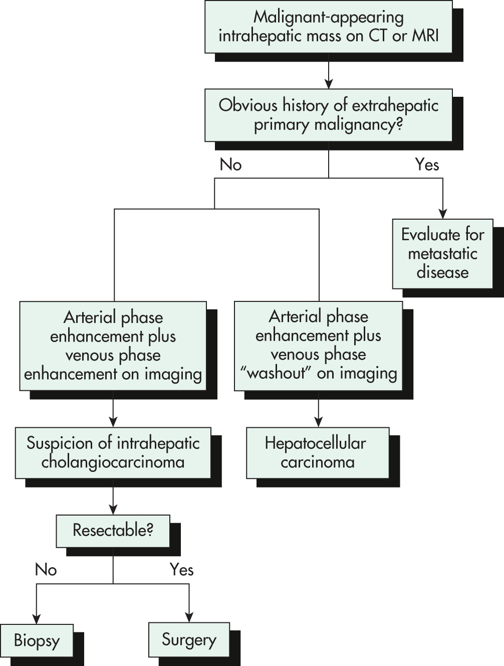
In cases of an intrahepatic mass lesion and in the absence of known extrahepatic primary malignancy, dynamic imaging with either computed tomography (CT) or magnetic resonance image (MRI) of the liver should be performed. Contrast enhancement throughout the arterial phase with "washout" in the portal venous phase indicates a hepatocellular carcinoma. Contrast enhancement throughout the arterial and portal venous phases should raise the suspicion of an intrahepatic cholangiocarcinoma; in such cases, the resectability of the tumor should be determined. If the lesion is deemed resectable, the patient should be referred for surgical resection without biopsy. If an intrahepatic cholangiocarcinoma is deemed unresectable, a biopsy should be performed to confirm the diagnosis and guide appropriate treatment.
(From Feldman M et al: Sleisenger and Fordtran’s gastrointestinal and liver disease, ed 11, Philadelphia, 2021, Elsevier.)
Figure E2 Algorithm for the diagnosis of perihilar cholangiocarcinoma.
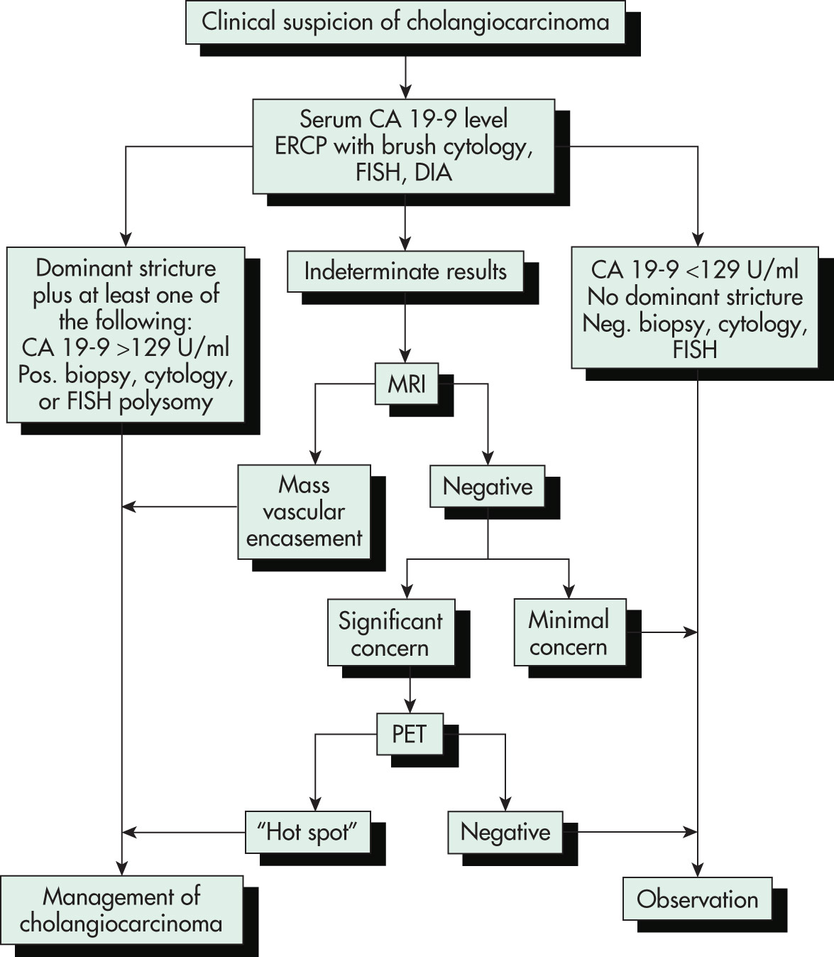
In cases of clinically suspected perihilar cholangiocarcinoma, a serum CA 19-9 level, ERCP, and conventional as well as molecular cytologic analysis of endoscopically obtained biliary brushings of malignant-appearing areas should be performed. If the results of these tests are normal or negative, close follow-up of the patient is recommended. Management of cholangiocarcinoma should be prompted by identification of a dominant stricture, a serum CA 19-9 level above 129 U/ml, or a biopsy or cytology result that is positive for carcinoma or polysomy. In indeterminate cases, gadolinium-enhanced MRI of the liver is recommended. If a mass lesion or vascular encasement is identified, management of cholangiocarcinoma should be initiated. If the MRI study is negative but clinical concern about cholangiocarcinoma persists, PET can be performed. If "hot spots" are identified on PET (positive result), treatment for cholangiocarcinoma should be initiated. If the result of the PET scan is negative, close follow-up of the patient is recommended. If MRI is negative and cholangiocarcinoma is considered unlikely, the patient can be followed expectantly. DIA, Digital image analysis; ERCP, endoscopic retrograde cholangiopancreatography; FISH, fluorescence in situ hybridization; MRI, magnetic resonance imaging; neg., negative; PET, positron emission tomography; pos., positive.
(From Feldman M et al: Sleisenger and Fordtran’s gastrointestinal and liver disease, ed 11, Philadelphia, 2021, Elsevier.)
Figure E3 Classification of cholangiocarcinoma.
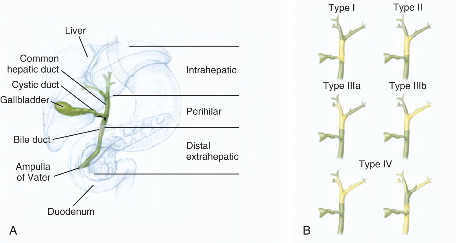
A, Anatomic classification of intrahepatic, perihilar, and extrahepatic cholangiocarcinoma. B, Bismuth-Corlette classification of hilar cholangiocarcinoma as types I to IV. Tumor is depicted in yellow and normal bile ducts in green.
(From Feldman M et al: Sleisenger and Fordtran’s gastrointestinal and liver disease, ed 11, Philadelphia, 2021, Elsevier.)
Laboratory Tests
- •Liver enzymes
- •International normalization ratio
- •Bilirubin
Blood work may reveal an "obstructive pattern" with predominant elevation of alkaline phosphatase and bilirubin.
- •Tumor markers:
- 1.CEA
- 2.CA19-9
It is important to note that these tumor markers may be elevated even in the setting of benign inflammation of the biliary tree, for example, during an episode of acute cholangitis. These markers are neither sensitive nor specific for cholangiocarcinoma, but if they are elevated at the time of diagnosis, they may be helpful in tracking response to therapy or in monitoring for disease recurrence.
Imaging StudiesAbdominal ultrasound:
- •Initial test of choice in patients presenting with nonspecific symptoms of obstruction or elevated liver enzymes
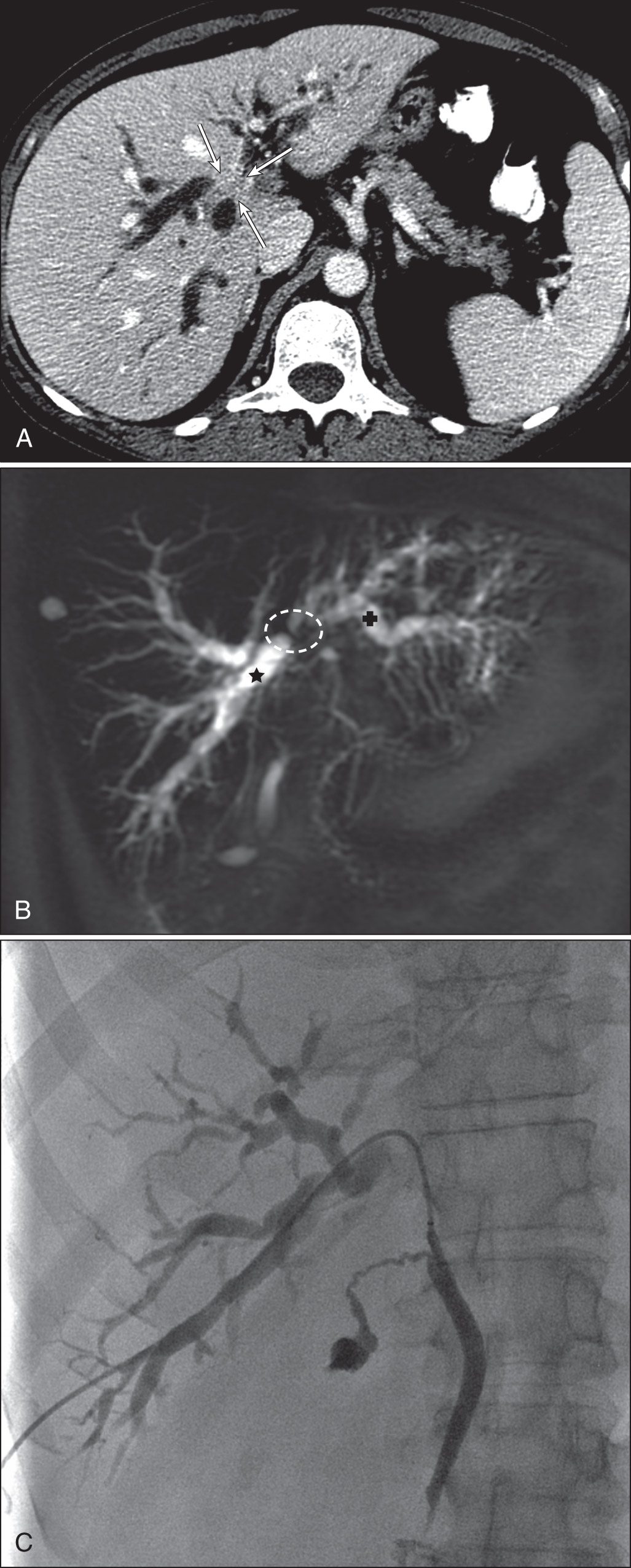
Computed tomography scan/MRI, magnetic resonance cholangiography (MRCP) (Fig. E4):
- •Improved visualization of the biliary tree to allow for identification of the tumor, level of obstruction, and extent of disease
Figure E4 Computed Tomography (CT) Scan (A), Magnetic Resonance Cholangiogram (B), and Image from Biliary Drainage (C) in a Patient with Hilar Cholangiocarcinoma
(A) The CT Scan Shows the Tumor (Arrows) at the Level Where the Right and Left Bile Ducts (Star and Cross, Respectively, in B) Would Be Expected to Join. (B) Magnetic Resonance Cholangiography Confirms This. (C) At the Time of Biliary Drainage, Contrast Opacifies Only the Right-Sided Bile Ducts. The Tumor is Obstructing the Duct to the Level of the Common Bile Duct at the Insertion of the Cystic Duct.
(From Niederhuber JE et al: Abeloff’s clinical oncology, ed 6, Philadelphia, 2020, Elsevier.)
Endoscopic retrograde cholangiopancreatography (ERCP) (Fig. E5):
- •Can be used to obtain brushings or biopsies of the biliary tree, but diagnostic yield is poor (35% to 86%), and there is a risk of cholangitis in patients with significant obstruction of bile drainage
Figure E5 Imaging of extrahepatic cholangiocarcinoma.
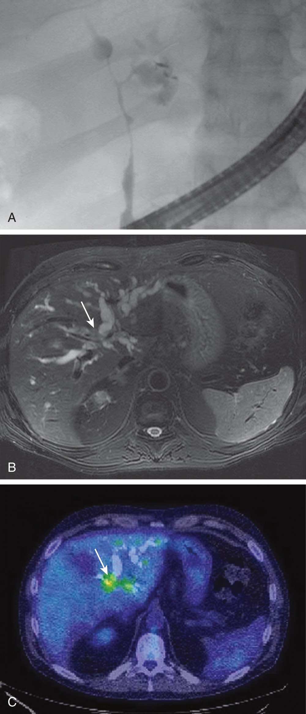
A, Film from endoscopic retrograde cholangiography in a patient with perihilar cholangiocarcinoma demonstrating dominant strictures of the biliary tract consistent with Bismuth-Corlette type IV. B, Gadolinium-enhanced magnetic resonance image with ferumoxide in the same patient. The arrow points to the biliary tumor seen on a T2-weighted image. C, Positron emission tomography/computed tomography scan of the same patient. The biliary tumor is seen as an enhancing region (arrow).
(From Feldman M et al: Sleisenger and Fordtran’s gastrointestinal and liver disease, ed 11, Philadelphia, 2021, Elsevier.)
Endoscopic ultrasound (EUS):
- •Allows local ultrasound visualization of tumor extent and regional lymphadenopathy, as well as diagnostic fine-needle aspiration of distal biliary duct tumors and involved lymph nodes


