section name header

Symptoms

Sudden, painless visual loss. Initially unilateral, but may rapidly become bilateral. Occurs in patients 55 years of age. Antecedent or simultaneous headache, jaw claudication (pain with chewing), scalp tenderness especially over the superficial temporal arteries (e.g., tenderness with hair combing), proximal muscle and joint aches (polymyalgia rheumatica), anorexia, weight loss, or fever may occur.

Signs

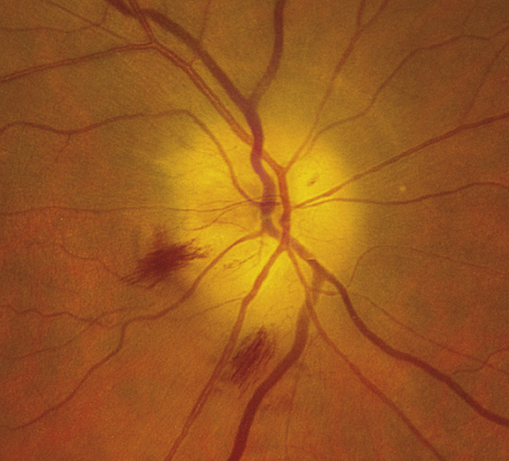
(See Figure 10.17.1.)

Critical

Afferent pupillary defect; visual loss (often counting fingers or worse); pale, swollen disc, at times with flame-shaped hemorrhages. Later, optic atrophy and cupping occur as the edema resolves. The ESR, CRP, and platelet count may be markedly increased.

Other

Visual field defect (commonly altitudinal or involving the central field); a palpable, tender, and often nonpulsatile temporal artery; and a central retinal artery occlusion or a cranial nerve palsy (especially a sixth cranial nerve palsy) may occur.

10-17.1 Chalky, pale, swollen optic disc with flame-shaped hemorrhages in giant cell arteritis.
Gervasio-ch010-image019

Differential Diagnosis

  • NAION: Patients may be younger. Visual loss often less severe, do not have the accompanying symptoms of GCA listed previously, and usually have a normal ESR and CRP. See 10.18, NONARTERITIC ISCHEMIC OPTIC NEUROPATHY.
  • Inflammatory optic neuritis: Younger age group. Pain with eye movements. Optic disc swelling, if present, is more hyperemic. See 10.14, OPTIC NEURITIS.
  • Compressive optic nerve tumor: Optic nerve pallor or atrophy may be seen; disc heme unlikely. Slowly progressive visual loss. Few to no symptoms common with GCA.
  • Central retinal vein occlusion: Severe visual loss may be accompanied by an afferent pupillary defect and disc swelling, but the retina shows diffuse retinal hemorrhages extending out to the periphery. See 11.8, CENTRAL RETINAL VEIN OCCLUSION.
  • Central retinal artery occlusion: Sudden, painless, severe visual loss with an afferent pupillary defect. No disc swelling. Retinal edema with a cherry-red spot frequently observed. See 11.6, CENTRAL RETINAL ARTERY OCCLUSION.

Work Up

Workup
  1. History: GCA symptoms present? Age is critical.
  2. Complete ocular examination, particularly pupillary assessment, dyschromatopsia evaluation (e.g., color plates), dilated retinal examination to rule out retinal causes of severe visual loss, and optic nerve evaluation.
  3. Immediate ESR (Westergren is the most reliable method), CRP (does not rise with age), and platelet count (may have thrombocytosis). A guideline for top-normal ESR calculation: men, age/2; women, (age + 10)/2. ESR may not be increased. CRP and platelet upper limit values are based on laboratory-specific standards.
  4. Perform a temporal artery biopsy if GCA is suspected.
NOTE:

The biopsy should be performed within 1 week after starting systemic steroids, but a positive result may be seen up to 1 month later. Biopsy is especially important in patients in whom steroids are relatively contraindicated (e.g., diabetics).

Treatment

  1. Systemic steroids should be given immediately once GCA is suspected. Methylprednisolone 250 mg i.v., q6h for 12 doses, and then switch to prednisone 80 to 100 mg p.o. daily. A temporal artery biopsy specimen should be obtained within 1 week of systemic steroid initiation.
  2. If the temporal artery biopsy is positive for GCA (or clinical presentation/therapeutic response warrants continued therapy), the patient must be maintained on prednisone, about 1 mg/kg initially. Oral steroids are tapered on an individual basis with the goal of using the lowest required dose necessary for disease suppression (see below).
  3. If the biopsy is negative on an adequate (2 to 3 cm) section, the likelihood of GCA is small. However, in highly suggestive cases, biopsy of the contralateral artery is performed.
  4. Steroids are usually discontinued if the disease is not found in adequate biopsy specimens, unless the clinical presentation is classic and a response to treatment has occurred.
NOTE:
  1. Without steroids (and occasionally on adequate steroids), the contralateral eye can become involved within 1 to 7 days.
  2. Concurrent antiulcer (e.g., proton-pump inhibitor [e.g., omeprazole 20 mg p.o. daily] or histamine type 2 receptor blocker [e.g., ranitidine 150 mg p.o. b.i.d.]) should be used for gastrointestinal prophylaxis while on steroids.
  3. A medication to help prevent osteoporosis should be used as directed by an internist, particularly given the long-term need for steroids.

Follow Up

  1. Patients suspected of having GCA must be evaluated and treated immediately.
  2. After the diagnosis is confirmed by biopsy, the initial oral steroid dosage is maintained until the symptoms resolve and lab values normalize. The dosage is then tapered slowly, repeating bloodwork with each dosage change and/or monthly (whichever appropriate based on disease stability) to ensure that the new steroid dosage is enough to suppress the disease.
  3. If the ESR or CRP increase or symptoms return, the dosage must be increased.
  4. Treatment should last at least 6 to 12 months. The smallest steroid dose that suppresses disease is used.

Tocilizumab, a novel humanized IL-6 receptor antagonist, has been recently approved by the FDA for long-term management of GCA. Addition of subcutaneous tocilizumab to standardized steroid regimens helps achieve remission faster and significantly reduces the dose and toxicity of corticosteroids.