Red eye, pain, blurred vision, double vision, eyelid and/or periorbital swelling, nasal congestion/discharge, sinus headache/pressure/congestion, tooth pain, infra- and/or supraorbital pain, or hypesthesia.
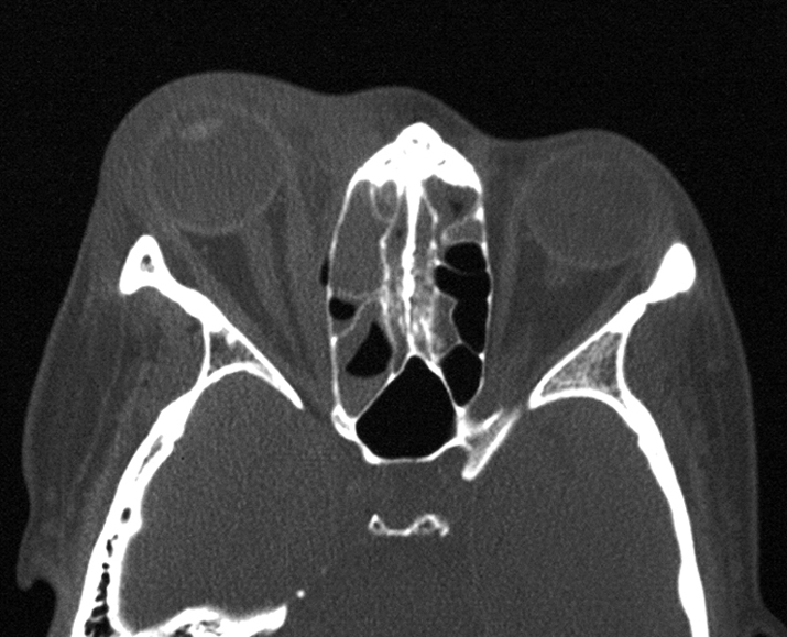
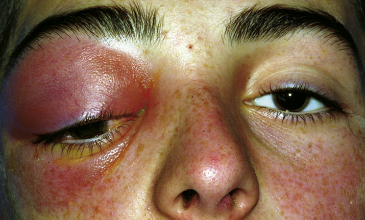
(See Figures 7.3.1.1 and 7.3.1.2.)
Critical
Eyelid edema, erythema, warmth, and tenderness. Conjunctival chemosis and injection, proptosis, and restricted extraocular motility with pain on attempted eye movement are usually present. Signs of optic neuropathy (e.g., afferent pupillary defect and dyschromatopsia) may be present in severe cases.
Other
Decreased vision, retinal venous congestion, optic disc edema, purulent discharge, decreased periorbital sensation, and fever. CT scan usually shows adjacent sinusitis (typically at least an ethmoid sinusitis) and possibly a subperiosteal orbital collection.
7-3.1.2 CT of right orbital cellulitis showing fat stranding and right ethmoiditis.

7-3.1.1 Orbital cellulitis.

Workup
See 7.1, ORBITAL DISEASE, for a nonspecific orbital workup.
- History: Trauma or surgery? Ear, nose, throat, or systemic infection? Tooth pain or recent dental abscess? Stiff neck or mental status changes? Diabetes or an immunosuppressive illness? Use of immunosuppressive agents?
- Complete ophthalmic examination to evaluate for orbital signs including afferent pupillary defect, restriction or pain with ocular motility, proptosis, increased resistance to retropulsion, elevated IOP, decreased color vision, decreased skin sensation, or an optic nerve or fundus abnormality.
- Check vital signs, mental status, and neck flexibility. Check for preauricular or cervical lymphadenopathy. Evaluate nasal passages for signs of eschar/fungal involvement in diabetic, acidotic, or immunocompromised patients.
- Imaging: CT scan of the orbits and paranasal sinuses (axial, coronal, and parasagittal views, with contrast if possible) to confirm the diagnosis and to rule out a retained foreign body, orbital or SPA, paranasal sinus disease, cavernous sinus thrombosis, or intracranial extension.
- Laboratory studies: CBC with differential and blood cultures.
- Explore and debride any penetrating wound, if present, and obtain a Gram stain and culture of any drainage (e.g., blood and chocolate agars, Sabouraud dextrose agar, and thioglycolate broth). Obtain CT before wound exploration to rule out skull base foreign body.
- Consult neurosurgery for suspected meningitis for management and possible lumbar puncture. If paranasal sinusitis is present, consider a consultation with otorhinolaryngology for possible surgical drainage. Consider an infectious disease consultation in atypical, severe, or unresponsive cases. If a dental source is suspected, oral maxillofacial surgery should be consulted urgently for assessment, since infections from this area tend to be aggressive, potentially vision threatening, and may spread into the cavernous sinus.
 NOTE: NOTE: |
Zygomycosis is an orbital, nasal, and sinus disease occurring in diabetic or otherwise immunocompromised patients. Typically associated with severe pain and external ophthalmoplegia. Profound visual loss may rapidly occur. Metabolic acidosis may be present. Sino-orbital zygomycosis is rapidly progressive and life threatening. See 10.10, CAVERNOUS SINUS AND ASSOCIATED SYNDROMES (MULTIPLE OCULAR MOTOR NERVE PALSIES). |
Re-evaluate at least twice daily in the hospital for the first 48 hours. Severe infections may require multiple daily examinations. Clinical improvement may take 24 to 36 hours.
- Progress is monitored by:
- Patient’s symptoms.
- Temperature and white blood cell (WBC) count.
- Visual acuity and evaluation of optic nerve function.
- Extraocular motility.
- Degree of proptosis and any displacement of the globe (significant displacement may indicate an abscess).
- C-reactive protein (CRP) has been found to be a helpful clinical marker in some studies. One study suggested initiating oral corticosteroids with antibiotic therapy at a threshold CRP of ≤4 mg/dL.
- Evaluate the cornea for signs of exposure.
- Check IOP.
- Examine the retina and optic nerve for signs of posterior compression (e.g., chorioretinal folds), inflammation, or exudative retinal detachment.
- If orbital cellulitis is clearly and consistently improving, then the regimen can be changed to oral antibiotics (depending on the culture and sensitivity results) to complete a 10- to 14-day course. We often use:
- Amoxicillin/clavulanate: 25 to 45 mg/kg/d p.o. in two divided doses for children and a maximum daily dose of 90 mg/kg/d; 875 mg p.o. q12h for adults;
- or
- Cefpodoxime: 10 mg/kg/d p.o. in two divided doses for children and a maximum daily dose of 400 mg; 200 mg p.o. q12h for adults;
- or
- If CA-MRSA is suspected, recommended oral treatment regimens include doxycycline 100 mg p.o. q12h (not in pregnant or nursing women and not in children younger than 8 years), one to two tablets trimethoprim/sulfamethoxazole 160/800 mg p.o. q12h, clindamycin 450 mg p.o. q6h, or linezolid 600 mg p.o. b.i.d. (only with approval from an infectious disease specialist, given current low resistance).
- The patient is examined every few days as an outpatient until the condition resolves and instructed to return immediately with worsening signs or symptoms.
 NOTE: NOTE: |
If clinical deterioration is noted after an adequate antibiotic load (three to four doses), a CT scan of the orbit and brain with contrast should be repeated to look for abscess formation (see 7.3.2, SUBPERIOSTEAL ABSCESS). If an abscess is found, surgical drainage may be required. Because radiographic findings may lag behind the clinical examination, clinical deterioration may be the only indication for surgical drainage. Other conditions that should be considered when the patient is not improving include cavernous sinus thrombosis, meningitis, resistant organism (HA-MRSA, CA-MRSA), aggressive organism (often from an undiagnosed odontogenic source), or a noninfectious etiology. |
 NOTE: NOTE: |
Medication noncompliance is an extremely common reason for recurrence or failure to improve. The oral antibiotic regimen should be individualized for ease of use and affordability. Effective generic alternatives to brand name antibiotics include doxycycline and trimethoprim/sulfamethoxazole. |
ReferencesYen MT, Yen KG. Effect of corticosteroids in the acute management of pediatric orbital cellulitis with subperiosteal abscess. Ophthalmic Plast Reconstr Surg. 2005;21:363-366.Davies BW, Smith JM, Hink EM, Durairaj VD. C-reactive protein as a marker for initiating steroid treatment in children with orbital cellulitis. Ophthalmic Plast Reconstr Surg. 2015;31:364-368.

