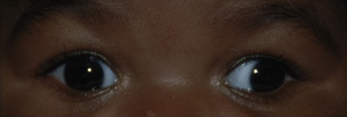
Pseudoexotropia: The patient appears to have an exodeviation, but no movement is noted on cover–uncover testing despite good vision in each eye. A wide interpupillary distance, a naturally large angle k (angle between the pupil center and the visual axis), or temporal dragging of the macula (e.g., from ROP, FEVR, toxocariasis, or other retinal disorders) may be responsible.
Types
- Exophoria: A latent exodeviation controlled by fusion under conditions of normal binocular vision. Usually asymptomatic, but prolonged strenuous visual activity may cause asthenopia.
- Intermittent exotropia: A manifest deviation in which one eye demonstrates exodeviation part of the time. The most common type of exodeviation in children. Onset is usually before age 5. Frequency often increases over time. Amblyopia is rare. Usually occurs when the patient is fatigued, sick, or not attentive. Patient often closes one eye or squints in bright sunlight. This is likely due to dissociation and breakdown of their binocular alignment.
- Clinical evaluation:
- Good control: One eye turns out at only after cover testing and the patient is able to regain fusion quickly without blinking or refixating when the cover is removed.
- Fair control: One eye turns out after cover testing and the patient can only regain fusion with blinking or refixating.
- Poor control: One eye turns out spontaneously and remains manifest for an extended period of time.
- Good, fair, or poor control can be seen in all four types of intermittent exotropia:
- Basic: Exodeviation is approximately the same with distance and near fixation.
- True divergence excess: Exodeviation that remains greater at distance than near after a period of monocular occlusion.
- Simulated divergence excess: Exodeviation that is initially greater with distance fixation than near that becomes approximately the same after an interval of monocular occlusion.
- Convergence insufficiency: Exodeviation is greater at near than distance. Distinct from isolated convergence insufficiency. See 13.4, CONVERGENCE INSUFFICIENCY.
- Constant exotropia: Encountered more often in older children. There are three types:
- Congenital exotropia: Also known as infantile exotropia. Presents before age 6 months with a large angle deviation. Uncommon in otherwise healthy infants and may be associated with a central nervous system or craniofacial disorder.
- Sensory-deprivation exotropia: An eye that does not see well for any reason may turn outward.
- Decompensated intermittent exotropia: A patient with long-standing intermittent exotropia that has decompensated.
- Consecutive exotropia: Follows a diagnosis of esotropia, most commonly after previous surgery for esotropia.
- Duane syndrome, type 2: Limitation of adduction of one eye, with globe retraction and narrowing of the palpebral fissure on attempted adduction. Rarely bilateral. See 8.6, STRABISMUS SYNDROMES. May present with head turn away from affected eye.
- Neuromuscular abnormalities:
- Dissociated horizontal deviation: A change in horizontal ocular alignment caused by a change in the balance of visual input from the two eyes. Not related to accommodation. Seen clinically as a spontaneous unilateral exodeviation or an exodeviation of greater magnitude in one eye during prism and alternate cover testing.
- Orbital disease (e.g., tumor, idiopathic orbital inflammatory syndrome): Proptosis and restriction of ocular motility are usually evident. See 7.1, ORBITAL DISEASE.
- Isolated convergence insufficiency: Usually occurs in patients >10 years old. Blurred near vision, asthenopia, or diplopia when reading. An exophoria at near fixation, but straight or small exophoria at distance fixation. Must be differentiated from convergence paralysis. See 13.4, CONVERGENCE INSUFFICIENCY.
- Convergence paralysis: Similar to convergence insufficiency, but with a relatively acute onset, an exotropia at near, and an inability to overcome base out prism. Often secondary to an intracranial lesion.
In all cases, correct significant refractive errors and treat amblyopia. See 8.7, AMBLYOPIA.
- Exophoria:
- No treatment necessary unless it progresses to intermittent exotropia.
- Intermittent exotropia:
- Good control: Correct refractive error and treat amblyopia if present. Follow patient closely.
- Fair control: Correct refractive error and treat amblyopia if present. Nonsurgical treatment may be indicated and include occlusion therapy with alternate daily patching to reduce suppression or additional minus lenses to stimulate accommodative convergence. Muscle surgery may be considered to maintain normal binocular vision.
- Poor control: Correct refractive error and treat amblyopia if present. Nonsurgical treatments as described above may be attempted. Muscle surgery is often indicated. Bifixation or peripheral fusion can occasionally be attained.
- Sensory-deprivation exotropia:
- Correct the underlying cause, if possible.
- Treat any amblyopia.
- Muscle surgery may be performed for manifest exotropia.
- When one eye has very poor vision, protective glasses (polycarbonate lens glasses) should be worn at all times to protect the good eye.
- Congenital exotropia:
- Muscle surgery within a year of onset, as in patients with congenital esotropia.
- Consecutive exotropia:
- Additional muscle surgery may be considered.
- Prism correction in glasses can be used.
- Over-minus or under-plus correction can stimulate accommodative convergence.
- Dissociated horizontal deviation:
- Muscle surgery may be considered.
- Duane syndrome: See 8.6, STRABISMUS SYNDROMES.
- Third cranial nerve palsy: See 10.5, ISOLATED THIRD CRANIAL NERVE PALSY.
- Convergence insufficiency: See 13.4, CONVERGENCE INSUFFICIENCY.
- Convergence paralysis:
- Base-in prisms at near to alleviate diplopia.
- Plus lenses if accommodation is also weakened.
