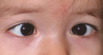
(See Figure 8.6.1.)
Figure 8.6.1: Esotropia.
Critical
One eye is turned inward off target of fixation. The nonfixating eye turns outward to fixate when examiner covers the originally fixating eye during the cover–uncover test. See Appendix 3, Cover/Uncover and Alternate Cover Tests.
Other
Amblyopia, overaction of the inferior oblique muscles, dissociated vertical deviation, and/or latent nystagmus may be present.
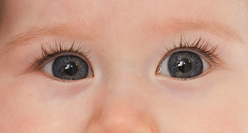
Pseudoesotropia: The eyes appear esotropic; however, there is no ocular misalignment detected during cover–uncover or corneal light reflex testing. Usually, the child has a wide nasal bridge, prominent epicanthal folds, or a small interpupillary distance (See Figure 8.6.2).
Figure 8.6.2: Pseudoesotropia.
See 8.8, Strabismus Syndromes.
Types
Comitant Esotropic Deviations
A manifest convergent misalignment of the eyes in which the measured angle of esodeviation is nearly constant in all fields of gaze at distance fixation.
Congenital (infantile) esotropia: Manifests by age 6 months. The angle of esodeviation is usually large (>40 prism diopters [PD]) and equal at distance and near fixation. Refractive error is usually normal for age (slightly hyperopic). Amblyopia is uncommon but may be present in those who do not cross-fixate. Prohibits development of binocular vision. Family history may be present. Latent nystagmus, inferior oblique overaction, and dissociated vertical deviation may develop as late findings. Congenital esotropia can occur in up to 30% of children with neurologic and developmental disorders (e.g., cerebral palsy, hydrocephalus); however, it is not necessary to perform a neurologic workup in the absence of other findings. Cycloplegic refraction should be considered first, but treatment is often surgical (in the absence of neurologic delays) when the deviation is stable.
Acquired nonaccommodative esotropia: Convergent misalignment of the eyes not corrected by hyperopic lenses that develops after 6 months of age. Typically starts as intermittent but can become constant over time. Esodeviation is comitant and usually smaller (20 to 35 PD) than in congenital esotropia. Patients may experience diplopia. Usually corrected with strabismus surgery once the angle of the esotropia becomes consistent. In children older than 6 years, this must be assumed to be posterior fossa pathology until proven otherwise and worked up with imaging emergently.
Accommodative esotropia: Convergent misalignment of the eyes associated with the accommodative reflex. May present at 6 months to 6 years of age with the average age of onset being 2.5 years. Subtypes of accommodative esotropia:
Refractive accommodative esotropia (normal accommodative convergence to accommodation [AC/A] ratio): These children have uncorrected hyperopia in the range of +3.00 to +10.00 D (average, +4.75 D). The measured angle of esodeviation is usually moderate (20 to 30 PD) and is relatively equal at distance and near fixation. Full hyperopic correction eliminates the esodeviation. Amblyopia is common at presentation.
Nonrefractive accommodative esotropia (high AC/A ratio): The measured angle of esodeviation is greater at near fixation than at distance fixation. The refractive error may range from normal for age (slight hyperopia) to high hyperopia (may be seen in conjunction with refractive-type accommodative esotropia) or even myopia. Amblyopia is common.
Partial or decompensated accommodative esotropia: Refractive and nonrefractive accommodative esotropias that is only partially corrected with full hyperopic correction, resulting in a residual esodeviation. When partial, the residual esodeviation is the nonaccommodative component.
Sensory-deprivation esotropia: An esodeviation that occurs in a patient with a monocular or binocular condition that results in persistent poor vision. Precipitating factors are numerous, including but not limited to cataract, corneal clouding, optic nerve disease, or retinal disorders.
Divergence insufficiency: A convergent ocular misalignment that is greater at distance fixation than at near. Although this can be a benign condition in older patients, called sagging eye syndrome, it must be differentiated from bilateral sixth cranial nerve palsies. See 10.8, Isolated Sixth Cranial Nerve Palsy.
Incomitant or Noncomitant Esodeviations
The measured angle of esodeviation differs in lateral gaze.
CNS pathology causing increased intracranial pressure: Acute and new onset of diplopia secondary to an acquired sixth cranial nerve palsy, which may be accompanied by nystagmus, headache, or other focal neurologic deficits depending on etiology.
Medial rectus restriction (e.g., thyroid disease, medial orbital wall fracture with entrapment).
Lateral rectus weakness (e.g., isolated sixth cranial nerve palsy, slipped or detached lateral rectus from trauma or previous surgery).
See 8.8, Strabismus Syndromes and 10.8, Isolated Sixth Cranial Nerve Palsy, for additional etiologies.
Other
Esophoria: Latent esodeviation controlled by fusion. Eyes are aligned under binocular conditions.
Intermittent esotropia: Esodeviation that is intermittently controlled by fusion. Becomes manifest spontaneously, especially with fatigue or illness.
In all cases, correct refractive errors of +2.00 D or more. In children, treat any underlying amblyopia (see 8.5, Amblyopia).
Congenital esotropia: Almost always requires strabismus surgery. However, prescribe glasses and initiate treatment of any underlying amblyopia prior to surgical intervention. Botulinum toxin may be useful in some cases.
Accommodative esotropia: Glasses must be worn full time.
If the patient is <6 years old, correct the hyperopia with the full cycloplegic refraction.
If the patient is >6 years old, attempts should be made to give as close to the full-plus refraction as possible, knowing that some may not tolerate the full prescription, initially. A short course of cycloplegics can improve acceptance of full hyperopic correction.
If the patient’s eyes are straight at distance with full correction, but still esotropic at near fixation (high AC/A ratio), treatment options include the following:
Bifocals (flat-top or executive type) +2.50 or +3.00 D add, with top of the bifocal at the lower pupillary border.
Extraocular muscle surgery targeting the near deviation only may be indicated. This typically requires posterior fixation sutures to the muscle to modify the surgical effect for near only.
Wearing full-plus distance glasses only.
|
 NOTE NOTEThere is no universal agreement on the treatment of patients with excess crossing at near only. |
Nonaccommodative, partially accommodative, or decompensated accommodative esotropia: Does not resolve with refraction alone. Muscle surgery is usually performed to correct the nonaccommodative deviation (residual esotropia that remains when glasses are worn). Botulinum toxin and prisms can be useful for some patients.
Sensory-deprivation esotropia.
Attempt to identify and correct the cause of poor vision.
Amblyopia treatment.
Give the full cycloplegic correction (in fixing eye) if the patient is <6 years of age, otherwise give as much plus as tolerated during manifest refraction.
Muscle surgery to correct the manifest esotropia.
All patients with low vision in one eye need to wear protective glasses (e.g., polycarbonate lenses) at all times.
At each visit, evaluate for amblyopia and measure the degree of deviation with prisms (with glasses worn).
If amblyopia is present, see 8.5, Amblyopia, for management.
In the absence of amblyopia, the child is reevaluated in 8 to 10 weeks after a new prescription is given. If no changes are made and the eyes are straight, the patient should be followed up several times a year when young, decreasing to annually when stable.
When a residual esotropia is present while the patient wears glasses, repeat cycloplegic refraction is required. If the eyes cannot be straightened with more plus power, then a decompensated accommodative esotropia has developed (see above under Comitant Esotropic Deviations).
Hyperopia often decreases slowly after 5 to 7 years of age, and the strength of the glasses may need to be reduced so as not to blur distance vision. If the strength of the glasses must be reduced to improve visual acuity and the esotropia returns, then this is a decompensated accommodative esotropia.