(See Figures 10.8.1 and 10.8.2. Sixth Cranial Nerve Palsy.)
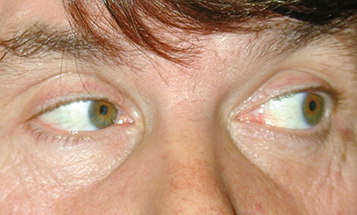
Deficient lateral movement of an eye with negative forced duction testing (see Appendix 6, Forced Duction Test and Active Force Generation Test).
Binocular horizontal diplopia, worse for distance than near, most pronounced in the direction of the paretic lateral rectus muscle.
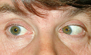
(See Figures 10.8.1 and 10.8.2. Sixth Cranial Nerve Palsy.)
Deficient lateral movement of an eye with negative forced duction testing (see Appendix 6, Forced Duction Test and Active Force Generation Test).
Differential Diagnosis of Limited Abduction
Thyroid eye disease: May have proptosis, eyelid lag, eyelid retraction, injection over the involved rectus muscles, and positive forced duction testing. See 7.2.1, Thyroid Eye Disease.
Myasthenia gravis: Variable symptoms with fatigability. Ptosis common. Positive ice test, rest test, or less commonly edrophonium chloride test. See 10.11, Myasthenia Gravis.
Idiopathic orbital inflammatory syndrome: Pain and proptosis are common. See 7.2.2, Idiopathic Orbital Inflammatory Syndrome.
Orbital trauma: Fracture causing medial rectus entrapment, positive forced duction testing. See 3.19, Orbital Blowout Fracture.
Duane syndrome, type 1: Congenital; narrowing of the palpebral fissure and retraction of the globe on adduction (usually no diplopia). See 8.8, Strabismus Syndromes.
Möbius syndrome: Congenital; bilateral facial paralysis present. See 8.8, Strabismus Syndromes.
Convergence spasm: Intermittent, variable episodes of convergence and miosis. May appear to have abduction deficit when assessing versions; however, ductions are full. Miotic pupils help to differentiate since pupils are not affected in an isolated sixth cranial nerve palsy.
Primary divergence insufficiency: Usually acquired and benign; esotropia and diplopia only at distance and single binocular vision at near. Symptoms may improve spontaneously without treatment or may be corrected with base-out prisms or surgery. If the history reveals sudden onset, trauma, infection (e.g., meningitis, encephalitis), MS, or malignancy, divergence paralysis should be considered and a neurologic workup with MRI of the brain and brainstem obtained. MRI rarely reveals pathology in true divergence insufficiency.
GCA: Less common; however, may occur with extraocular muscle ischemia in patients age ≥55 years. May be associated with systemic symptoms. See 10.17, Arteritic Ischemic Optic Neuropathy (Giant Cell Arteritis).
Vasculopathic (e.g., diabetes, hypertension, other atherosclerotic risk factors), trauma, idiopathic.
Increased intracranial pressure, cavernous sinus mass (e.g., meningioma, aneurysm, metastasis), MS, sarcoidosis, vasculitis, after myelography or LP, stroke (usually with other neurologic deficits), meningeal inflammation/infection (e.g., Lyme disease, neurosyphilis), and GCA.
Benign and usually self-limited after viral infection or vaccination, trauma, increased intracranial pressure (e.g., obstructive hydrocephalus), pontine glioma, and Gradenigo syndrome (petrositis causing sixth and often seventh cranial nerve involvement, with or without eighth and fifth cranial nerve involvement on the same side; associated with complicated otitis media).
History: Do the symptoms fluctuate during the day? Cancer, diabetes, or thyroid disease? Symptoms of GCA (in the appropriate age group)?
Complete neurologic and ophthalmic examinations; pay careful attention to the function of the other cranial nerves and the appearance of the optic disc. Because of the risk of corneal damage, it is especially important to evaluate the fifth cranial nerve. Corneal sensation (supplied by the first division) can be tested by touching a wisp of cotton or a tissue to the corneas before applying topical anesthetic. Ophthalmoscopy looking for papilledema is required because increased intracranial pressure from any cause can result in unilateral or bilateral sixth cranial nerve palsies.
Check blood pressure, fasting blood sugar, and hemoglobin A1c.
Immediate ESR, CRP, and platelet count if GCA is suspected. See 10.17, Arteritic Ischemic Optic Neuropathy (Giant Cell Arteritis).
An occlusion patch may be placed over one eye or fogging plastic tape applied to one spectacle lens to relieve symptomatic diplopia. In patients <11 years, patching is avoided, and these patients are monitored closely for the development of amblyopia. See 8.5, Amblyopia.
Prisms in glasses may be fit acutely for temporary relief or for chronic stable deviations (e.g., after stroke). Consider strabismus surgery for a stable deviation that persists >6 months.