section name header

General Information

Malignant melanoma of the iris can occur as a localized or diffuse pigmented (melanotic) or nonpigmented (amelanotic) lesion.

Signs

Critical

Unilateral brown or translucent iris mass lesion exhibiting slow growth. It is more common in the inferior half of the iris and in light-skinned individuals. Rare in Black patients (see Figure 5.14.1).

Figure 5.14.1: Iris melanoma.

Rapuano9781975243722-ch005_f026.jpg

Other

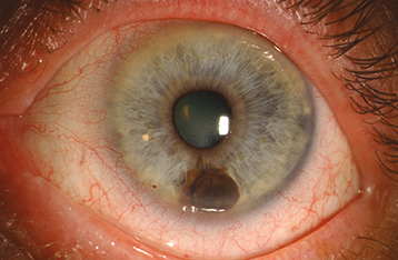
A localized melanoma is usually >3 mm in diameter at the base and >1 mm in depth with a variable prominent feeding vessel. Can produce a sector cortical cataract, ectropion iridis, spontaneous hyphema, seeding of tumor cells into the anterior chamber, or direct invasion of tumor into the trabecular meshwork and secondary glaucoma. A diffuse melanoma causes progressive darkening of the involved iris, loss of iris crypts, angle invasion, and increased IOP. Focal iris nodules can be present.

Differential Diagnosis

Melanotic Masses

Amelanotic Masses

Diffuse Lesions

Workup

  1. History: Previous cancer, ocular surgery, or trauma? Weight loss? Anorexia?

  2. Slit-lamp examination: Carefully evaluate the irides. Check IOP.

  3. Gonioscopy.

  4. Dilated fundus examination using indirect ophthalmoscopy.

  5. Transillumination of iris mass (helps differentiate epithelial cysts that transmit light from pigmented lesions that do not).

  6. Photograph the lesion and accurately draw it in the chart, including dimensions. UBM and AS-OCT can be helpful.

Treatment/Follow-Up

  1. Observe the patient with periodic examinations and photographs every 3 to 12 months, depending on the suspicion of malignancy.

  2. Surgical resection is indicated if growth is documented, the tumor interferes with vision, or it produces intractable glaucoma.

  3. Diffuse iris melanoma with secondary glaucoma may require enucleation.

NOTE

Avoid filtering surgery for glaucoma associated with possible iris melanoma because of the high risk of tumor dissemination.