Usually asymptomatic, unless acute angle-closure glaucoma develops. See 9.4, Acute Angle Closure Attack.
Acute angle-closure glaucoma associated with pupillary block: The central anterior chamber depth is decreased, but more pronounced peripheral shallowing with iris bombé, giving the iris a convex appearance. See 9.4, Acute Angle Closure Attack.
Aqueous misdirection syndrome: Marked diffuse shallowing of the anterior chamber both centrally and peripherally, often after cataract extraction or glaucoma surgery. See 9.16, Aqueous Misdirection Syndrome/Malignant Glaucoma.
For other disorders, see 9.4, Acute Angle Closure Attack.
Types
Plateau iris configuration: Because of the anatomic configuration of the angle, acute angle-closure glaucoma can develop from only a mild degree of pupillary block. These angle closure attacks may be treated with a laser PI to break any component of pupillary block but this is not curative.
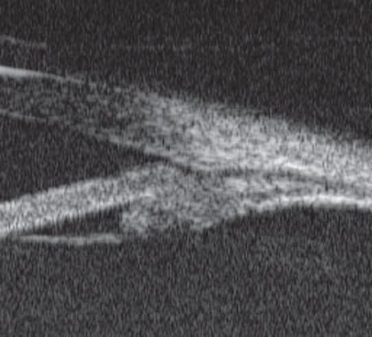
Plateau iris syndrome: The peripheral iris can bunch up in the anterior chamber angle and obstruct aqueous outflow without any element of pupillary block. The plateau iris syndrome is present when the angle closes and the IOP rises after dilation, despite a patent PI, and in the absence of phacomorphic glaucoma. UBM findings are characterized by an anteriorly rotated ciliary body. See Figure 9.12.1.
Slit-lamp examination: Specifically check for the presence of a patent PI and the critical signs listed previously.
Gonioscopy of both anterior chamber angles. Double hump sign on indentation gonioscopy is critical where the iris drapes over the lens more centrally near the pupil, then falls back over the zonular area, and is again forward and appositional in the angle.
If acute angle closure is present:
Treat medically. See 9.4, Acute Angle Closure Attack.
A laser PI is performed within 1 to 3 days if the angle closure attack can be broken medically. If the attack cannot be controlled, a laser or surgical PI may need to be done as an emergency procedure. Consider a laser iridoplasty to break an acute attack not responsive to medical treatment and PI.
One week after the laser PI, gonioscopy should be repeated prior to dilating the eye with a weak mydriatic (e.g., tropicamide 0.5%). If the IOP increases or a spontaneous angle closure episode occurs, plateau iris syndrome is diagnosed and can be treated with an iridoplasty. Second-line therapy includes chronic instillation of a weak miotic agent (e.g., pilocarpine 0.5% to 1% t.i.d. to q.i.d.).
If the patients IOP does respond to a laser PI (e.g., plateau iris configuration), then a prophylactic laser PI may be indicated in the contralateral eye within 1 to 2 weeks.
If acute angle closure is not present:
Laser PI to relieve any pupillary block component; also done to prove pupillary block is not the primary mechanism.
Check gonioscopy every 4 to 6 months to evaluate the angle.
Most do well with close observation alone. Perform iridoplasty if new PAS or further narrowing of the angle develops.
If the angle continues to develop new PAS or becomes narrower despite iridoplasty, then consider lens extraction. Can consider ECP at the time of phacoemulsification to shrink ciliary processes. If uncontrolled IOP, treat as CACG (see 9.5, Chronic Angle Closure Glaucoma).
Similar to performing a PI in acute angle closure. Reevaluate in 1 week, 1 month, and 3 months, and then yearly if no problems have developed.
Patients with a plateau iris configuration without previous acute angle closure are examined every 6 months.
Every examination should include IOP measurement and gonioscopy looking for PAS formation, narrowing angle recess, or increasing angle closure. The PI should be examined for patency. Dilation should cautiously be performed periodically (approximately every 2 years) to ensure that the PI remains adequate to prevent angle closure. If the patient needs more frequent dilation due to retinal pathology, consider cataract surgery to help open the angle.