section name header

Symptoms

Tearing, irritation, redness, and mucous discharge. May be asymptomatic.

Signs

(see Figure 6.2.1.)

Figure 6.2.1: Ectropion.

Rapuano9781975243722-ch006_f002.jpg

Critical

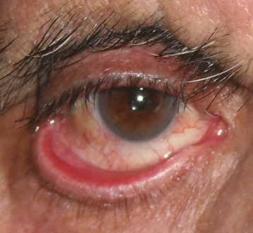
Outward turning of the eyelid margin, which can lead to significant corneal exposure. In extreme cases, an exposed cornea can develop erosion, ulceration, perforation and permanent vision loss.

Other

Superficial punctate keratopathy (SPK) from corneal exposure; conjunctival injection and thickening and eventual keratinization from chronic dryness. Eyelid scarring may be seen in cicatricial cases. Facial hemiparesis and lagophthalmos may be seen in paralytic cases.

Etiology

Workup

  1. History: Previous surgery, trauma, chemical burn, or CN VII palsy?

  2. External examination: Check orbicularis oculi function and assess horizontal eyelid laxity and punctal location. Look for an eyelid tumor, scarring, herniated orbital fat, etc. If there is concomitant CN VII palsy and CN VIII deficit (hearing loss), consider CT or MRI brain to rule out acoustic neuroma. Consider imaging for isolated CNVII palsy if it has not been worked up or etiology other than Bell palsy is suspected.

  3. Slit-lamp examination: Evaluate for exposure keratopathy and conjunctival inflammation.

Treatment

  1. Treat exposure keratopathy with lubricating agents. See 4.5, Exposure Keratopathy.

  2. Treat an inflamed, exposed eyelid margin with warm compresses and antibiotic ointment (e.g., bacitracin or erythromycin q.i.d.). A short course of combination antibiotic–steroid ointment (e.g., neomycin/polymyxin B/dexamethasone) may be helpful if close follow-up is ensured.

  3. Taping the eyelids into position may be a temporizing measure.

  4. Definitive treatment may require surgery. Surgical options should address the underlying etiology for ectropion:

    • Involutional ectropion: Lateral tarsal strip involves horizontally shortening the lower eyelid and resecuring to the periosteum. Other options include the modified-Bick, Bick, and lateral canthoplasty.

    • Punctal or medial ectropion: Medial spindle procedure, medial canthoplasty.

    • Paralytic ectropion: Lateral tarsal strip coupled with cheek lift. A temporary tarsorrhaphy can be used to protect ocular surface if nerve recovery is anticipated.

    • Mechanical ectropion: Address etiologic source and proceed with appropriate reconstruction of the eyelid.

    • Cicatricial ectropion: Release of cicatricial tethering, lengthening the lower eyelid with either anterior or posterior lamellar grafts. In some cases, cheek or midface lift can offer further vertical support of the lower eyelid. Medical management of any present underlying dermatologic condition is essential to prevent recurrence.

  5. Surgery may be delayed for 3 to 6 months in patients with idiopathic CN VII palsy (Bell palsy) because the ectropion may resolve spontaneously with nerve recovery (see 10.9, Isolated Seventh Nerve Palsy). Corneal exposure may make the repair more urgent.

Follow-Up

Patients with corneal or conjunctival exposure are examined as needed based on the severity of signs and symptoms. If the tissues are relatively healthy, follow-up is not urgent. Patients using topical steroids need to be followed up routinely for steroid-induced side effects.