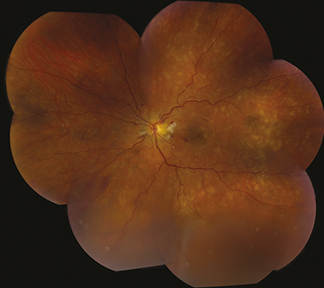
(See Figure 11.10.1.)
Figure 11.10.1: Chronic hypertensive retinopathy with arteriolar narrowing and arteriovenous nicking.

Generalized or localized retinal arteriolar narrowing, almost always bilateral.
Chronic HTN: Arteriovenous crossing changes (AV nicking), retinal arteriolar sclerosis (copper or silver wiring), CWSs, flame-shaped hemorrhages, arterial macroaneurysms, central or branch occlusion of an artery or vein. Rarely, neovascular complications can develop.
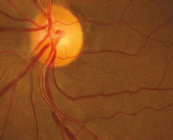
Acute (malignant) HTN or accelerated HTN: Hard exudates often in a macular star configuration, retinal edema, CWSs, flame-shaped hemorrhages, optic nerve head edema. Rarely serous RD or VH. Areas of focal chorioretinal atrophy (from previous choroidal infarcts [Elschnig spots]) are a sign of past episodes of acute HTN.
(See Figure 11.10.2.)
Diabetic retinopathy: Hemorrhages are usually dot-blot and microaneurysms are common; vessel attenuation is less common. See 11.12, Diabetic Retinopathy.
Collagen vascular disease: May show multiple CWSs, but few to no other fundus findings characteristic of HTN.
Radiation retinopathy: History of irradiation. Most commonly occurs within a few years, but can develop at any time.
CRVO or BRVO: Unilateral, multiple hemorrhages, venous dilation, and tortuosity. May be the result of HTN. See 11.8, Central Retinal Vein Occlusion or 11.9, Branch Retinal Vein Occlusion.
Complete ocular examination, particularly dilated fundus examination.
Refer patient for medical evaluation. The urgency depends on the blood pressure reading and whether the patient is symptomatic. A systolic blood pressure ≥180 mm Hg, a diastolic blood pressure ≥110 mm Hg or the presence of chest pain, difficulty breathing, headache, change in mental status, or blurred vision with optic disc swelling requires immediate medical attention including sending patient to the emergency department.
Patients may need workup for secondary causes of HTN such as those listed above.