(see Figure 6.7.1.)
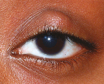
Visible or palpable, well-defined, subcutaneous nodule in the eyelid. In some cases, a nodule cannot be identified.
Blocked meibomian gland orifice, eyelid swelling and erythema, focal tenderness. Associated with blepharitis, seborrheic dermatitis, and acne rosacea. May also note lesion coming to a head or draining mucopurulent material.
Definitions
Chalazion: Focal, tender, or nontender granulomatous inflammation within the eyelid secondary to obstruction of a meibomian gland or gland of Zeis. Typically not due to infection. May be acute, subacute, or chronic.
Hordeolum (i.e., stye): Acute, tender infection; can be external (abscess of a gland of Zeis or Moll on eyelid margin) or internal (abscess of the meibomian gland). Usually involves Staphylococcus species and occasionally evolves into preseptal cellulitis.
Preseptal cellulitis: Eyelid and periocular erythema, edema, and warmth. See 6.9, Preseptal Cellulitis.
Forniceal foreign body: Eyelid swelling, particularly in soft contact lens wearers or those with a history of trauma. See 3.4, Corneal and Conjunctival Foreign Bodies.
Basal cell carcinoma: Slow-growing eyelid mass, typically painless, that may appear as pearly nodule or with ulceration. See 6.8, Malignant Tumors of the Eyelid.
Sebaceous carcinoma: Suspect in older patients with recurrent chalazia, eyelid thickening, madarosis, or chronic unilateral blepharitis. See 6.8, Malignant Tumors of the Eyelid.
Pyogenic granuloma: Benign, deep-red, pedunculated conjunctival lesion often associated with chalazia, hordeola, trauma, or surgery. May be excised or treated with a topical antibioticsteroid combination such as neomycin/polymyxin B/dexamethasone q.i.d. for no more than 1 to 2 weeks. Intraocular pressure must be monitored if topical steroids are used.
History: Previous ocular surgery or trauma? Previous chalazia or eyelid lesions?
External examination: Palpate involved eyelid for a nodule. Look for rosacea.
Slit-lamp examination: Evaluate meibomian glands for inspissation and evert the eyelid. Assess for madarosis, poliosis, and ulceration to rule out other etiologies.
Warm compresses for at least 10 minutes q.i.d. with gentle massage over the lesion.
Consider a short course of a topical antibiotic for hordeolum (e.g., bacitracin, tobramycin, or erythromycin ointment b.i.d. for 1 to 2 weeks) or a short course of topical antibiotic/steroid for chalazion (e.g., neomycin/polymyxin B/dexamethasone ointment b.i.d. for 1 to 2 weeks). Consider chronic low-dose doxycycline 20 to 50 mg p.o. daily to b.i.d. for its anti-inflammatory properties (e.g., for multiple or recurrent chalazia and/or ocular rosacea).
If a hordeolum worsens, consider incision and drainage and management as per preseptal cellulitis (see 6.9, Preseptal Cellulitis).
If the chalazion fails to resolve after 3 to 4 weeks of medical therapy and the patient desires surgical intervention, incision and curettage may be performed. Alternatively, an intralesional steroid injection may be performed. Options include 0.2 to 1.0 mL of dexamethasone sodium phosphate 4 mg/mL or betamethasone sodium phosphate/betamethasone acetate 6 mg/mL that may or may not be mixed with 2% lidocaine and epinephrine. Total dosage depends on the lesion size. Injection may be repeated in 1 to 2 weeks for persistent lesions. It is recommended that recurrent or atypical chalazia, be sent for pathology upon removal.