Tenderness, redness, warmth, and swelling of the eyelid and periorbital area. Often there is a history of local skin abrasions, penetrating injury/trauma, hordeolum, or insect bites. Can be associated with sinusitis, which can indicate increased risk for orbital abscess formation and postseptal inflammation. May complain of fever or chills.
(see Figures 6.9.1 and 6.9.2.)
Figure 6.9.1: Preseptal cellulitis.
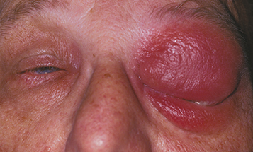
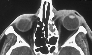
Figure 6.9.2: CT of preseptal cellulitis.
Critical
Eyelid erythema, tense edema, warmth, and tenderness. No proptosis, no optic neuropathy, no extraocular motility restriction, usually little to no conjunctival injection, and no pain with eye movement (unlike orbital cellulitis). The patient may not be able to open the eye because of eyelid edema. Visual changes such as blurred vision or monocular diplopia attributed to swollen eyelids.
Other
Tightness of eyelid skin or fluctuant eyelid edema. The eye itself may be slightly injected but is relatively uninvolved.
Adjacent infection (e.g., hordeolum, dacryocystitis, or sinusitis).
Trauma (e.g., puncture wound, laceration, insect bite).
Organisms
S. aureus and Streptococcus are most common, but H. influenzae should be considered in nonimmunized children. Suspect anaerobes if foul-smelling discharge or necrosis is present, or if there is a history of an animal or human bite. Consider a viral cause if preseptal cellulitis is associated with a vesicular skin rash (e.g., herpes simplex or varicella zoster).
Depends upon disease severity. If hospital admission is needed or there is concern for orbital progression, then daily follow up is necessary until clear and consistent improvement is demonstrated, then every 2 to 7 days until the condition has totally resolved. If preseptal cellulitis progresses despite antibiotic therapy, the patient is admitted to the hospital and a repeat (or initial) orbital CT scan is obtained. For patients already on p.o. antibiotics, switch to i.v. antibiotics (see 7.3.1, Orbital Cellulitis).