section name header

Symptoms

Irritation, redness, and decreased vision (due to associated astigmatism or visual axis obscuration); may be asymptomatic

Signs

Critical

One of the following, almost always located at the 3-o’clock or 9-o’clock perilimbal position.
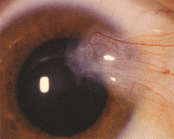
Other

Either lesion may be highly vascularized and injected or may be associated with SPK or delle (thinning of the adjacent cornea secondary to drying). An iron line (Stocker line) may be seen in the corneal epithelium just beyond the leading edge of a pterygium, indicative of chronicity.

Differential Diagnosis

NOTE

Atypical pterygia (e.g., calcifications, leukoplakia, atypical elevation or location, irregular feeder vessels, and rapid growth) require imaging or biopsy to rule out ocular surface squamous neoplasia or melanoma.

Etiology

Elastotic degeneration of deep conjunctival layers resulting in fibrovascular tissue proliferation. Related to sunlight/ultraviolet exposure, aging, environmental insults (e.g., dust, wind), and chronic irritation. More common in individuals from equatorial regions.

Workup

Slit-lamp examination to identify the lesion and evaluate the adjacent corneal integrity and thickness. Check for corneal astigmatism, which is often irregular but may be oriented with the rule.

Treatment

  1. Protect eyes from sun, dust, and wind (e.g., ultraviolet-blocking sunglasses or goggles if appropriate).

  2. Lubrication with artificial tears, preferably preservative free, four to eight times per day to reduce ocular irritation.

  3. For an inflamed pterygium or pinguecula:

    • Mild: Artificial tears q.i.d.

    • Moderate to severe: A mild topical steroid (e.g., fluorometholone 0.1%, fluorometholone acetate 0.1%, or loteprednol 0.2% to 0.5% q.i.d.), a nonsteroidal anti-inflammatory drop (e.g., ketorolac 0.4% to 0.5% q.i.d.), or a topical antihistamine ± mast cell stabilizer (e.g., bepotastine, ketotifen, olopatadine, cetirizine) may be used to decrease symptoms. Long-term corticosteroid therapy is discouraged.

  4. If a delle is present, then apply artificial tear ointment q2h. See 4.24, Delle.

  5. Surgical removal is indicated when:

    • The pterygium threatens the visual axis, induces significant astigmatism, or restricts ocular motility.

    • The patient is experiencing excessive irritation not relieved by the aforementioned treatment.

    • The lesion is interfering with contact lens wear.

    • The lesion is visually apparent and causing cosmetic concerns.

    • Consider removal prior to cataract or refractive surgery.

NOTE

Pterygia can recur after surgical excision. Bare sclera dissection with a conjunctival autograft or amniotic membrane graft reduces the recurrence rate. Intraoperative application of an antimetabolite (e.g., mitomycin C) also reduces recurrence. Antimetabolites are more commonly reserved for excision of recurrent pterygia, as these medications are associated with an increased risk of corneoscleral thinning or necrosis. 

Follow-Up

  1. Asymptomatic, stable patients may be checked every 1 to 2 years.

  2. Pterygia should be measured periodically (every 3 to 12 months, initially) to determine the rate at which they are growing toward the visual axis.

  3. If treating with a topical steroid, check after a few weeks to monitor inflammation and IOP. Taper and discontinue the steroid drop over several weeks once the inflammation has abated.