- Left ventricular end-diastolic pressure (LVEDP) is defined as the pressure in the ventricles at the end of diastole. The measurement is used in clinical practice as a means to approximate the left ventricular end-diastolic volume (LVEDV) or preload (LVEDP LVEDV).
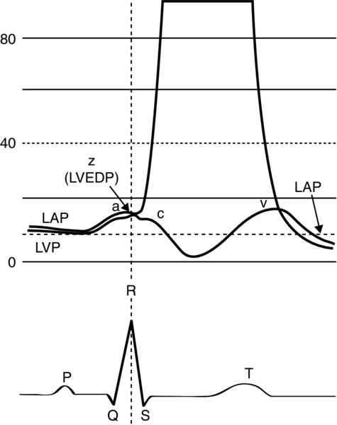
LVEDP can be ideally determined by left ventricular catheterization in the catheterization laboratory. It is measured at the Z-point, the point on the left ventricular pressure trace where the slope of the ventricular pressure upstroke changes (approximately 50 ms after the EKG Q wave, generally coincides with the R wave) (1).
FIGURE 1. The LVEDP is measured at the Z point.
It is the point on the ventricular pressure upstroke where the slope changes. It occurs approximately 50 ms after the Q wave usually corresponds with the R wave.
- In most clinical situations, LVEDP is determined by Swan Ganz catheterization, by floating a balloon-tipped catheter into the pulmonary artery. Although it does not enter the left ventricle, when the balloon is inflated (wedged), it isolates the catheter tip brings it in continuity with the blood column in the left atrium LV during end diastole.
- Measurements: The pulmonary artery wedge pressure (PAWP) is equal to the LVEDP, given that there is no mitral valve pathology the pulmonary artery catheter is in the West zone 3 of the lung (where pulmonary artery pressure > pulmonary venous pressure > pulmonary alveolar pressure).
- Waveforms: Left atrial tracings are similar to central venous pressure (CVP) tracing from the right atrium, with “a”, “c”, “v” waves.
- During an ideal ventricular diastole, the pressures in all cardiac chambers equilibrate: CVP = RVEDP = PADP = PAWP = LAP = LVEDP; where CVP is central venous pressure, RVEDP is right ventricular end diastolic pressure, PADP is pulmonary artery diastolic pressure, PAWP is pulmonary artery wedge pressure, LAP is left atrial pressure.
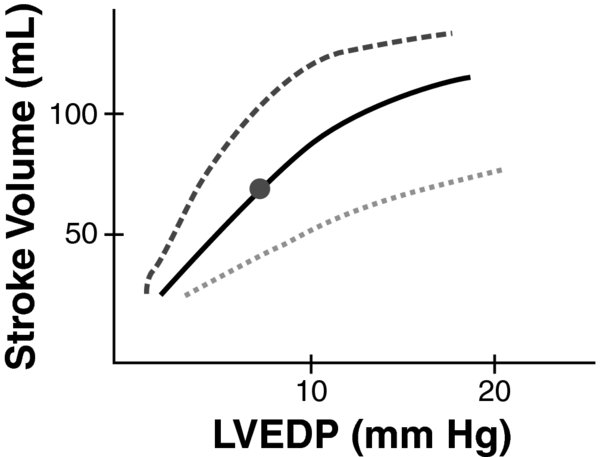
- Frank Starling principle states that the force of cardiac contraction is directly proportional to end-diastolic muscle fiber length at any given level of intrinsic contractility or inotropy.
- Increasing the venous return of the left ventricle increases the volume preload thereby LVEDP (increases stroke volume).
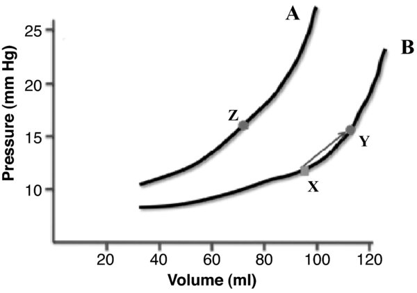
- Flat portion of diastolic filling: A significant increase in filling volume or preload results in a small increase in filling pressure.
- Steep portion of diastolic filling: In comparison, there is a significant increase in filling pressure with the same volume towards the end of diastole.
Left-shifting describes abnormal decreases in compliance (e.g., sepsis, shock, myocardial ischemia, or fibrotic chambers). Additionally, there is a paradoxical increase in filling pressures with a decrease in filling volume.
FIGURE 2. The ventricular diastolic pressure-volume relationship forms a curvilinear line the slope reflects wall compliance.
Compliance is dynamic changes with chamber volume, thus affecting the ability of using the LVEDP to approximate the LVEDV. Curve A represents normal compliance. Curve B represents a right shift or increased compliance, as can occur with dilated cardiomyopathy, where a change in the volume (x$\rightarrow$y) results in a smaller increase in pressure.
FIGURE 3. Frank Starling curve.
Changes in venous return correlate to changes in LVEDP/LVEDV that affect the stroke volume (SV). The inotropic state affects the stroke volume at a given preload (dashes–higher inotropy, dots–decreased inotropy).
- PAWP is taken at the pulmonary artery (distal tip of pulmonary artery catheter) with the balloon inflated occluding the branch of the pulmonary artery.
- The ability of proximal pressures (CVP, PAWP, etc.) to accurately reflect the LVEDP is dependent on unobstructed continuity of blood flow during diastole. Any anatomical or physiological condition that impairs this will result in inaccurate downstream pressure estimation.
- Conditions in which PAWP underestimates LVEDP:
- Decreased left ventricular compliance (e.g., MI, LVH). The mean left atrial pressure (LAP) is less than LVEDP, there is an increased end-diastolic “a” wave.
- Aortic regurgitation (AR): The mitral valve closes before the end of diastole due to run-off from the aorta (LAP LVEDP).
- Pulmonary regurgitation: Bidirectional run-off of the pulmonary artery flow (PADP LVEDP).
- Decreased pulmonary vascular bed
- Conditions in which PAWP overestimates LVEDP:
- Positive end-expiratory pressure (PEEP): The pulmonary artery catheter may become lodged in lung zone 1 or 2. Pulmonary venous pressure readings are actually lower than airway pressure, leading to a falsely elevated PAWP.
- Pulmonary hypertension: Increases in the pulmonary vascular resistance will record a higher PADP which does not reflect left ventricular pressures (PADP > mPAWP).
- Mitral stenosis: There is obstruction to the flow of blood through the mitral valve, which results in a higher mean LAP thereby overestimation of the LVEDP.
- Mitral regurgitation: A retrograde systolic “v” wave or regurgitant systolic flow raises the mean atrial pressure.
- Increased compliance. In dilated cardiomyopathy, the left ventricle is very dilated without any appreciable increase in ventricular wall thickness. This will result in increased compliance even though the LVEDV may be very high, the corresponding LVEDP elevation might not be significant.
- Elevated LVEDP is an independent risk factor of mortality in cardiac surgery, independent of left ventricular ejection fraction (2,3).
- Atrial kick: Normally provides 20% contribution to the LVEDV. In LVH, this may increase to 50% the “a” wave may be prominent provide a better estimate of LVEDP than PAWP (4).
- Mitral valve: Both stenosis regurgitation can overestimate the LVEDP.
- Perioperative presentation of cardiogenic versus hypovolemic shock can be differentiated by using the LVEDP (CVP/PAWP) as a surrogate marker of preload. A low CVP, or PAWP, is consistent with hypovolemia, a high CVP/PAWP would indicate cardiogenic shock (e.g., MI, CHF). This can be clinically important in deciding when to give fluids versus other interventions.
- The goal of all-fluid resuscitation is to increase preload “recruit” stroke volume to increase end-organ perfusion, as per the Starling mechanism.
- Markers of LVEDP (CVP/PAWP) can be trended over time with improvement or worsening of cardiac output.
- Clinically, signs of increased cardiac output changes would relate to increased urine output, decreased capillary refill time, improvement in mental status blood pressure.
- Other more sophisticated monitors which can measure cardiac output changes with preload changes are esophageal Doppler monitor, echocardiography, systolic pressure variations.
- Myocardial compliance: PAWP measurements are dependent on myocardial compliance. Multiple studies on ICU patients have shown the failure of PAWP in acute illness to correlate with LVEDV (5).
- Clinical use: CVP or PAWP alone is rarely used to guide therapy. In situations of shock, after the patient has been given IV fluids to raise the CVP to around 12 mm Hg (adequate preload), without improvement in blood pressure or cardiac output, other methods of assessment such as bedside echocardiography may be used for cardiac assessment.
- The American Society of Anesthesiologist Practice Guidelines recommends PAC for high-risk surgical patients only (6).
- Suggested clinical indications for monitoring LVEDV LVEDP are severe sepsis trauma, high-risk cardiac surgery, pulmonary hypertension, abdominal compartment syndrome, therapy with PEEP.


