▶Chronic paronychia is common among children who are thumb-suckers, nail-biters, or nail pickers.
▶Candida species usually are responsible.
▶Non-tender erythematous swelling of the skin surrounding the nail (Figure 35C.1).
▶Loss of the cuticle in affected digits is common.
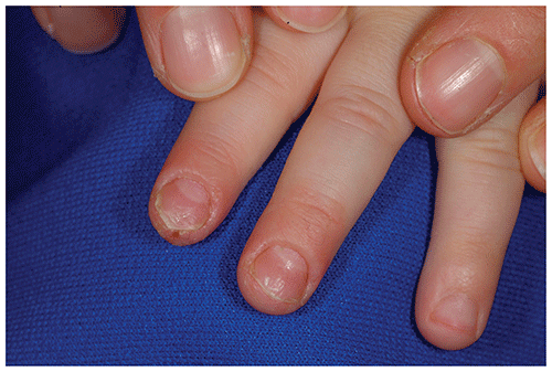
▶Associated nail dystrophy may be present, most often presenting as pits or transverse ridges; yellow debris and separation of the nail plate from the nail bed may be present (Figure 35C.2).
Figure 35C.1. Chronic Paronychia Caused by Candida. There is Periungual Erythema and Loss of the Cuticle.
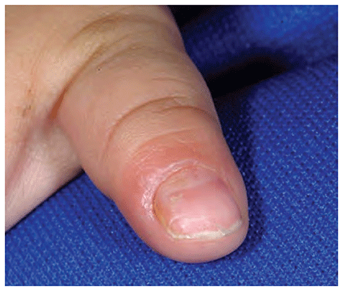
Figure 35C.2. Chronic Paronychia Caused by Candida. In Addition to Periungual Erythema and Cuticle Loss, Note the Dystrophy of the Nail Plates.
Look-alikes
| Disorder | Differentiating Features |
|---|
| Acute paronychia | Acute onset with painful swelling, erythema, and purulent exudate. Culture often positive for Staphylococcus aureus.
|
| Tinea unguium (onychomycosis) | Skin surrounding nail usually typical. When toenails infected, usually evidence of associated tinea pedis (eg, fissuring, scaling, maceration between the digits). Nail usually thickened and white to yellow, with debris under the nail plate.
|
| Herpetic whitlow | |
| Blistering dactylitis | Acute onset of a bulla affecting distal portion of a digit. Located on digit but not limited to proximal or lateral nail folds. Culture of blister fluid often yields group A β-hemolytic streptococci or S aureus.
|
| Chronic mucocutaneous candidiasis | Multiple digits involved. Associated recurrent candidal mucocutaneous infections. May have associated immunologic or endocrinologic variations.
|
▶The diagnosis is usually made clinically.
▶If uncertainty exists, the diagnosis may be confirmed by microscopic observation of budding yeast or pseudohyphae in a potassium hydroxide preparation performed on scrapings of the affected area, or by fungal culture (see Figure 35A.2).
▶Treatment is complicated by predisposing behaviors that cause trauma or moisture (eg, nail-biting, thumb-sucking). When possible, these factors should be addressed.
▶Topical nystatin or an imidazole antifungal agent applied during the day and under occlusion at night (care must be taken to secure occlusive dressings to prevent aspiration by young patients).
▶Oral fluconazole (or other appropriate agent) may be indicated in severe or persistent cases.
▶Provide positive feedback for substitute behaviors.
▶Use of noxious agents to control thumb-sucking should be considered second-line therapy.
▶Involvement of multiple nails along with recurrent mucosal or skin infection may indicate the presence of chronic mucocutaneous candidiasis.