▶Uncommon infection that may be observed at birth or within a week of delivery.
▶Candida albicans (occasionally a non-albicans Candida species) is acquired during passage through a colonized birth canal or by ascending infection before delivery.
▶Eruption presents as papules and pustules superimposed on an erythematous base (Figure 35D.1) or as diffuse erythema with scaling (Figure 35D.2).
▶Presence of papules and pustules on the palms and soles is characteristic.
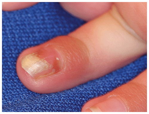
▶Nail dystrophy with yellow discoloration may be present (Figure 35D.3).
▶Any body surface may be involved.
▶Most full-term neonates experience a benign course, but very low-birth-weight neonates are at higher risk for invasive disease.
Figure 35D.1. Congenital Candidiasis is Characterized by Erythematous Papules, Pustules, and Scaling.
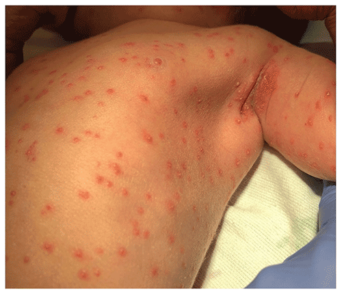
Figure 35D.2. Congenital Candidiasis in a Preterm Neonate with Skin of Color. Note Diffuse Faint Erythema with Marked Scaling and Hyperkeratosis. Such Preterm Neonates Have a Higher Risk for Systemic Dissemination of the Fungal Infection. Reproduced from Jani S, Ariss R, Chawla S. A Preterm Infant with a Characteristic Erythematous and Scaly Rash after Birth. Neoreviews. 2020;21(7):e495–e498.
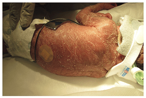
Figure 35D.3. Nail Dystrophy in Congenital Candidiasis. There is Yellow Discoloration, Onycholysis, and Mild Surrounding Paronychia (Ie, Inflammation of Proximal and Lateral Nail Folds).
Look-alikes
In each of the disorders listed herein, a potassium hydroxide preparation would fail to demonstrate the pseudohyphae and spores that would be observed in candidal infection.
| Disorder | Differentiating Features |
|---|
| Erythema toxicum | Discrete, blotchy, erythematous macules or patches, each with a central papule, vesicle, or pustule. Typically not present at birth. Vesicular/pustular fluid contains eosinophils.
|
| Transient neonatal pustular melanosis | Pustules (without erythema) or ruptured pustules, which appear as small freckle-like hyperpigmented macules surrounded by a rim of scale. Pustular fluid contains neutrophils.
|
| Miliaria rubra | |
| Neonatal cephalic pustulosis (neonatal acne) | |
| Staphylococcal folliculitis | |
| Scabies | Occurs rarely during first month after birth. Generalized eruption; may have vesicles but usually will be accompanied by erythematous papules or nodules and burrows. Mineral oil preparation of scrapings of lesions will reveal mites, eggs, or fecal material.
|
| Neonatal herpes simplex virus infection | Typically clustered vesicles on an erythematous base (although solitary vesicles occasionally occur). Lesions concentrated on head, particularly at sites of trauma (eg, those caused by a scalp electrode). Neonates may have signs of sepsis (in disseminated disease) or seizures or coma (in central nervous system disease). Direct fluorescent examination, viral culture, or polymerase chain reaction (skin lesions, cerebrospinal fluid) will confirm diagnosis.
|
| Infantile acropustulosis | Usually begins in first months (not in first days) after birth. Vesicles or pustules that are limited to the hands and feet, including the palms and soles, wrists, and ankles. Eruption lasts for 5 to 10 days and reappears every 2 to 4 weeks.
|
| Incontinentia pigmenti | Vesicles on an erythematous base appear at birth or within the first 2 weeks after birth. Lesions arranged in a linear fashion on the extremities or in a swirled pattern on the trunk (along the Blaschko lines).
|
| Eosinophilic pustular folliculitis | Papules and pustules typically located on the scalp. Exhibits a chronic, intermittent course. Severe pruritus is usually present.
|
▶The diagnosis is made by performing a potassium hydroxide preparation on skin scrapings (see Figure 35A.2) and fungal culture of specimens from the skin, placenta, or umbilical cord.
▶Topical application of an antifungal agent such as nystatin or clotrimazole.
▶In neonates with diffuse skin involvement, oral fluconazole may accelerate resolution.
▶In the rare patient who has evidence of (or risk factors for) systemic infection, complete evaluation for this possibility and parenteral antifungal therapy are required.
▶The prognosis for term neonates with cutaneous congenital candidiasis is excellent.
▶Low-birth-weight neonates (or those born to mothers with a history of an indwelling device [eg, cervical cerclage, intrauterine device]) are at increased risk for systemic involvement (eg, infection of blood, lungs, central nervous system, urinary tract).
▶Consider consultation by a dermatologist when the diagnosis is in doubt or lesions fail to respond to appropriate therapy.
▶When systemic disease is likely, immediate consultation with a pediatric infectious disease specialist is warranted.