▶Paronychia (ie, inflammation of the periungual nail folds) occurs when the cuticle becomes disrupted by maceration or injury and pathogens enter the space.
▶Paronychia occurs more frequently in individuals who often have their hands in water or in children with a habit of finger sucking or nail-biting. In addition, trauma to the periungual folds is another risk factor for its development.
▶Staphylococcus aureus is the agent primarily responsible for acute paronychia. Paronychia with a green discoloration may indicate the presence of Pseudomonas. Candida species most often result in chronic paronychia (Chapter 35C).
▶Acute paronychia typically lasts less than 6 weeks, while chronic paronychia (Chapter 35C) refers to paronychia lasting more than 6 weeks.
▶Some medications, including oral retinoids, epidermal growth factor receptor inhibitors, chemotherapy agents, and antiretrovirals, can increase the risk for paronychia.
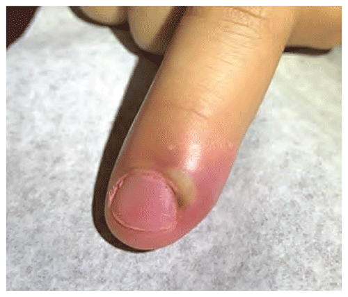
▶Periungual folds show erythema, swelling, and tenderness (Figures 23.1 and 23.2).
▶Purulent drainage is commonly present.
▶Although it is most common to have only 1 nail involved, multiple nail involvement can be seen, especially with medication-induced paronychia.
▶Patients with chronic infections may have dermatitis of the surrounding areas (ie, fingers, hands).
▶Paronychia of the toenails can be seen with ingrown toenails, retronychia (inward growth of the nail plate at the proximal nail fold), or congenital malalignment of the great toenails.
Figure 23.1. Acute Paronychia with Inflammation, Pustule Formation, and Crusting of the Periungual Fold.
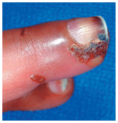
Figure 23.2. Acute Paronychia with Loculated Pus and Surrounding Erythema.
Look-alikes
| Disorder | Differentiating Features |
|---|
| Chronic paronychia | Problem long-standing (not acute). Usually without symptoms. Swelling and erythema of proximal and lateral nail folds with loss of cuticle; purulent drainage absent. May have associated nail dystrophy (eg, ridging, pitting).
|
| Felon | A bacterial infection of the digital pulp space of the finger (also typically caused by Staphylococcus aureus, less often by streptococci). Characterized by severe pain, swelling, erythema in the pad of the distal fingertip.
|
| Herpes simplex virus infection (ie, herpetic whitlow) | Usually presents as discrete, deep-seated, often clustered vesicles with surrounding erythema. Usually very painful. Regional lymphadenopathy may be present. Recurrent lesions can be associated with prodromal symptoms. Viral culture or polymerase chain reaction testing will confirm herpes simplex virus.
|
| Blistering distal dactylitis | Usually presents as a tender, deep-seated blister on the volar surface of the distal finger pad. Bacterial culture reveals group A β-hemolytic streptococci (or occasionally S aureus).
|
| Psoriasis | |
| Trauma | |
▶The condition is often diagnosed based on the clinical features.
▶Gram stain and culture of the drainage can identify the organisms.
▶Bacterial culture usually reveals S aureus, although group A β-hemolytic streptococci and rarely other organisms may be etiologic.
▶An oral antistaphylococcal antibiotic (eg, cephalexin) usually is effective and also provides coverage against group A β-hemolytic streptococci. Failure of treatment may indicate presence of methicillin-resistant S aureus, and changing therapy to clindamycin, doxycycline (in children >8 years), trimethoprim-sulfamethoxazole, or another appropriate agent (based on bacterial culture and sensitivity testing) should be considered.
▶Topical antibiotic ointment (eg, mupirocin, retapamulin) may be used in mild cases, but the condition often requires systemic therapy.
▶Warm soaks may hasten resolution.
▶Drainage and culture of purulent pockets occasionally is necessary.
▶Preventive strategies include the following:
■Institute drying measures, including minimizing exposure to water and wearing gloves for wet work.
■Avoid trauma, when feasible.
▶Acute paronychia usually resolves completely without long-term sequelae.
▶Mechanical factors or exposures may result in recurrence.
▶Permanent nail ridging or dystrophy may result with severe infections.
▶Consider referral to a dermatologist or infectious disease specialist for patients who have severe or extensive involvement or in whom standard treatment does not work.