▶Present at birth.
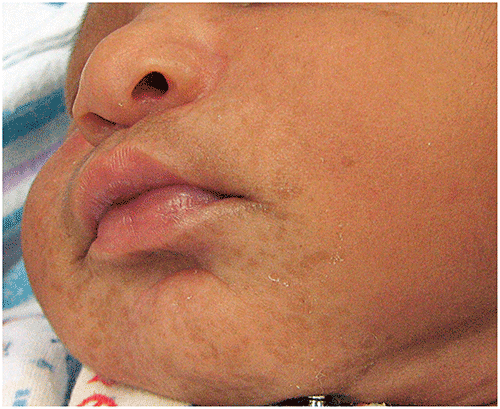
▶May present as pustules without surrounding erythema (Figure 108.1) or ruptured pustules that appear as small (several millimeters) hyperpigmented macules, often with a rim of surrounding scale (Figures 108.2 and 108.3).
▶Lesions may occur at any location, but the forehead, chin, neck, and trunk most often are affected; the palms and soles occasionally are involved.
▶Pustules resolve in several days; hyperpigmented macules resolve in 3 to 4 months.
Figure 108.1. Pustules Without Surrounding Erythema May be Observed in Neonates Who Have Transient Neonatal Pustular Melanosis.
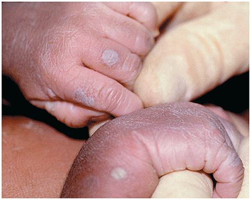
Figure 108.2. Hyperpigmented Macules, Some with a Rim of Scale, are Seen in Transient Neonatal Pustular Melanosis.
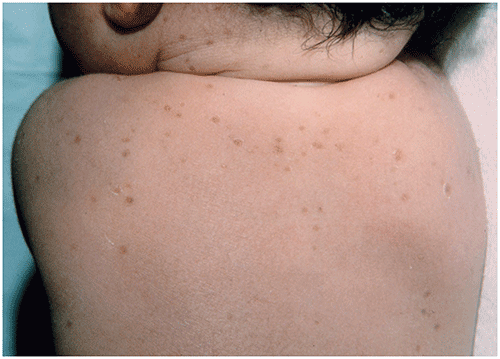
Figure 108.3. Transient Neonatal Pustular Melanosis. Hyperpigmented Macules on the Chin. Note the Collarettes of Scale Present in Some Areas.
Look-alikes
The differential diagnosis includes miliaria, staphylococcal folliculitis, and congenital candidiasis (although these disorders produce lesions that exhibit erythema); infantile acropustulosis (lesions typically are pruritic and limited to the hands and feet); and congenital herpes simplex virus infection (lesions often are clustered, include erythema, and lack hyperpigmentation).
|
|
▶The diagnosis is made clinically. If uncertainty exists, a Wright stain of vesicular fluid will reveal a predominance of neutrophils. Gram stain reveals no organisms.
▶Gram stain and bacterial culture of pustular fluid will exclude staphylococcal folliculitis or pustulosis, the conditions with which transient neonatal pustular melanosis is most often confused. A potassium hydroxide preparation will exclude congenital cutaneous candidiasis.
▶Resolves spontaneously; does not recur.
▶Hyperpigmented macules may take 3 to 6 months to resolve.
▶In view of the age of onset and typical clinical appearance, referral rarely is necessary.