▶Inflammatory reaction of the skin caused by physical contact with an irritating substance.
▶Occurs in any individual exposed to a sufficient amount of the offending agent.
▶Can be exacerbated by local physical factors (eg, diapering) and individual susceptibility (eg, diminished skin barrier function, as in atopic dermatitis).
▶Common forms of irritant contact dermatitis include:
■Irritant diaper dermatitis: most common presentation of irritant contact dermatitis in infancy (occurs in up to 20% of all infants); caused by friction, moisture, maceration, and occlusion. (See also Chapter 101, Diaper Dermatitis.)
■Dry skin dermatitis (ie, asteatotic eczema or winter eczema): caused by low relative humidity and aggravated by soaps, excessive bathing or washing, and alcohol-containing lotions and solutions such as hand sanitizers.
■Lip-licking dermatitis and thumb-sucking dermatitis: caused by wetting the skin frequently with saliva.
■Berries, tomatoes, and citrus fruits are common causes of irritant contact dermatitis of the perioral skin in infants and toddlers.
▶Irritant contact dermatitis is more common in children with underlying atopic dermatitis, presumably related to their diminished skin barrier function.
Irritant Diaper Dermatitis
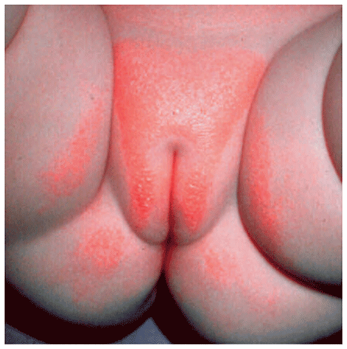
▶Affects convex surfaces of buttocks, upper inner thighs (Figure 5.1).
▶Characteristically spares creases and folds.
▶Sharply marginated erythema that becomes more deeply red with a “glazed” appearance.
▶May have dermatitis along the diaper margins (“tidemark dermatitis”).
Figure 5.1. Irritant Diaper Dermatitis. Erythematous Patches Sparing the Skinfolds.
Asteatotic Eczema
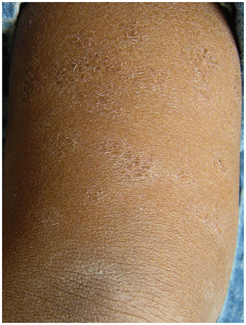
▶Dry, rough skin with white, sometimes rectangular, scaling and variable erythema (Figure 5.2).
▶Often associated with keratosis pilaris.
Figure 5.2. Asteatotic Eczema is Characterized by Dry, Rough Skin with White Rectangular Scaling.
Look-alikes (See also Chapter 101, Diaper Dermatitis.)
| Disorder | Differentiating Features |
|---|
| Irritant Diaper Dermatitis |
|---|
| Candidiasis | Erythematous patches that involve convexities and inguinal creases. Satellite papules and pustules. Scaling at margins of involved areas.
|
| Seborrheic dermatitis | Salmon-pink patches with greasy scale that involve the convexities and inguinal creases. Involvement of scalp, face, postauricular creases, or chest may be present.
|
| Bullous impetigo | Flaccid blisters filled with clear or purulent fluid. Blisters rupture rapidly, leaving round or oval crusted erosions with a rim of scale (collarettes).
|
| Folliculitis | |
| Intertrigo | |
| Jacquet erosive dermatitis | |
| Perianal bacterial dermatitis | |
| Langerhans cell histiocytosis | Erythematous to brown papules and plaques, often with erosions; typically involving skin creases. Erosions, petechiae, and hemorrhagic papules often present at examination, classically involving the scalp. Seborrheic dermatitis-like eruption may be present and is poorly responsive to typical treatments. Lymphadenopathy is often present; associated hepatosplenomegaly may be present.
|
| Nutritional or metabolic disorders (eg, zinc deficiency, cystic fibrosis, biot-independent multiple carboxylase deficiency) | Sharply marginated plaques with shiny, peeling scale. Eruption often located in a periorificial (eg, perioral, perianal) and acral distribution. Secondary bacterial and/or candidal infections are common. Unresponsive to typical therapies. May have associated diarrhea, alopecia, or failure to thrive.
|
| Asteatotic Eczema |
|---|
| Nummular eczema | Well-defined, coin-shaped, scaly, pink plaques; may appear more pigmented in darker skin types. History of atopic dermatitis often present.
|
| Allergic contact dermatitis | Rash located at site of antigen exposure. Characteristic (eg, linear) or geographic patterns may be present. History of exposure to antigen.
|
| Atopic dermatitis | Family history of atopic disorders (atopic dermatitis, allergic rhinitis, and/or asthma) usually present. Eruption located in typical areas (eg, antecubital and popliteal fossae in a toddler or school-aged child).
|
▶Irritant diaper dermatitis: the diagnosis is made clinically based on the typical appearance and distribution of the eruption.
▶Asteatotic eczema: the diagnosis is made clinically based on the appearance of lesions; a history of using harsh, drying soaps; and its occurrence during times of low environmental humidity. Concomitant keratosis pilaris may be present.
General Principles for All Types of Irritant Contact Dermatitis
▶Decrease or eliminate contact with the irritant when feasible.
▶Restore skin barrier function with emollients.
▶Decrease inflammation with anti-inflammatory measures (typically topical corticosteroids).
▶Treat secondary infection, if present.
Irritant Diaper Dermatitis
▶Remove or minimize contactants (urine and feces) from skin surface by changing diapers frequently.
▶Decrease skin maceration by considering superabsorbent disposable diapers.
▶Specific measures include:
■Gently cleanse with tap water or cotton balls or pads soaked in mineral oil; avoid scrubbing skin or using soaps; if diaper wipes are used, ensure that they are free of fragrance and allergens to decrease risk of secondary allergic contact dermatitis (ACD).
■Use emollient ointments or barrier creams to protect skin surface (eg, zinc oxide ointment or paste, petrolatum).
■Selectively use a low-potency topical corticosteroid (eg, 1% to 2.5% hydrocortisone ointment or cream) for active inflammation in a thin layer before application of emollient or barrier ointments.
■Selectively use anti-candidal creams (eg, nystatin, an imidazole antifungal agent) if there is evidence of secondary candidiasis; apply a thin layer before application of emollient or barrier ointments.
■Do not use combination topical therapies that contain potent topical steroids (eg, betamethasone-clotrimazole).
Asteatotic Eczema
▶Restore skin barrier function and diminish trans-epidermal water loss.
▶Apply emollients and moisturizers 1 to 2 times a day and after water exposure.
▶Eliminate use of soaps and alcohol-based lotions and solutions as much as possible; when improved, reintroduce soaps that are superfatted or contain emollient ingredients.
▶Selectively use low- or mid-potency topical corticosteroids for more severe cases.
▶The prognosis for irritant dermatitis is excellent, provided appropriate treatment is instituted.
▶Recurrences are common.
▶Uncertainty about the diagnosis.
▶Failure for therapy to work.